. 2024 Sep 16;25(18):9974.
doi: 10.3390/ijms25189974.

Identification of Tumor Suppressive miR-144-5p Targets: FAM111B Expression Accelerates the Malignant Phenotypes of Lung Adenocarcinoma

Affiliations

Identification of Tumor Suppressive miR-144-5p Targets: FAM111B Expression Accelerates the Malignant Phenotypes of Lung Adenocarcinoma

Yuya Tomioka et al. Int J Mol Sci. .

Abstract

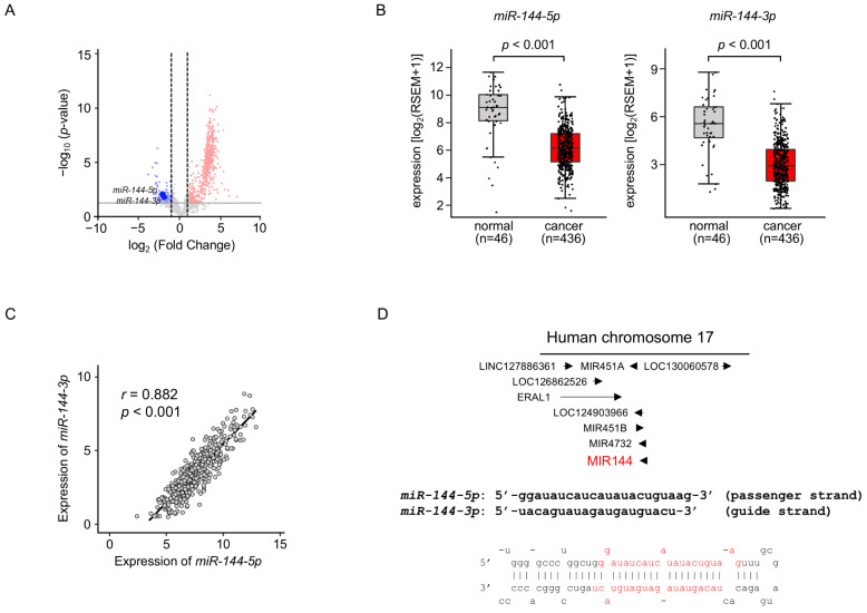
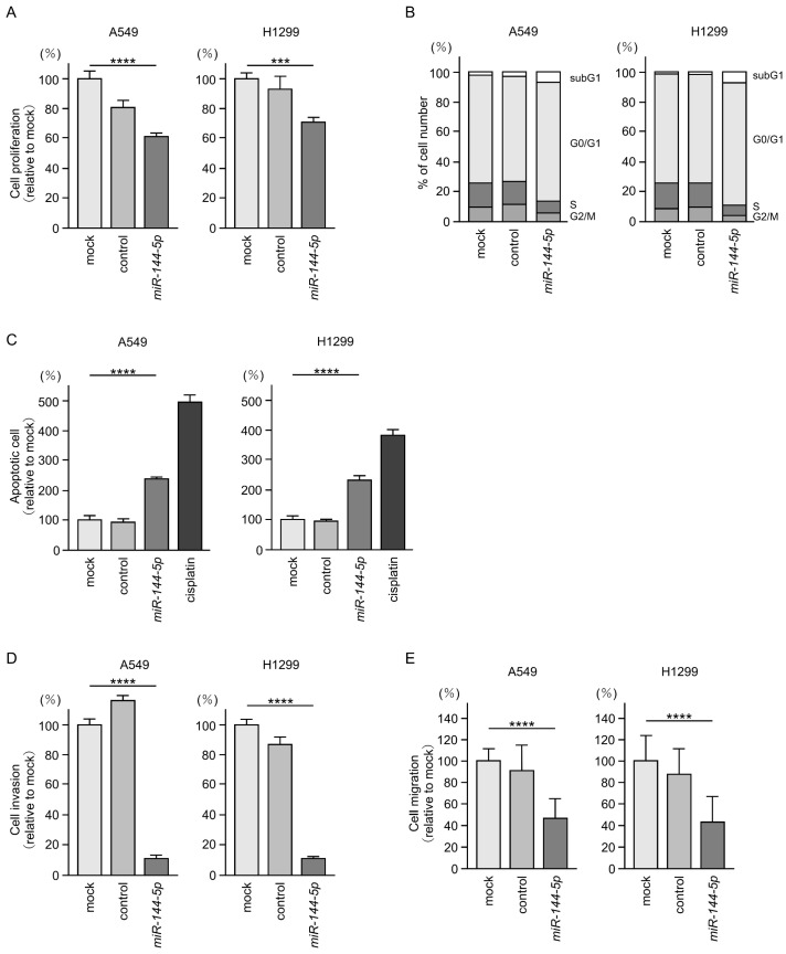
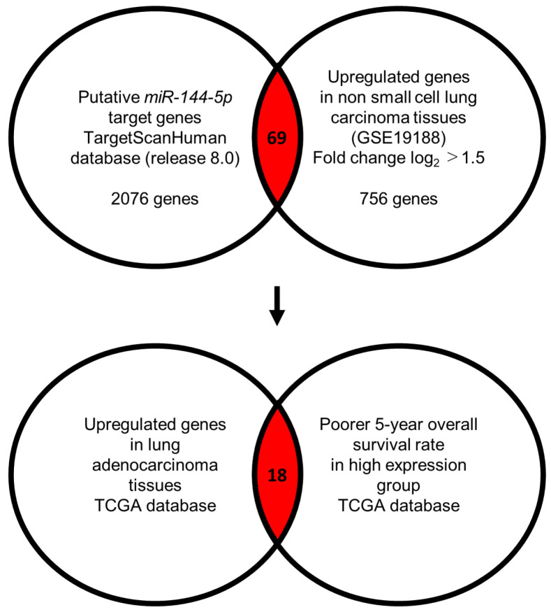
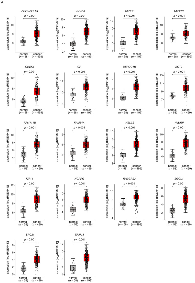
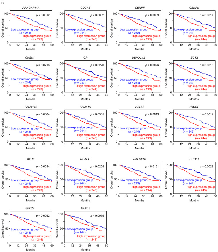
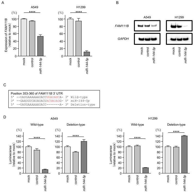
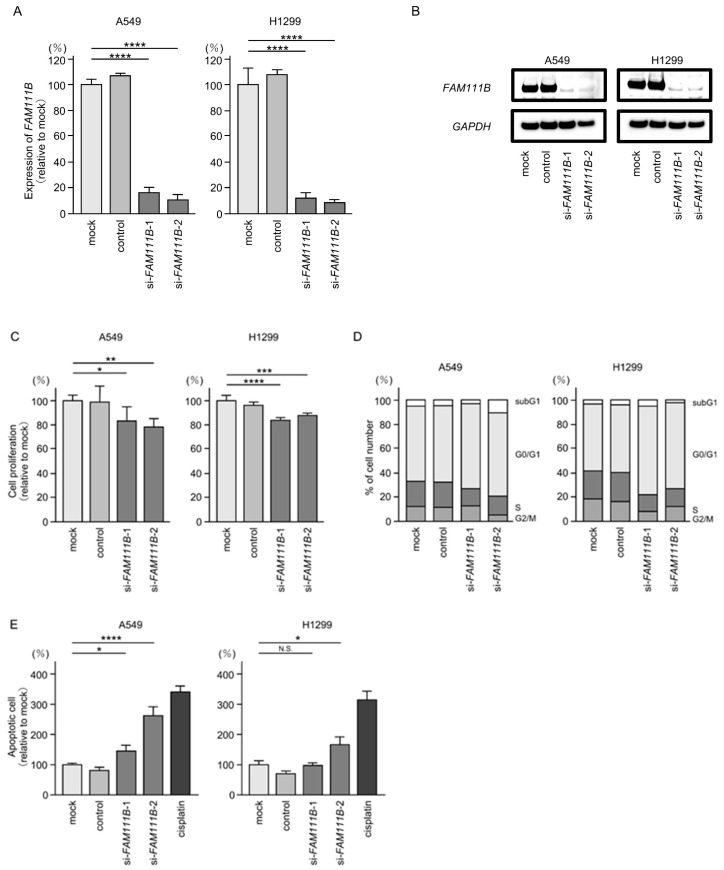
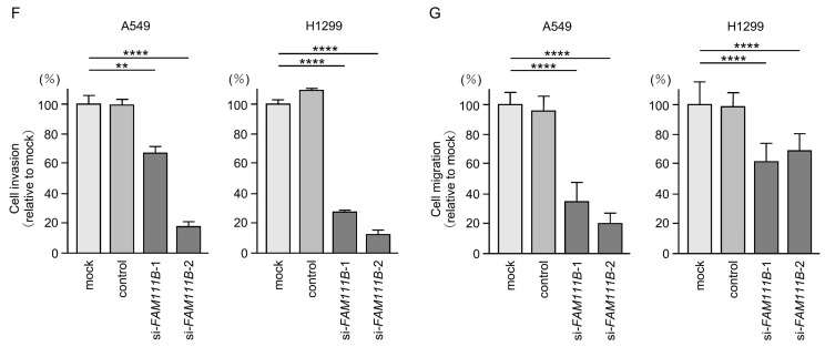
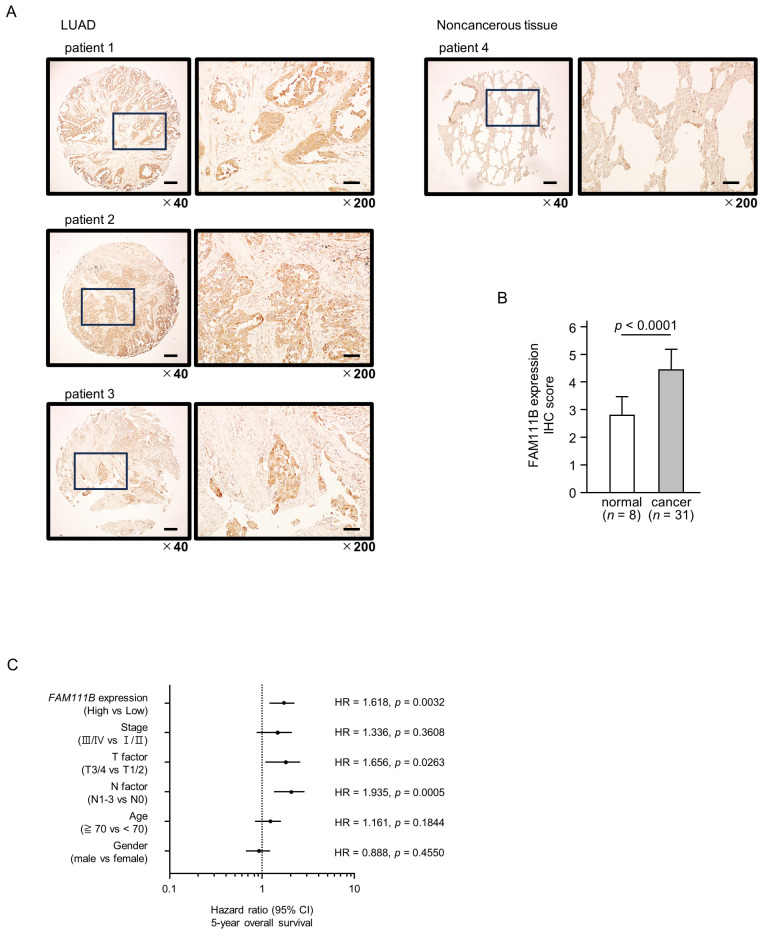
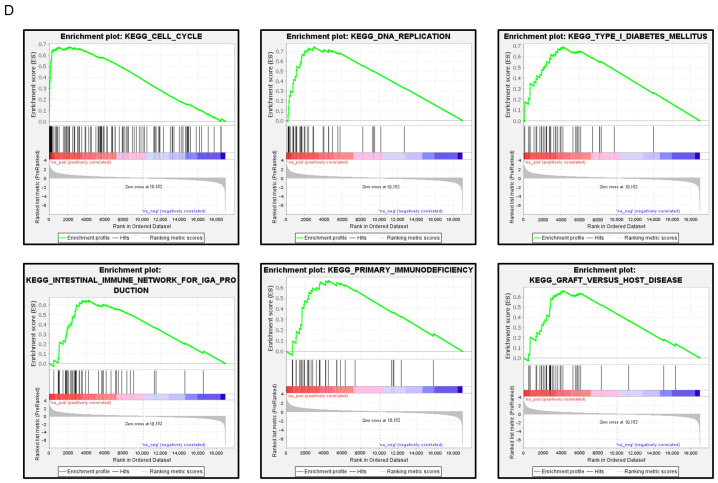
Accumulating evidence suggests that the passenger strands microRNAs (miRNAs) derived from pre-miRNAs are closely involved in cancer pathogenesis. Analysis of our miRNA expression signature of lung adenocarcinoma (LUAD) and The Cancer Genome Atlas (TCGA) data revealed that miR-144-5p (the passenger strand derived from pre-miR-144) was significantly downregulated in LUAD tissues. The aim of this study was to identify therapeutic target molecules controlled by miR-144-5p in LUAD cells. Ectopic expression assays demonstrated that miR-144-5p attenuated LUAD cell aggressiveness, e.g., inhibited cell proliferation, migration and invasion abilities, and induced cell cycle arrest and apoptotic cells. A total of 18 genes were identified as putative cancer-promoting genes controlled by miR-144-5p in LUAD cells based on our in silico analysis. We focused on a family with sequence similarity 111 member B (FAM111B) and investigated its cancer-promoting functions in LUAD cells. Luciferase reporter assay showed that expression of FAM111B was directly regulated by miR-144-5p in LUAD cells. FAM111B knockdown assays showed that LUAD cells significantly suppressed malignant phenotypes, e.g., inhibited cell proliferation, migration and invasion abilities, and induced cell cycle arrest and apoptotic cells. Furthermore, we investigated the FAM111B-mediated molecular networks in LUAD cells. Identifying target genes regulated by passenger strands of miRNAs may aid in the discovery of diagnostic markers and therapeutic targets for LUAD.

Keywords: FAM111B; lung adenocarcinoma; miR-144-5p; microRNA; passenger strand; tumor-suppressor.


Conflict of interest statement

The authors declare no conflicts of interest.

Figures

Figure 1
Figure 1
Expression levels of miR-144-5p and miR-144-3p in LUAD clinical specimens (A) Volcano plot showing the miRNA expression signature obtained through miRNA sequencing (GEO accession number: GSE230229). The log2 fold change (FC) in expression is plotted on the x-axis and the log10 p-value is on the y-axis. The red and blue dots represent the upregulated (log2 FC > 1.0 and p < 0.05) miRNAs and downregulated (log2 FC < −1.0 and p < 0.05), respectively. (B) Validation of miR-144-5p and miR-144-3p expression levels in LUAD clinical specimens. The expression levels of both miRNAs were markedly reduced in cancer tissues. (p < 0.001). (C) Positive correlations (Spearman’s rank test) between the expression levels of miR-144-5p and miR-144-3p in clinical specimens (r = 0.882, p < 0.001). (D) The chromosomal position of pre-miR-144 within the human genome. The mature sequences of miR-144-5p (passenger strand) and miR-144-3p (guide strand) are shown.
Figure 2
Figure 2
Antitumor functions of miR-144-5p in LUAD cells (A549 and H1299). (A) Cell proliferation was evaluated using XTT assay. Cancer cell viability was analyzed 72 h after transient transfection of miRNAs. (B) At 72 h after transient transfection with miR-144-5p, cell cycle status evaluated using flow cytometry. (C) At 72 h after transient transfection with miR-144-5p, apoptotic cells was evaluated using flow cytometry with Annexin V-FITC- and PI-PerCP-Cy5-5-A-stained cells. Cisplatin (30 µM) was used as a positive control for induction of apoptosis. (D) At 72 h after seeding miR-144-5p-transfected cells into the chambers, cell invasion was evaluated using Matrigel invasion assays. (E) At 72 h after seeding miR-144-5p transfected cells into the chambers, cell migration assessed using a membrane culture system. ***, p < 0.001; ****, p < 0.0001.
Figure 3
Figure 3
Flowchart for identification of miR-144-5p targets in LUAD cell. To identify putative targets of miR-144-5p in LUAD cells, we used two datasets: the TargetScanHuman database (release 8.0) and our original mRNA expression profile (Upregulated genes in non-small cell lung carcinoma tissues; GEO accession number: GSE19188). A total of 69 genes were identified as candidate targets of miR-144-5p. Furthermore, we searched for genes that were associated with the prognosis of LUAD patients using two databases: OncoLnc (http://www.oncolnc.org, accessed on 17 May 2024) and GEPIA (http://gepia2.cancer-pku.cn/#analysis, accessed on 17 May 2024). Among the miR-144-5p target genes, 18 genes were upregulated in LUAD tissues, and closely associated with poor prognosis in LUAD patients.
Figure 4
Figure 4
Expression levels and 5-year overall survival rate of the 18 target genes regulated by miR-144-5p in LUAD (A) The expression levels of the 18 target genes of miR-144-5p (ARHGAP11A, CDCA3, CENPF, CENPN, CHEK1, CP, DEPDC1B, ECT2, FAM111B, FAM64A, HELLS, HJURP, KIF11, NCAPG, RALGPS, SGOL1, SPC24, TRIP13) in LUAD clinical specimens were assessed using the TCGA-LUAD dataset. All genes were upregulated in LUAD tissues (n = 499) compared with normal tissues (n = 58) (p < 0.001). (B) Kaplan–Meier curves of the 5-year overall survival rates based on expression of the 18 target genes (ARHGAP11A, CDCA3, CENPF, CENPN, CHEK1, CP, DEPDC1B, ECT2, FAM111B, FAM64A, HELLS, HJURP, KIF11, NCAPG, RALGPS, SGOL1, SPC24, TRIP13) are shown. Lower expression levels of all 18 genes were significantly associated with poorer overall survival in LUAD patients. The patients (n = 487) were divided into high and low-expression groups based on the median gene expression level. The red and blue lines denote the high and low expression groups, respectively.
Figure 4
Figure 4
Expression levels and 5-year overall survival rate of the 18 target genes regulated by miR-144-5p in LUAD (A) The expression levels of the 18 target genes of miR-144-5p (ARHGAP11A, CDCA3, CENPF, CENPN, CHEK1, CP, DEPDC1B, ECT2, FAM111B, FAM64A, HELLS, HJURP, KIF11, NCAPG, RALGPS, SGOL1, SPC24, TRIP13) in LUAD clinical specimens were assessed using the TCGA-LUAD dataset. All genes were upregulated in LUAD tissues (n = 499) compared with normal tissues (n = 58) (p < 0.001). (B) Kaplan–Meier curves of the 5-year overall survival rates based on expression of the 18 target genes (ARHGAP11A, CDCA3, CENPF, CENPN, CHEK1, CP, DEPDC1B, ECT2, FAM111B, FAM64A, HELLS, HJURP, KIF11, NCAPG, RALGPS, SGOL1, SPC24, TRIP13) are shown. Lower expression levels of all 18 genes were significantly associated with poorer overall survival in LUAD patients. The patients (n = 487) were divided into high and low-expression groups based on the median gene expression level. The red and blue lines denote the high and low expression groups, respectively.
Figure 5
Figure 5
MiR-144-5p expression directly regulated FAM111B in LUAD cells. (A) Expression level of FAM111B mRNA is markedly reduced by ectopic expression of miR-144-5p in LUAD cells (A549 and H1299). Total RNA was isolated 72 h after miRNA transfection and quantified by real-time PCR. GAPDH was used as an internal control. (B) Significant reduction of the FAM111B protein level by ectopic expression of miR-144-5p in LUAD cells (A549 and H1299). Proteins were isolated 72 h after miR-144-5p transfection and quantified by Western blotting. GAPDH was used as an internal control. (C) Putative miR-144-5p binding sites in the 3′UTR of the FAM111B gene were detected using the TargetScanHuman database (release 8.0). (D) Dual luciferase reporter assays revealed reduced luminescence activity after co-transfection of miR-144-5p with a vector containing the miR-144-5p binding site (wild-type) in LUAD cells (A549 and H1299). In contrast, no luminescence activity was observed after co-transfection of miR-144-5p with a vector lacking the miR-144-5p binding site (deletion-type) in LUAD cells. ****, p < 0.0001.
Figure 6
Figure 6
Effects of knockdown of FAM111B by siRNAs in LUAD cells (A549 and H1299) (A) The inhibitory effects of two different siRNAs targeting FAM111B (siFAM111B-1 and siFAM111B-2) expression were examined. FAM111B- mRNA levels were effectively inhibited by each siRNA in LUAD cells (A549 and H1299). (B) FAM111B protein levels were effectively inhibited by two siRNAs (siFAM111B-1 and siFAM111B-2) in LUAD cells (A549 and H1299). (C) Cell proliferation was evaluated using XTT assays 72 h after siRNA transfection into LUAD cells. (D) At 72 h after transient transfection with siFAM111B-1 and siFAM111B-2, cell cycle status was evaluated using flow cytometry. (E) At 72 h after transient knockdown of FAM111B, apoptotic cells were evaluated using flow cytometry with Annexin V-FITC- and PI-PerCP-Cy5-5-A-stained cells. Cisplatin (30 µM) was used as a positive control for induction of apoptosis. (F) At 72 h after seeding FAM111B-knockdown cells into the chambers, cell invasion assessed using Matrigel invasion assays. (G) At 72 h after seeding FAM111B-knockdown cells into the chambers, cell migration was assessed using a membrane culture system. *, p < 0.05; **, p < 0.01; ***, p < 0.001; ****, p < 0.0001; N.S., not significant.
Figure 6
Figure 6
Effects of knockdown of FAM111B by siRNAs in LUAD cells (A549 and H1299) (A) The inhibitory effects of two different siRNAs targeting FAM111B (siFAM111B-1 and siFAM111B-2) expression were examined. FAM111B- mRNA levels were effectively inhibited by each siRNA in LUAD cells (A549 and H1299). (B) FAM111B protein levels were effectively inhibited by two siRNAs (siFAM111B-1 and siFAM111B-2) in LUAD cells (A549 and H1299). (C) Cell proliferation was evaluated using XTT assays 72 h after siRNA transfection into LUAD cells. (D) At 72 h after transient transfection with siFAM111B-1 and siFAM111B-2, cell cycle status was evaluated using flow cytometry. (E) At 72 h after transient knockdown of FAM111B, apoptotic cells were evaluated using flow cytometry with Annexin V-FITC- and PI-PerCP-Cy5-5-A-stained cells. Cisplatin (30 µM) was used as a positive control for induction of apoptosis. (F) At 72 h after seeding FAM111B-knockdown cells into the chambers, cell invasion assessed using Matrigel invasion assays. (G) At 72 h after seeding FAM111B-knockdown cells into the chambers, cell migration was assessed using a membrane culture system. *, p < 0.05; **, p < 0.01; ***, p < 0.001; ****, p < 0.0001; N.S., not significant.
Figure 7
Figure 7
FAM111B expression and its clinical significance in LUAD. (A) Immunohistochemical staining of FAM111B. (B) Cancer tissues showed strong immunostaining, in contrast to the weak staining observed in noncancerous tissues. The data are means and standard errors of the means. Mann–Whitney U-tests. Scale bar: 200 µm (low magnification); 50 µm (high magnification). (C) Forest plot showing the results of multivariate Cox proportional hazards regression analysis of the 5-year overall survival rate. A significantly lower overall survival rate was observed in patients with high FAM111B expression. The data were sourced from TCGA-LUAD datasets. (D) FAM111B-mediated pathways identified by gene set enrichment analysis. The “cell cycle”, “DNA replication” pathways were enriched in patients with high FAM111B expression.
Figure 7
Figure 7
FAM111B expression and its clinical significance in LUAD. (A) Immunohistochemical staining of FAM111B. (B) Cancer tissues showed strong immunostaining, in contrast to the weak staining observed in noncancerous tissues. The data are means and standard errors of the means. Mann–Whitney U-tests. Scale bar: 200 µm (low magnification); 50 µm (high magnification). (C) Forest plot showing the results of multivariate Cox proportional hazards regression analysis of the 5-year overall survival rate. A significantly lower overall survival rate was observed in patients with high FAM111B expression. The data were sourced from TCGA-LUAD datasets. (D) FAM111B-mediated pathways identified by gene set enrichment analysis. The “cell cycle”, “DNA replication” pathways were enriched in patients with high FAM111B expression.

Similar articles

Cited by

References

    1. Siegel R.L., Miller K.D., Wagle N.S., Jemal A. Cancer statistics, 2023. CA Cancer J. Clin. 2023;73:17–48. doi: 10.3322/caac.21763. - DOI - PubMed
    1. Schabath M.B., Cote M.L. Cancer Progress and Priorities: Lung Cancer. Cancer Epidemiol. Biomark. Prev. 2019;28:1563–1579. doi: 10.1158/1055-9965.EPI-19-0221. - DOI - PMC - PubMed
    1. Pirker R. Conquering lung cancer: Current status and prospects for the future. Pulmonology. 2020;26:283–290. doi: 10.1016/j.pulmoe.2020.02.005. - DOI - PubMed
    1. Tan A.C., Tan D.S.W. Targeted Therapies for Lung Cancer Patients with Oncogenic Driver Molecular Alterations. J. Clin. Oncol. 2022;40:611–625. doi: 10.1200/JCO.21.01626. - DOI - PubMed
    1. Reck M., Remon J., Hellmann M.D. First-Line Immunotherapy for Non-Small-Cell Lung Cancer. J. Clin. Oncol. 2022;40:586–597. doi: 10.1200/JCO.21.01497. - DOI - PubMed