. 2024 Sep 18;25(18):10045.
doi: 10.3390/ijms251810045.

OXGR1-Dependent (Pro)Renin Receptor Upregulation in Collecting Ducts of the Clipped Kidney Contributes to Na+ Balance in Goldblatt Hypertensive Mice

Affiliations

OXGR1-Dependent (Pro)Renin Receptor Upregulation in Collecting Ducts of the Clipped Kidney Contributes to Na+ Balance in Goldblatt Hypertensive Mice

Pilar Cárdenas et al. Int J Mol Sci. .

Abstract

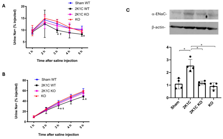
The two-kidney, one-clip (2K1C) Goldblatt rodent model elicits a reduction in renal blood flow (RBF) in the clipped kidney (CK). The reduced RBF and oxygen bio-ability causes the accumulation of the tricarboxylic cycle intermediary, α-ketoglutarate, which activates the oxoglutarate receptor-1 (OXGR1). In the kidney, OXGR1 is abundantly expressed in intercalated cells (ICs) of the collecting duct (CD), thus contributing to sodium transport and electrolyte balance. The (pro)renin receptor (PRR), a member of the renin-angiotensin system (RAS), is a key regulator of sodium reabsorption and blood pressure (BP) that is expressed in ICs. The PRR is upregulated in 2K1C rats. Here, we tested the hypothesis that chronic reduction in RBF in the CK leads to OXGR1-dependent PRR upregulation in the CD and alters sodium balance and BP in 2K1C mice. To determine the role of OXGR1 in regulating the PRR in the CDs during renovascular hypertension, we performed 2K1C Goldblatt surgery (clip = 0.13 mm internal gap, 14 days) in two groups of male mice: (1) mice treated with Montelukast (OXGR1 antagonist; 5 mg/Kg/day); (2) OXGR1-/- knockout mice. Wild-type and sham-operated mice were used as controls. After 14 days, 2K1C mice showed increased systolic BP (SBP) (108 ± 11 vs. control 82 ± 5 mmHg, p < 0.01) and a lower natriuretic response after the saline challenge test. The CK group showed upregulation of erythropoietin, augmented α-ketoglutarate, and increased PRR expression in the renal medulla. The CK of OXGR1 knockout mice and mice subjected to the OXGR1 antagonist elicited impaired PRR upregulation, attenuated SBP, and better natriuretic responses. In 2K1C mice, the effect of reduced RBF on the OXGR1-dependent PRR upregulation in the CK may contribute to the anti-natriuretic and increased SBP responses.

Keywords: 2K1C; kidney; prorenin; renal blood flow; tricarboxylic cyclic acid pathway.


Conflict of interest statement

The authors declare no conflicts of interest.

Figures

Figure 1
Figure 1
Control sham-operated and mice with 2K1C surgery were housed in metabolic cages for urine collection. A silver clip with an internal diameter of 0.13 mm was placed around the left artery for 14 days (A). After 14 days of chronic reduction in blood flow, clipped kidneys showed reduced size and weight as compared to sham mice (B). Intrarenal levels of α-Ketoglutarate in the clipped kidney were significantly augmented as compared to control (sham) kidneys (C). Reduced blood supply for 14 days was also evidenced by the increased abundance of erythropoietin (EPO) protein in the clipped kidney as compared to sham-operated mice, * p < 0.05; versus sham group (D). Chronic reduction in renal blood flow caused the upregulation of juxtaglomerular (JG) renin, even at day 14 in the clipped kidney (CK). In contrast, renin was not augmented in the non-clipped kidney (NCK). Collecting duct (CD) renin was augmented in both clipped and non-clipped kidneys (E). As previously demonstrated by our group, medullary prorenin and renin protein bands can be detected in medullary tissues. Immunoblotting showed that prorenin and renin bands are upregulated in the clipped and non-clipped kidneys after 14 days (F). Co-localization of OXGR1 and PRR in medullary collecting ducts was confirmed by immunofluorescence using specific antibodies—OXGR1 (Alexa Fluor 488, Green color) and PRR (Alexa Fluor 594, red color), leading to merge-yellow color (G), scale bar 50 µm. Specific controls with omission of the first or secondary antibody and nuclei staining with DAPI demonstrated the specificity of the experiment. In previous protocols using whole trunk blood collection, we showed that plasma renin content (PRC) is augmented in 2K1C mice, as compared to sham mice (H). No changes were observed in blood creatinine. * p < 0.05; ** p < 0.01 versus sham group, n = 5 for (B,C,F). For (H,I) n = 4, p < 0.05 versus sham group, scale bar 50 µm. For F and H * p < 0.05 versus sham group.
Figure 2
Figure 2
OXGR1 antagonist Montelukast (ML) attenuated the increases in systolic blood pressure in 2K1C (A), and blunted the increases in diastolic blood pressure in 2K1C mice (B). ML mice were not different from controls. Clipped kidneys (CKs) and kidneys from 2K1C + ML mice showed an enlarged Bowman’s capsule; 2K1C + ML mice showed hyaline substances (C). 2K1C surgery led to a reduction in the size of the CKs; 2K1C + ML mice showed no observable size difference compared to 2K1C alone (D). Goldblatt surgery did not change water intake (E), urine flow (F), or 24 h sodium excretion (G) after 14 days. To verify kidney injury signs shown in (C), we performed albumin measurements in urine. Data show that, after 2 weeks of 2K1C surgery, there was an increased urinary albumin/creatinine ratio in the 2K1C and 2K1C + ML groups as compared to the sham surgery group (* p < 0.05), while ML treatment did not have any effect (H). ** p < 0.01 versus sham group in B. scale bar 50 μm.
Figure 3
Figure 3
Upregulation of PRR and intrarenal Ang II in the clipped kidney was blunted by OXGR1 antagonist Montelukast (ML). PRR protein levels (A) and mRNA abundance (B) were increased in the clipped kidney, while ML treatment for 14 days prevented this effect. * p < 0.05 versus sham group.
Figure 4
Figure 4
Mice were subjected to a saline load challenge to test the ability of the mice to excrete a saline load on day 10. The sodium excretion was significantly lower in 2K1C mice at 3 and 5 h post-injection as compared with wild-type (sham WT) mice (A). Similar results were observed when analyzed by cumulated sodium excretion (B). Co-treatment with Montelukast (ML) was able to prevent this effect. Since ML prevented PRR upregulation and augmentation of intrarenal Ang II levels, the expression of ENaC might likely be altered. 2K1C surgery caused an increased ENaC expression in the clipped kidney that was blunted by ML (C). * p < 0.05 2K1C versus sham; p < 0.05 versus 2K1C + ML mice.
Figure 5
Figure 5
Increases in systolic (A) and diastolic (B) blood pressure were prevented in OXGR1 knockout (KO) mice subjected to 2K1C surgery. KO mice did not differ from controls regarding diastolic or systolic blood pressure. The enlarged distance from Bowman’s capsule distance to glomeruli was observed in the left clipped kidney (L) of 2K1C and 2K1C KO mice, and the same comparative image from 2K1C in Figure 2 is shown (C). The reduction in clipped-kidney size was also observed in KO mice subjected to the 2K1C surgery and did not differ from wild-type mice (D). No changes were observed in water intake (E), urine flow (F), and 24 h sodium excretion (G) among the groups after 14 days. Kidney injury signs showed in C were confirmed by the results showing increased urinary albumin/creatinine ratio in 2K1C and 2K1C KO mice as compared to sham surgery group (* p < 0.05). No changes were observed in KO mice (H). Scale bar 50 μm.
Figure 6
Figure 6
Upregulation of PRR in the clipped kidneys of 2K1C mice was not evidenced in OXGR1 knockout (KO) mice. PRR protein levels (A) and mRNA abundance (B) were increased in the clipped kidneys but not in OXGR1 KO mice. * indicates statistical differences (p < 0.05 between the groups compered).
Figure 7
Figure 7
The ability to excrete a saline load was evaluated in wild-type (WT) mice with sham or 2K1C surgery and in OXGR1 knockout (KO) mice. Urine was collected for 5 h in metabolic cages. Sodium excretion was expressed as the amount of the sodium load excreted during the 5 h observation period. The reduced sodium excretion evidenced in the 2K1C group at 3 and 5 h was not observed in 2K1C KO mice (A); the same results were observed for accumulated sodium excretion (B). As described before, 2K1C surgery caused an increased ENaC expression in the clipped kidneys that was not observed in KO mice (C). * p < 0.05 2K1C versus sham; p < 0.05 versus 2K1C + ML group.
Figure 8
Figure 8
Proposed hypothetical model showing the effect of chronic partial obstruction of the left renal artery by using the 2-kidney, 1-clip, 2K1C model. Increased synthesis and release of renin from juxtaglomerular (JG) cells promotes systemic renin–angiotensin system (RAS) activation and enhance distal tubular sodium (Na+) reabsorption through basolateral/blood Ang II type 1 receptor (AT1R) via epithelial Na+ channel (ENaC). Chronic reductions in oxygen bio-ability were evidenced by increased levels of erythropoietin at 14 days. Systemic RAS activation also promotes upregulation of prorenin and renin in principal cells of the collecting duct via AT1R and binding of prorenin or renin to the PRR in the neighbor intercalated cell. Reduced blood flow leads to the accumulation of α-ketoglutarate. We propose that, during reduced blood flow, α-ketoglutarate activates the collecting duct OXGR1 receptor, leading to the upregulation of PRR enhancing the activity of renin and prorenin, stimulating intra-tubular Ang II formation and activation of AT1R promoting Na+ reabsorption.

Similar articles

Cited by

References

    1. Matavelli L.C., Huang J., Siragy H.M. (Pro)renin receptor contributes to diabetic nephropathy by enhancing renal inflammation. Clin. Exp. Pharmacol. Physiol. 2010;37:277–282. doi: 10.1111/j.1440-1681.2009.05292.x. - DOI - PMC - PubMed
    1. Siragy H.M., Huang J. Renal (pro)renin receptor upregulation in diabetic rats through enhanced angiotensin AT1 receptor and NADPH oxidase activity. Exp. Physiol. 2008;93:709–714. doi: 10.1113/expphysiol.2007.040550. - DOI - PMC - PubMed
    1. Tang J., Wysocki J., Ye M., Valles P.G., Rein J., Shirazi M., Bader M., Gomez R.A., Sequeira-Lopez M.S., Afkarian M., et al. Urinary Renin in Patients and Mice With Diabetic Kidney Disease. Hypertension. 2019;74:83–94. doi: 10.1161/HYPERTENSIONAHA.119.12873. - DOI - PMC - PubMed
    1. Nurun N.A.H.M., Uddin N.M., Nakagawa T., Iwata H., Ichihara A., Inagami T., Suzuki F. Role of “handle” region of prorenin prosegment in the non-proteolytic activation of prorenin by binding to membrane anchored (pro)renin receptor. Front. Biosci-Landmrk. 2007;12:4810–4817. doi: 10.2741/2429. - DOI - PubMed
    1. Redublo Quinto B.M., Camargo de Andrade M.C., Ronchi F.A., Santos E.L., Alves Correa S.A., Shimuta S.I., Pesquero J.B., Mortara R.A., Casarini D.E. Expression of angiotensin I-converting enzymes and bradykinin B-2 receptors in mouse inner medullary-collecting duct cells. Int. Immunopharmacol. 2008;8:254–260. doi: 10.1016/j.intimp.2007.09.013. - DOI - PubMed

MeSH terms

LinkOut - more resources