. 2024 Sep 18;25(18):10050.
doi: 10.3390/ijms251810050.

Staphylococcus aureus Co-Infection in COVID-19 Patients: Virulence Genes and Their Influence on Respiratory Epithelial Cells in Light of Risk of Severe Secondary Infection

Affiliations

Staphylococcus aureus Co-Infection in COVID-19 Patients: Virulence Genes and Their Influence on Respiratory Epithelial Cells in Light of Risk of Severe Secondary Infection

Lidia Piechowicz et al. Int J Mol Sci. .

Abstract

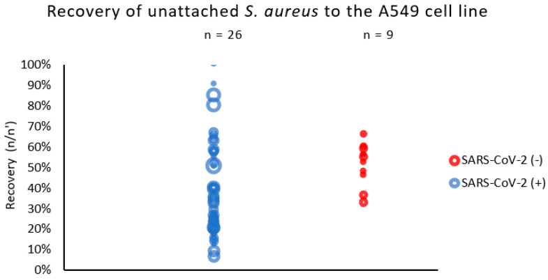
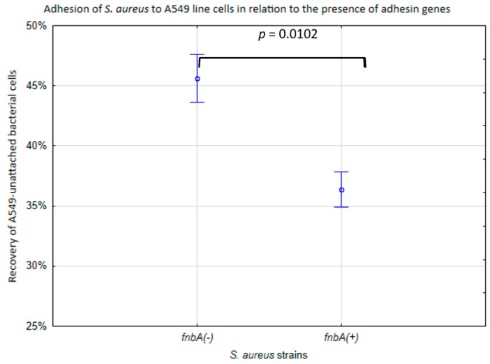
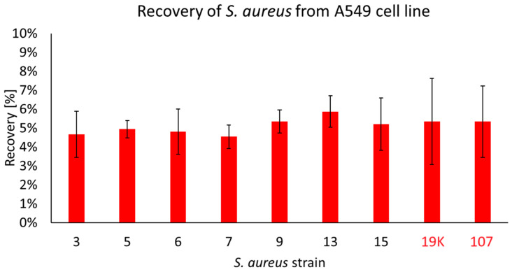
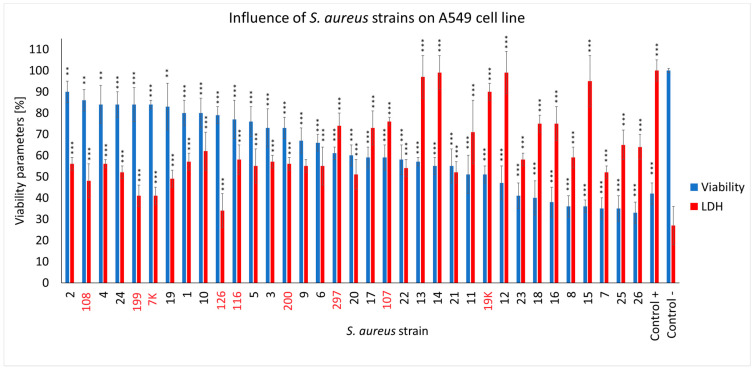
Pandemics from viral respiratory tract infections in the 20th and early 21st centuries were associated with high mortality, which was not always caused by a primary viral infection. It has been observed that severe course of infection, complications and mortality were often the result of co-infection with other pathogens, especially Staphylococcus aureus. During the COVID-19 pandemic, it was also noticed that patients infected with S. aureus had a significantly higher mortality rate (61.7%) compared to patients infected with SARS-CoV-2 alone. Our previous studies have shown that S. aureus strains isolated from patients with COVID-19 had a different protein profile than the strains in non-COVID-19 patients. Therefore, this study aims to analyze S. aureus strains isolated from COVID-19 patients in terms of their pathogenicity by analyzing their virulence genes, adhesion, cytotoxicity and penetration to the human pulmonary epithelial cell line A549. We have observed that half of the tested S. aureus strains isolated from patients with COVID-19 had a necrotizing effect on the A549 cells. The strains also showed greater variability in terms of their adhesion to the human cells than their non-COVID-19 counterparts.

Keywords: COVID-19; SARS-CoV-2; Staphylococcus aureus; adhesion.


Conflict of interest statement

The authors declare no conflicts of interest.

Figures

Figure 1
Figure 1
Adhesion of S. aureus strains measured by % of recovered bacteria not adhered to respiratory epithelial cells (A549 line). n/n′—ratio of counts per 60 s of sample flow before and after exposition to epithelial cells (A549). The circle represents the result (median) of a single strain; the size of the circle represents the relative initial inoculum of the sample.
Figure 2
Figure 2
Adhesion of the S. aureus cell line in relation to the presence of adhesin genes. % of recovery of unattached A549 bacterial cells to the initial bacterial suspension. The vertical bars represent standard errors. The statistical analysis was performed using the Kruskal–Wallis test.
Figure 3
Figure 3
The percentage of COVID-19 (black numbers) and non-COVID-19 (red numbers) S. aureus cells recovered from the inside of A549 pulmonary epithelial cells. The arithmetic mean of the triplicates, including the error bars that represents the SD. The statistical analysis was performed using a t-test. No statistical differences were found between the strains recovered from the inside of A549 cells.
Figure 4
Figure 4
The cell viability and LDH release assay for the A549 cells infected with different S. aureus strains (COVID-19 strains are marked in black, and non-COVID-19 strains are marked in red) after 4 h of incubation compared to the uninfected cells and the lysis-buffer-treated positive control. The mean values from the three independent experiments are shown, with the error bars representing the SD. The statistical analysis was performed using the Kruskal–Wallis test, followed by Dunn’s multiple comparison test for the values with a nonparametric distribution, with *** p < 0.001 and ** p < 0.01.

Similar articles

Cited by

References

    1. Joseph C., Togawa Y., Shindo N. Bacterial and Viral Infections Associated with Influenza. Influenza Other Respir. Viruses. 2013;7:105–113. doi: 10.1111/irv.12089. - DOI - PMC - PubMed
    1. Mulcahy M.E., McLoughlin R.M. Staphylococcus aureus and Influenza A Virus: Partners in Coinfection. mBio. 2016;7:10-1128. doi: 10.1128/mBio.02068-16. - DOI - PMC - PubMed
    1. Borgogna T.R., Hisey B., Heitmann E., Obar J.J., Meissner N., Voyich J.M. Secondary Bacterial Pneumonia by Staphylococcus aureus Following Influenza a Infection Is SaeR/S Dependent. J. Infect. Dis. 2018;218:809–813. doi: 10.1093/infdis/jiy210. - DOI - PMC - PubMed
    1. Morgene M.F., Botelho-Nevers E., Grattard F., Pillet S., Berthelot P., Pozzetto B., Verhoeven P.O. Staphylococcus aureus Colonization and Non-Influenza Respiratory Viruses: Interactions and Synergism Mechanisms. Virulence. 2018;9:1354–1363. doi: 10.1080/21505594.2018.1504561. - DOI - PMC - PubMed
    1. Cusumano J.A., Dupper A.C., Malik Y., Gavioli E.M., Banga J., Berbel Caban A., Nadkarni D., Obla A., Vasa C.V., Mazo D., et al. Staphylococcus aureus Bacteremia in Patients Infected with COVID-19: A Case Series. Open Forum Infect. Dis. 2020;7:ofaa518. doi: 10.1093/ofid/ofaa518. - DOI - PMC - PubMed

Substances

LinkOut - more resources