. 2024 Sep 19;25(18):10054.
doi: 10.3390/ijms251810054.

Characterization of Neutrophil Functional Responses to SARS-CoV-2 Infection in a Translational Feline Model for COVID-19

Affiliations

Characterization of Neutrophil Functional Responses to SARS-CoV-2 Infection in a Translational Feline Model for COVID-19

Sachithra Gunasekara et al. Int J Mol Sci. .

Abstract

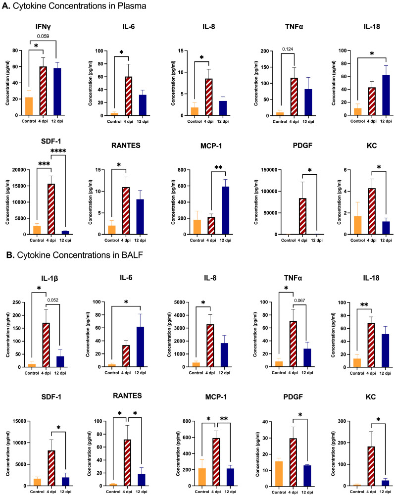
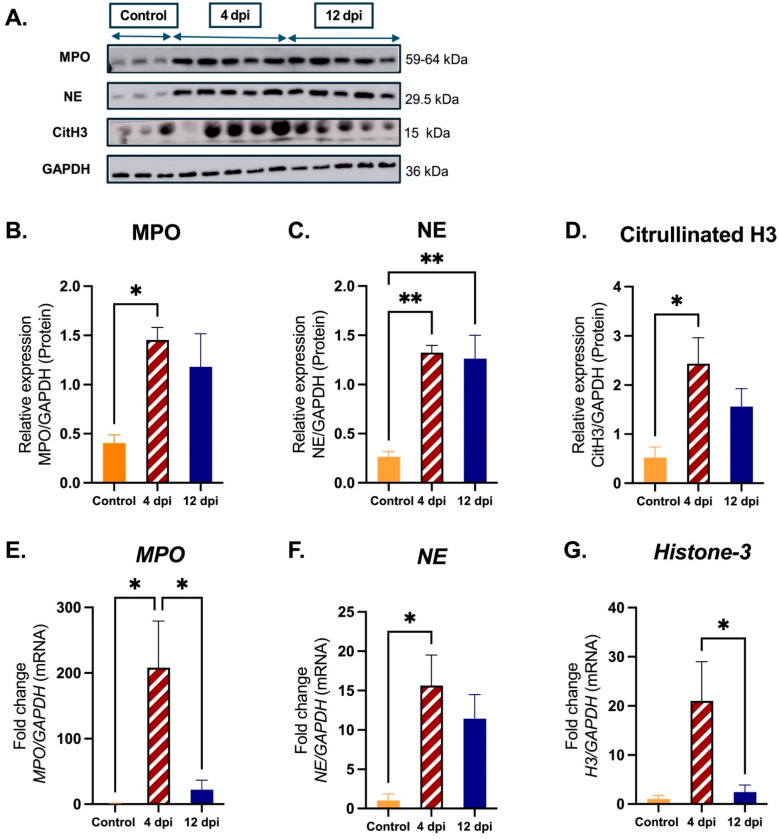
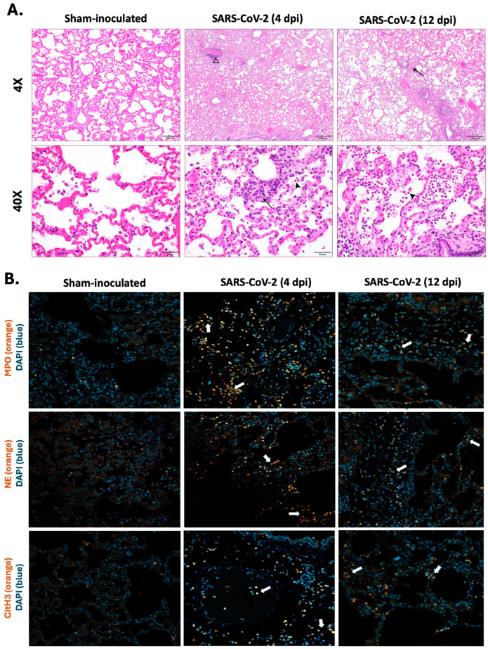
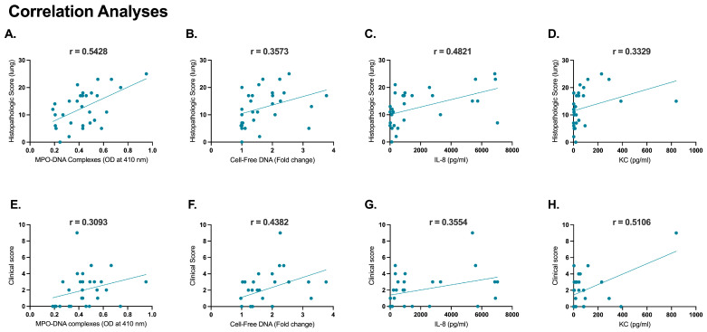
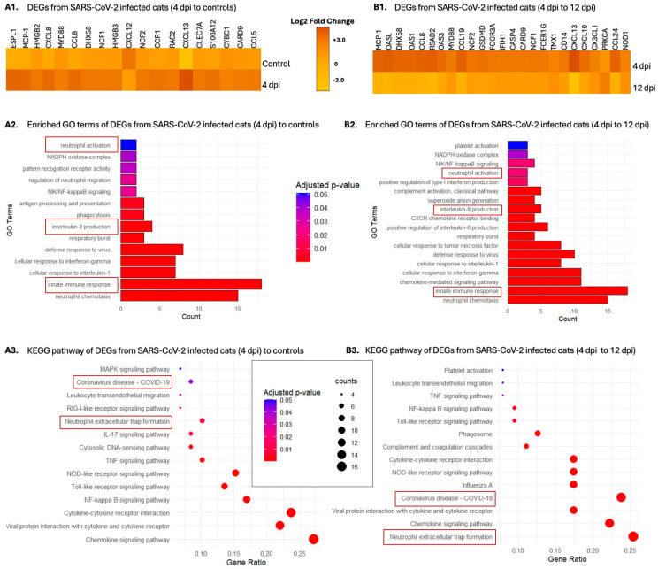
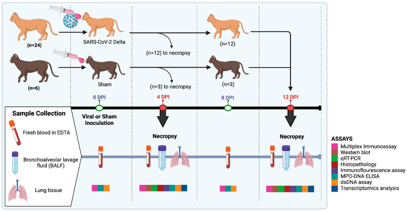
There is a complex interplay between viral infection and host innate immune response regarding disease severity and outcomes. Neutrophil hyperactivation, including excessive release of neutrophil extracellular traps (NETs), is linked to exacerbated disease in acute COVID-19, notably in hospitalized patients. Delineating protective versus detrimental neutrophil responses is essential to developing targeted COVID-19 therapies and relies on high-quality translational animal models. In this study, we utilize a previously established feline model for COVID-19 to investigate neutrophil dysfunction in which experimentally infected cats develop clinical disease that mimics acute COVID-19. Specific pathogen-free cats were inoculated with SARS-CoV-2 (B.1.617.2; Delta variant) (n = 24) or vehicle (n = 6). Plasma, bronchoalveolar lavage fluid, and lung tissues were collected at various time points over 12 days post-inoculation. Systematic and temporal evaluation of the kinetics of neutrophil activation was conducted by measuring markers of activation including myeloperoxidase (MPO), neutrophil elastase (NE), and citrullinated histone H3 (citH3) in SARS-CoV-2-infected cats at 4 and 12 days post-inoculation (dpi) and compared to vehicle-inoculated controls. Cytokine profiling supported elevated innate inflammatory responses with specific upregulation of neutrophil activation and NET formation-related markers, namely IL-8, IL-18, CXCL1, and SDF-1, in infected cats. An increase in MPO-DNA complexes and cell-free dsDNA in infected cats compared to vehicle-inoculated was noted and supported by histopathologic severity in respiratory tissues. Immunofluorescence analyses further supported correlation of NET markers with tissue damage, especially 4 dpi. Differential gene expression analyses indicated an upregulation of genes associated with innate immune and neutrophil activation pathways. Transcripts involved in activation and NETosis pathways were upregulated by 4 dpi and downregulated by 12 dpi, suggesting peak activation of neutrophils and NET-associated markers in the early acute stages of infection. Correlation analyses conducted between NET-specific markers and clinical scores as well as histopathologic scores support association between neutrophil activation and disease severity during SARS-CoV-2 infection in this model. Overall, this study emphasizes the effect of neutrophil activation and NET release in SARS-CoV-2 infection in a feline model, prompting further investigation into therapeutic strategies aimed at mitigating excessive innate inflammatory responses in COVID-19.

Keywords: COVID-19; SARS-CoV-2; feline; neutrophil; neutrophil extracellular traps.


Conflict of interest statement

The authors declare no conflicts of interest.

Figures

Figure 1
Figure 1
Comparison of cytokine concentrations in plasma and BALF of SARS-CoV-2-infected cats. Plasma cytokine concentrations were measured in SARS-CoV-2-infected cats 4 dpi (n = 12) and 12 dpi (n = 12) and in sham-inoculated controls (n = 6) using multiplex immunoassay: (A) IFN-γ, IL-8, TNF-α, IL-18, SDF-1, RANTES, MCP-1, PDGF, and KC show significant alterations between study groups. (B) Cytokine concentrations in BALF of SARS-CoV-2-infected 4 dpi cats (n = 12), 12 dpi cats (n = 12), and sham-inoculated controls (n = 6) were measured using multiplex immunoassay. Significant differences were observed in levels of IL-1β, IL-8, TNF-α, IL-18, SDF-1, RANTES, MCP-1, PDGF, and KC among the study groups. One-way ANOVA statistical analysis was performed. Data are represented as the mean ± SEM. * p < 0.05, ** p < 0.01, *** p < 0.001, **** p < 0.0005.
Figure 2
Figure 2
Expression of NET-related markers in lung tissues from SARS-CoV-2-infected cats. Lung tissues were collected from SARS-CoV-2-infected cats at 4 dpi (n = 12) and 12 dpi (n = 12), and from sham-inoculated controls (n = 6). For Western blot analyses: (A) samples from randomly selected SARS-CoV-2-infected 4 dpi cats (n = 5), SARS-CoV-2-infected 12 dpi cats (n = 5), and sham-inoculated controls (n = 3) were analyzed for protein expression of NET-related markers (MPO, NE, and citrullinated H3); objective quantification and comparisons were carried out using densitometric analysis of MPO, NE, or CitH3 protein expression relative to GAPDH: (B) significantly increased MPO protein was quantified in 4 dpi cats versus controls, (C) significantly increased NE protein was quantified in 4 and 12 dpi cats relative to controls, and (D) significantly increased CitH3 protein was quantified in 4 dpi cats relative to controls. For mRNA expression of NET-related markers, samples from SARS-CoV-2-infected 4 dpi cats (n = 12), 12 dpi cats (n = 12), and sham-inoculated controls (n = 6) were analyzed using real-time quantitative PCR analysis. Fold changes from 0 dpi were analyzed between study groups for MPO, NE, and Histone-3: (E) The fold change in mRNA expression of MPO relative to GAPDH was significantly higher in 4 dpi relative to controls and reduced 12 dpi relative to 4 dpi (F) The fold change in mRNA expression of NE relative to GAPDH was significantly higher in 4 dpi compared to controls. (G) The fold change in mRNA expression of Histone H3 relative to GAPDH indicates significant reduction in expression from 4 dpi to 12 dpi. Statistical comparisons were performed using one-way ANOVA analysis. Data are represented as the mean ± SEM. * p < 0.05, ** p < 0.01.
Figure 3
Figure 3
Quantification of NET-specific markers in plasma and BALF in SARS-CoV-2-infected cats. Plasma was collected from SARS-CoV-2-infected cats (n = 12) and sham-inoculated controls (n = 3) at 0 dpi, 4 dpi, 8 dpi, and 12 dpi. BALF was collected from SARS-CoV-2-infected cats at 4 dpi (n = 6), and 12 dpi (n = 12), and from sham-inoculated controls (n = 12). (A) MPO-DNA complexes in plasma were measured using MPO-DNA ELISA. Results indicate significant increases in these complexes in infected cat plasma relative to sham-inoculated controls, most markedly at 4 and 8 dpi. (B) MPO-DNA complexes in BALF were measured using MPO-DNA ELISA and indicate significant increases in complexes in infected cats relative to controls at both 4 and 12 dpi. (C) Cell-free DNA in plasma measured using Quanti-pico dsDNA assay showed a significant increase in infected cats from inoculation to 4 dpi. (D) Cell-free DNA in BALF measured using Quanti-pico dsDNA assay showed significant increase in DNA detection at 12 dpi relative to control cats. Statistical comparisons were performed using two-way ANOVA analysis and one-way ANOVA analysis. Data are represented as the mean ± SEM * p < 0.05, ** p < 0.01.
Figure 4
Figure 4
Visualization of neutrophil infiltration with NET-related markers in lung tissues of SARS-CoV-2-infected cats. Histopathological analysis and immunofluorescence staining were performed on lung tissues collected from SARS-CoV-2-infected cats at 4 dpi (n = 12) and 12 dpi (n = 12) and sham-inoculated controls (n = 6). (A) Representative images of histopathological lesions in infected cats; 4 dpi cats showed increased perivascular infiltration of inflammatory cells (open arrowhead) including neutrophils (arrow/arrowhead), while 12 dpi cats showed an intense neutrophil infiltration (arrow/arrowhead). (B) Representative images for MPO, NE, and citrullinated H3 in the lungs of SARS-CoV-2-infected 4 dpi cats, SARS-CoV-2-infected 12 dpi cats, and sham-inoculated controls. NETs were identified by co-localization of DNA (blue) with MPO/NE/citrullinated H3 (orange). White arrows show activated neutrophils with an indication of NET formation and release within infected lungs of 4 dpi and 12 dpi cats. Magnification: (A) 4x, scale bar = 500 µm; 40×, scale bar = 50 µm; (B) 20x, scale bar = 50 µm.
Figure 5
Figure 5
Correlation analyses of histopathology scores and clinical scores with neutrophil activation and NET-related markers. Positive correlations were evident following the correlation analysis conducted for histopathological scores from right cranial lung lobes from SARS-CoV-2-infected 4 dpi cats (n = 12), 12 dpi cats (n = 12), and sham-inoculated controls (n = 6) with NET-specific markers: (A) MPO-DNA complexes (r = 0.5428, p = 0.001), (B) cell-free DNA (r = 0.3573, p = 0.0526); and neutrophil activation associated markers: (C) IL-8 (r = 0.4831, p = 0.0069) and (D) KC (r = 0.3329, p = 0.0723). Positive correlations were evident following the correlation analysis conducted for the clinical scores of these cats with NET-specific markers revealed positive correlations: (E) MPO-DNA complexes (r = 0.3093, p = 0.0962), (F) cell-free DNA (r = 0.4382, p = 0.01); and neutrophil activation-associated markers: (G) IL-8 (r = 0.3554, p = 0.0539) and (H) KC (r = 0.5106, p = 0.0039). Statistical comparisons were performed using Pearson coefficient correlation analysis. Data are represented using the Pearson correlation coefficient (r).
Figure 6
Figure 6
Heatmap and enrichment analysis of differentially expressed genes in SARS-CoV-2-infected cats. The figure includes heatmaps and bar plots illustrating the enriched gene ontology (GO) terms and Kyoto Encyclopedia of Genes and Genomes (KEGG) pathways associated with differentially expressed genes in SARS-CoV-2-infected cats. (A1,B1) Heatmaps showing the expression levels of differentially expressed genes across various conditions and time points revealed significant increases of neutrophil activation-related genes at 4 dpi compared to both sham-inoculated controls and 12 dpi SARS-CoV-2-infected cats. (A2,B2) Bar plots of enriched GO terms suggest significant upregulation of neutrophil activation and recruitment pathways with an overall increase of innate immune response in 4 dpi cats compared to both sham-inoculated controls and 12 dpi SARS-CoV-2-infected cats. (A3,B3) Bubble plot of top enriched KEGG pathways supporting the association of DEGs from 4 dpi cats with COVID-19 and neutrophil trap formation compared to DEGs from sham-inoculated controls and 12 dpi SARS-CoV-2-infected cats.
Figure 7
Figure 7
Experimental design. This diagram illustrates the experimental timeline and procedures throughout the study. The study consisted of a total of 30 cats, 24 of which were inoculated with SARS-CoV-2 (Delta variant), and 6 of which were sham-inoculated. Twelve SARS-CoV-2-infected and 3 sham-inoculated were euthanized and samples collected 4 DPI. The remaining twelve infected and 3 sham-inoculated cats were euthanized for sample collection 12 DPI. Blood, tissues, and bronchoalveolar lavage were all collected for analysis. Image made in Biorender.

Similar articles

Cited by

References

    1. Lai C.-C., Shih T.-P., Ko W.-C., Tang H.-J., Hsueh P.-R. Severe acute respiratory syndrome coronavirus 2 (SARS-CoV-2) and coronavirus disease-2019 (COVID-19): The epidemic and the challenges. Int. J. Antimicrob. Agents. 2020;55:105924. doi: 10.1016/j.ijantimicag.2020.105924. - DOI - PMC - PubMed
    1. Aleem A., Akbar Samad A.B., Vaqar S. StatPearls. StatPearls Publishing; Treasure Island, FL, USA: 2021. Emerging variants of SARS-CoV-2 and novel therapeutics against coronavirus (COVID-19) - PubMed
    1. Cascella M., Rajnik M., Aleem A., Dulebohn S.C., Di Napoli R. StatPearls. StatPearls Publishing; Treasure Island, FL, USA: 2020. Features, evaluation, and treatment of coronavirus (COVID-19) - PubMed
    1. Stokol T., McAloose D., Terio K.A., Salguero F.J. Severe Acute Respiratory Syndrome-Coronavirus-2 (SARS-CoV-2): A Perspective Through the Lens of the Veterinary Diagnostic Laboratory. Front. Vet. Sci. 2020;7:576267. doi: 10.3389/fvets.2020.576267. - DOI - PMC - PubMed
    1. Cesta M.C., Zippoli M., Marsiglia C., Gavioli E.M., Cremonesi G., Khan A., Mantelli F., Allegretti M., Balk R. Neutrophil activation and neutrophil extracellular traps (NETs) in COVID-19 ARDS and immunothrombosis. Eur. J. Immunol. 2023;53:e2250010. doi: 10.1002/eji.202250010. - DOI - PMC - PubMed