. 2024 Sep 19;25(18):10060.
doi: 10.3390/ijms251810060.

HRS Facilitates Newcastle Disease Virus Replication in Tumor Cells by Promoting Viral Budding

Affiliations

HRS Facilitates Newcastle Disease Virus Replication in Tumor Cells by Promoting Viral Budding

Yu Chen et al. Int J Mol Sci. .

Abstract

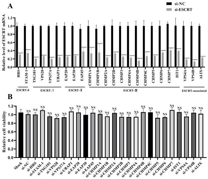
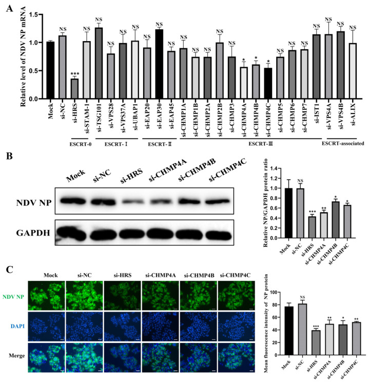
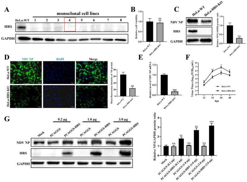
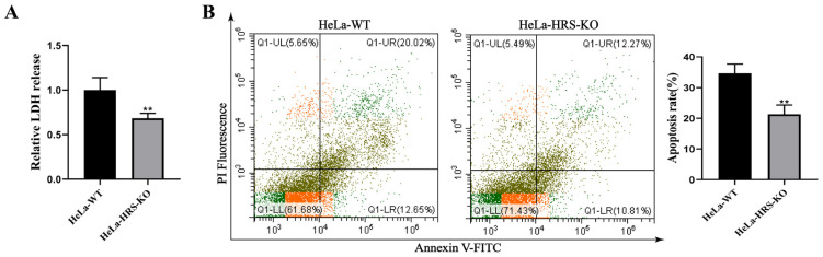
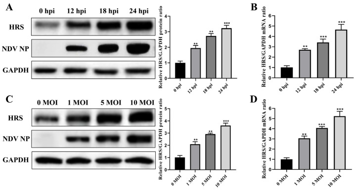
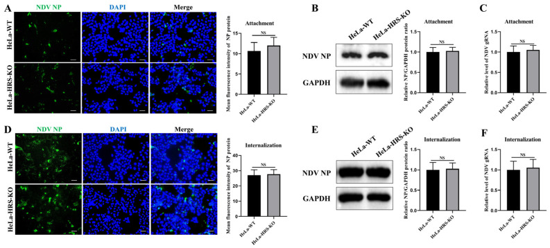
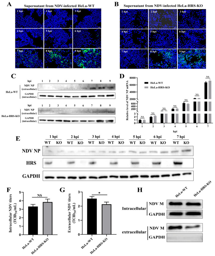
Newcastle disease virus (NDV) is a highly pathogenic avian infectious disease agent and also a promising oncolytic virus with broad application prospects. The Endosomal Sorting Complex Required for Transport (ESCRT) machinery has been increasingly recognized for its crucial role in the life cycles of enveloped viruses, influencing processes such as viral entry, replication, and budding. In this study, we employed an RNA interference screening approach to identify key ESCRT components that regulate NDV replication in tumor cells. qPCR, immunofluorescence, and Western blot assays demonstrated that knockdown of HRS, CHMP4A, CHMP4B, and CHMP4C significantly impaired NDV replication in HeLa cells, with HRS exhibiting the most pronounced inhibitory effect. Additionally, HRS knockout significantly inhibited viral budding and suppressed NDV-induced cell death in HeLa cells. Notably, NDV infection was shown to significantly upregulate HRS gene and protein expression in a time-dependent manner. In conclusion, this study systematically identifies critical ESCRT components involved in NDV replication within tumor cells, with a particular focus on the role of HRS in promoting NDV's replication by promoting viral budding, offering new insights for the development of NDV-based oncolytic therapies.

Keywords: ESCRT; HRS; NDV; viral budding; viral replication.


Conflict of interest statement

The authors declare no conflicts of interest.

Figures

Figure 1
Figure 1
Assessment of siRNA knockdown efficiency and cytotoxicity. (A) Detection of siRNA knockdown efficiency. HeLa cells were transfected with siRNAs targeting ESCRT subunits or si-NC for 48 h. The mRNA levels of the specified ESCRT subunits were quantified by qPCR and normalized to GAPDH using the 2−ΔΔCt method. (B) siRNA cytotoxicity assessment. HeLa cells cultured in 96-well plates were transfected with the indicated siRNAs, and cell viability was assessed using a CCK-8 assay at 48 h post-transfection (hpt). Data are presented as mean ± standard deviations (SDs) from three independent experiments. Statistical analysis was performed using a two-way ANOVA for the qPCR assay and a one-way ANOVA for the CCK-8 assay. NS means no significant difference, **** p < 0.0001.
Figure 2
Figure 2
Identification of key ESCRT subunits regulating NDV replication. (AC) HeLa cells were transfected with the indicated siRNAs or mock transfected for 48 h and then infected with the Herts/33 strain at a multiplicity of infection (MOI) of 1 for 12 h. (A) The mRNA levels of the NP gene were quantified by qPCR and normalized to GAPDH using the 2−ΔΔCt method. (B) The NP protein levels were detected by Western blot. The relative intensity of the NP protein was quantified using GAPDH as a normalization control, as shown in the right panel. (C) The NP protein was detected by immunofluorescence assay (IFA). The total fluorescence intensity of the NP protein was calculated using ImageJ software (version number: 1.53s) and displayed in the right panel. Scale bars represent 50 µm. Error bars represent SDs from triplicate analyses of three independent experiments. Representative images for Western blot and IFA are shown. Statistical significance was assessed using one-way ANOVA. NS means no significant difference, * p < 0.05, ** p < 0.01, *** p < 0.001.
Figure 3
Figure 3
HRS positively regulates NDV replication in HeLa cells. (A) Validation of HRS knockout cell lines. HeLa cell lines with HRS gene knockout were created using CRISPR/Cas9 technology, as detailed in the Materials and Methods section. The knockout of HRS was confirmed by Western blot. HeLa-WT were used as a control. The red box highlights the cell line used in this study, named HeLa-HRS-KO. (B) Cytotoxicity evaluation of HRS knockout. The cell viability of HeLa-WT and HeLa-HRS-KO cells was assessed using a CCK-8 assay. (CF) Impact of HRS knockout on NDV replication. HeLa-WT and HeLa-HRS-KO cells were infected with the Herts/33 strain at an MOI of 1. (C) The viral NP protein was detected by Western blot, and its relative intensity was quantified using ImageJ software and normalized to GAPDH, as shown in the right panel. (D) The viral NP protein was detected by IFA. The numbers of NP-expressing (green) and DAPI-stained (blue) cells were counted using ImageJ software. The infection rate was calculated as the ratio of NP-expressing cells to DAPI-stained cells, shown in the right panel. Scale bars represent 50 µm. (E) The mRNA levels of the NO gene were quantified by qPCR and normalized to GAPDH using the 2−ΔΔCt method. (F) Extracellular progeny virus titers in the cell supernatant were measured using a TCID50 assay. (G) Effect of HRS overexpression on NDV replication. HeLa-HRS-KO cells were transfected with the PCAGGS-HRS plasmid or vector control at specified doses for 48 h. The cells were then infected with the Herts/33 strain at an MOI of 1. The viral NP protein was detected by Western blot, and its relative intensity was quantified using ImageJ software and normalized to GAPDH, as shown in the right panel. Error bars represent SDs from triplicate analyses of three independent experiments. Representative images for Western blot and IFA are shown. Statistical significance was assessed using Student’s t tests for (BE), one-way ANOVA for (G) and two-way ANOVA for (F). NS means no significant difference, * p < 0.05, ** p < 0.01, *** p < 0.001.
Figure 4
Figure 4
Knockout of HRS significantly suppresses NDV-induced cell death of HeLa cells. (A) Detection of LDH release in NDV-infected HeLa-WT or HeLa-HRS-KO cells. HeLa-WT and HeLa-HRS-KO cells were infected with the Herts/33 strain at an MOI of 1. The abundance of LDH was detected at 12 h post-infection (hpi). (B) Detection of apoptosis in NDV-infected HeLa-WT or HeLa-HRS-KO cells. HeLa-WT and HeLa-HRS-KO cells were infected with the Herts/33 strain at an MOI of 1. The apoptosis level was measured by AnnexinV-FITC/PI staining using flow cytometry at 12 hpi, and quantitation of the apoptosis rate was shown in the right panel. Error bars represent SDs from triplicate analyses of three independent experiments. Representative images for apoptosis detection are shown. Statistical significance was assessed using Student’s t tests for (A,B). ** p < 0.01.
Figure 5
Figure 5
NDV infection significantly upregulates the expression of HRS. (A,B) HeLa cells were infected with Herts/33 at an MOI of 1. The protein and mRNA levels of endogenous HRS were then detected by Western blot (A) and qPCR (B) at indicated time points (0, 12, 18, and 24 hpi). (C,D) HeLa cells were infected with Herts/33 at indicated MOIs (0.01, 0.1, and 1). The protein and mRNA levels of endogenous HRS were then detected by Western blot (C) and qPCR (D) at 12 hpi. The grayscale of HRS was quantified using ImageJ software and normalized to GAPDH, as shown in the right panel of corresponding image. The mRNA levels of the HRS gene were also normalized to GAPDH using the 2−ΔΔCt method. Error bars represent SDs from triplicate analyses of three independent experiments. Representative images for Western blot are shown. Statistical significance was assessed using one-way ANOVA. ** p < 0.01, *** p < 0.001.
Figure 6
Figure 6
HRS does not affect NDV attachment and internalization in HeLa cells. (AC) Knockout of HRS does not affect NDV attachment. HeLa-WT and HeLa-HRS-KO cells were infected with Herts/33 at an MOI of 10 and cultured at 4 °C for 1 h. After adsorption, unbound viruses were extensively washed away with ice-cold phosphate-buffered saline (PBS). (A) Cell-attached NDV virions were assessed with a mouse anti-NP monoclonal antibody (green) by IFA. (B) NP protein of cell-attached NDV virions was quantified by Western blot. (C) Genomic RNA of cell-attached NDV virions was quantified by qPCR. (DF) Knockout of HRS does not affect NDV internalization. HeLa-WT and HeLa-HRS-KO cells were infected with Herts/33 at an MOI of 10 and incubated at 4 °C for 1 h. Unbound NDV virions were washed away with ice-cold PBS, and the cells were switched to 37 °C for 1 h. Internalized virions were quantified by IFA (D), Western blot (E), and qPCR (F) as described above. Total fluorescence intensity of NP protein in (A,D) was calculated using ImageJ software and shown in the right panel. Relative intensity of NP protein in (B,E) was normalized to GAPDH and shown in the right panel. Viral genomic RNA was also normalized to GAPDH using the 2−ΔΔCt method. Error bars represent SDs from triplicate analyses of three independent experiments. Representative images for IFA and Western blot are shown. Scale bars for IFA images represent 50 µm. Statistical significance was assessed using Student’s t tests. NS means no significant difference.
Figure 7
Figure 7
HRS promotes NDV replication in HeLa cells by enhancing viral budding. (A,B) Determination of the first life cycle of NDV. HeLa-WT and HeLa-HRS-KO cells were infected with Herts/33 at an MOI of 1, and the cellular supernatant was harvested every 1 h until 9 hpi. Fresh DF-1 cells grown in 24-well plates were infected with the collected supernatants for 12 h. IFA was performed to detect the fluorescence signal of the virus stained with a mouse anti-NP monoclonal antibody (green). Nuclei were stained with DAPI (blue). Scale bars represent 50 µm. (C) The proteins in the supernatants were concentrated using TCA precipitation. Proteins from each sample were used for Western blot to detect the abundance of progeny virus with a mouse anti-NP monoclonal antibody. The protein level of intracellular GAPDH was used as a control. (D,E) HRS does not impact viral RNA replication or protein translation. HeLa-WT and HeLa-HRS-KO cells were infected with Herts/33 at an MOI of 1, and cells were harvested every 1 h until 7 hpi. (D) The NP mRNA levels in the cells were detected by qPCR and normalized to GAPDH using the 2−ΔΔCt method. (E) The NP protein levels in the cells were detected by Western blot. (F,G) HRS promotes NDV replication by enhancing viral budding. The HeLa-WT and HeLa-HRS-KO cells were infected with Herts/33 at an MOI of 1 for 7 h. The infected cells and supernatants were harvested separately for detection of the intracellular (F) and extracellular (G) progeny virus titers by assessing TCID50. (H) Evaluation of the impact of HRS knockout on NDV M protein-mediated budding. HeLa-WT and HeLa-HRS-KO cells were transfected with plasmids expressing Flag-tagged M protein for 48 h. The cells and culture supernatants were collected, and the supernatants were purified and concentrated as described in the Materials and Methods section. M protein expression in the cells and culture supernatants was then detected by Western blot. Error bars represent SDs from triplicate analyses of three independent experiments. Representative images for IFA and Western blot are shown. Statistical significance was assessed using two-way ANOVA for (D) and Student’s t tests for (F,G). NS means no significant difference, * p < 0.05.

Similar articles

References

    1. Alexander D.J. Newcastle disease and other avian paramyxoviruses. Rev. Sci. Tech. 2000;19:443–462. doi: 10.20506/rst.19.2.1231. - DOI - PubMed
    1. Chen Y., Zhu S., Liao T., Wang C., Han J., Yang Z., Lu X., Hu Z., Hu J., Wang X., et al. The HN protein of Newcastle disease virus induces cell apoptosis through the induction of lysosomal membrane permeabilization. PLoS Pathog. 2024;20:e1011981. doi: 10.1371/journal.ppat.1011981. - DOI - PMC - PubMed
    1. Ginting T.E., Christian S., Larasati Y.O., Suryatenggara J., Suriapranata I.M., Mathew G. Antiviral interferons induced by Newcastle disease virus (NDV) drive a tumor-selective apoptosis. Sci. Rep. 2019;9:15160. doi: 10.1038/s41598-019-51465-6. - DOI - PMC - PubMed
    1. Zhang D., Ding Z., Xu X. Pathologic Mechanisms of the Newcastle Disease Virus. Viruses. 2023;15:864. doi: 10.3390/v15040864. - DOI - PMC - PubMed
    1. Zamarin D., Palese P. Oncolytic Newcastle disease virus for cancer therapy: Old challenges and new directions. Future Microbiol. 2012;7:347–367. doi: 10.2217/fmb.12.4. - DOI - PMC - PubMed

MeSH terms

Substances

LinkOut - more resources