. 2024 Sep 19;25(18):10071.
doi: 10.3390/ijms251810071.

Strongly ROS-Correlated, Time-Dependent, and Selective Antiproliferative Effects of Synthesized Nano Vesicles on BRAF Mutant Melanoma Cells and Their Hyaluronic Acid-Based Hydrogel Formulation

Affiliations

Strongly ROS-Correlated, Time-Dependent, and Selective Antiproliferative Effects of Synthesized Nano Vesicles on BRAF Mutant Melanoma Cells and Their Hyaluronic Acid-Based Hydrogel Formulation

Silvana Alfei et al. Int J Mol Sci. .

Abstract

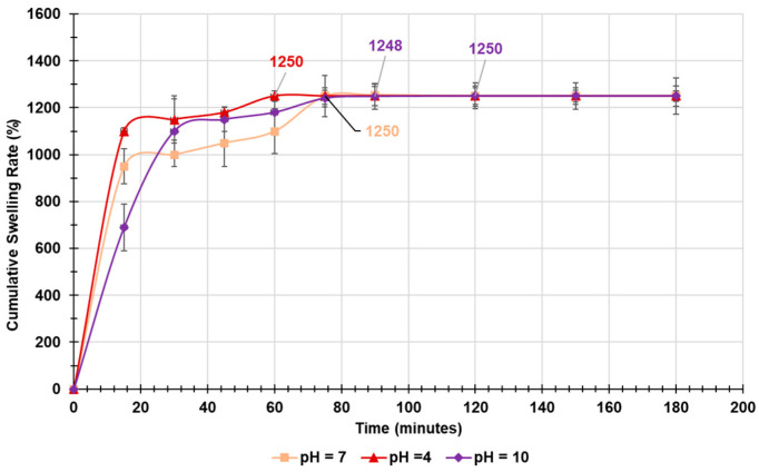
Cutaneous metastatic melanoma (CMM) is the most aggressive form of skin cancer with a poor prognosis. Drug-induced secondary tumorigenesis and the emergency of drug resistance worsen an already worrying scenario, thus rendering urgent the development of new treatments not dealing with mutable cellular processes. Triphenyl phosphonium salts (TPPSs), in addiction to acting as cytoplasmic membrane disruptors, are reported to be mitochondria-targeting compounds, exerting anticancer effects mainly by damaging their membranes and causing depolarization, impairing mitochondria functions and their DNA, triggering oxidative stress (OS), and priming primarily apoptotic cell death. TPP-based bola amphiphiles are capable of self-forming nanoparticles (NPs) with enhanced biological properties, as commonly observed for nanomaterials. Already employed in several other biomedical applications, the per se selective potent antibacterial effects of a TPP bola amphiphile have only recently been demonstrated on 50 multidrug resistant (MDR) clinical superbugs, as well as its exceptional and selective anticancer properties on sensitive and MDR neuroblastoma cells. Here, aiming at finding new molecules possibly developable as new treatments for counteracting CMM, the effects of this TPP-based bola amphiphile (BPPB) have been investigated against two BRAF mutants CMM cell lines (MeOV and MeTRAV) with excellent results (even IC50 = 49 nM on MeOV after 72 h treatment). With these findings and considering the low cytotoxicity of BPPB against different mammalian non-tumoral cell lines and red blood cells (RBCs, selectivity indexes up to 299 on MeOV after 72 h treatment), the possible future development of BPPB as topical treatment for CMM lesions was presumed. With this aim, a biodegradable hyaluronic acid (HA)-based hydrogel formulation (HA-BPPB-HG) was prepared without using any potentially toxic crosslinking agents simply by dispersing suitable amounts of the two ingredients in water and sonicating under gentle heating. HA-BPPB-HA was completely characterized, with promising outcomes such as high swelling capability, high porosity, and viscous elastic rheological behavior.

Keywords: biodegradability; cutaneous metastatic melanoma (CMM); cytotoxicity studies; high porosity; high swelling; hydrogel formulation; low hemolytic effects; mitochondria-targeting molecules; nanosized bola amphiphiles vesicles; triphenyl phosphonium (TPP) groups.


Conflict of interest statement

The authors declare no conflicts of interest.

Figures

Figure 1
Figure 1
Chemical structure of a typical unit of HA in its sodium salt form.
Figure 2
Figure 2
Chemical structure of BPPB here evaluated as new possible agent against CMM.
Scheme 1
Scheme 1
Synthetic route to achieve BPPB.
Figure 3
Figure 3
Cell viability was evaluated in MeOV CMM cells exposed to increasing concentrations of BPPB (0.1–2.0 µM) for 24 (blue bars), 48 (green bars), and 72 h (pink bars). Control (Ctr) refers to a concentration of 0 µM. Bar graphs summarize quantitative data of means ± S.D. of four independent experiments run in triplicate. Significance refers exclusively to control (*). Specifically, p > 0.05 no symbols; **** p < 0.0001; *** p < 0.001 (0.0002) (one-way ANOVA followed by Dunnet’s multi-comparisons test). Black dots in the image represent the number of independent experiments.
Figure 4
Figure 4
Cell viability was evaluated in MeTRAV CMM cells exposed to increasing concentrations of BPPB (0.1–2.0 µM) for 24 (blue bars), 48 (green bars), and 72 h (pink bars). Control (Ctr) refers to concentration of 0 µM. Bar graphs summarize quantitative data of means ± S.D. of four independent experiments run in triplicate. Significance refers exclusively to control (*). Specifically, p > 0.05 no symbols; **** p < 0.0001 (one-way ANOVA followed by Dunnet’s multi-comparisons test). Black dots in the image represent the number of independent experiments.
Figure 5
Figure 5
IC50 values of BPPB towards MeOV (blue trace) and MeTRAV (red trace) as functions of exposure timing.
Figure 6
Figure 6
Selectivity of BPPB towards MeOV (blue trace) and MeTRAV (red trace) as functions of time of exposure in relation to its cytotoxic effects on MRC-5 cells (A) and its hemolytic effects on RBCs (B).
Figure 7
Figure 7
H2O2 production was analyzed in MeOV cells exposed to increasing concentrations of BPPB (0.1–2 µM) at 24 h (blue bars), 48 h (green bars), and 72 h (pink to purple bars). Control (Ctr) refers to concentration 0 µM. Bar graphs summarize quantitative data of means ± SD of three independent experiments. Significance refers to control (*). Specifically, p > 0.05 ns; p < 0.01 ** p < 0.001 ***, and p < 0.0001 ****. Black dots in the image represent the number of independent experiments.
Figure 8
Figure 8
H2O2 production was analyzed in MeTRAV cells exposed to increasing concentrations of BPPB (0.1–2 µM) at 24 h (blue bars), 48 h (green bars), and 72 h (pink to purple bars). Control (Ctr) refers to concentration 0 µM. Bar graphs summarize quantitative data of means ± SD of three independent experiments. Significance refers to control (*). Specifically, p > 0.05 ns; p < 0.05 *, p < 0.01 ** p < 0.001 ***, and p < 0.0001 ****. Black dots in the image represent the number of independent experiments.
Figure 9
Figure 9
Dispersion graph of the ratios DCFH positive cells (%)/viable cells (%) observed upon administration of max concentration of BPPB (2.0 µM) to both cells lines vs. exposure timing.
Figure 10
Figure 10
Dispersion graphs of the values of ratios DCFH positive cells (%)/viable cells (%) vs. cell viability reported in logarithmic scale concerning MeOV cell line (A) and MeTRAV (B) after 24 (light blue indicators and tendency line), 48 (green indicators and tendency line), and 72 h (fuchsia indicators and tendency line) of treatment.
Figure 11
Figure 11
ATR-FTIR spectra of BPPB (blue line), HA (fuchsia line), and HA-BPPB-HG (dark purple line).
Figure 12
Figure 12
PCA results reported as PC1 vs. PC2. The red cross refers to the score zero on both PCs.
Figure 13
Figure 13
Cross-sectional SEM images of HA-BPPB-HG. Reported images were acquired at 250× (A) and 274× (B) magnification. Bar scale corresponds to 100 µm.
Figure 14
Figure 14
Plots of the cumulative swelling rate values of HA-BPPB-HG vs. time at pH = 4 (red line), 7 (orange line), and pH = 10 (purple line).
Figure 15
Figure 15
Cumulative mass loss percentage curves of developed HA-BPPB-HG at pH = 4 (red line), pH = 7 (orange line), and pH = 10 (purple line).
Figure 16
Figure 16
Cumulative release percentage curves of BPPB from HA-BPPB-HG at pH = 4 (red line) and pH = 7 (purple line).

Similar articles

Cited by

References

    1. Boutros A., Croce E., Ferrari M., Gili R., Massaro G., Marconcini R., Arecco L., Tanda E.T., Spagnolo F. The Treatment of Advanced Melanoma: Current Approaches and New Challenges. Crit. Rev. Oncol. Hematol. 2024;196:104276. doi: 10.1016/j.critrevonc.2024.104276. - DOI - PubMed
    1. Hodi F.S., Chiarion -Sileni V., Lewis K.D., Grob J.-J., Rutkowski P., Lao C.D., Cowey C.L., Schadendorf D., Wagstaff J., Dummer R., et al. Long-Term Survival in Advanced Melanoma for Patients Treated with Nivolumab plus Ipilimumab in CheckMate 067. J. Clin. Oncol. 2022;40:9522. doi: 10.1200/JCO.2022.40.16_suppl.9522. - DOI
    1. Garbe C., Eigentler T.K., Keilholz U., Hauschild A., Kirkwood J.M. Systematic Review of Medical Treatment in Melanoma: Current Status and Future Prospects. Oncologist. 2011;16:5–24. doi: 10.1634/theoncologist.2010-0190. - DOI - PMC - PubMed
    1. Natarelli N., Aleman S.J., Mark I.M., Tran J.T., Kwak S., Botto E., Aflatooni S., Diaz M.J., Lipner S.R. A Review of Current and Pipeline Drugs for Treatment of Melanoma. Pharmaceuticals. 2024;17:214. doi: 10.3390/ph17020214. - DOI - PMC - PubMed
    1. Akbani R., Akdemir K.C., Aksoy B.A., Albert M., Ally A., Amin S.B., Arachchi H., Arora A., Auman J.T., Ayala B., et al. Genomic Classification of Cutaneous Melanoma. Cell. 2015;161:1681–1696. doi: 10.1016/j.cell.2015.05.044. - DOI - PMC - PubMed

MeSH terms

LinkOut - more resources