. 2024 Sep 19;25(18):10080.
doi: 10.3390/ijms251810080.

Tissue Kallikrein-1 Suppresses Type I Interferon Responses and Reduces Depressive-Like Behavior in the MRL/lpr Lupus-Prone Mouse Model

Affiliations

Tissue Kallikrein-1 Suppresses Type I Interferon Responses and Reduces Depressive-Like Behavior in the MRL/lpr Lupus-Prone Mouse Model

Priyanka S Bhoj et al. Int J Mol Sci. .

Abstract

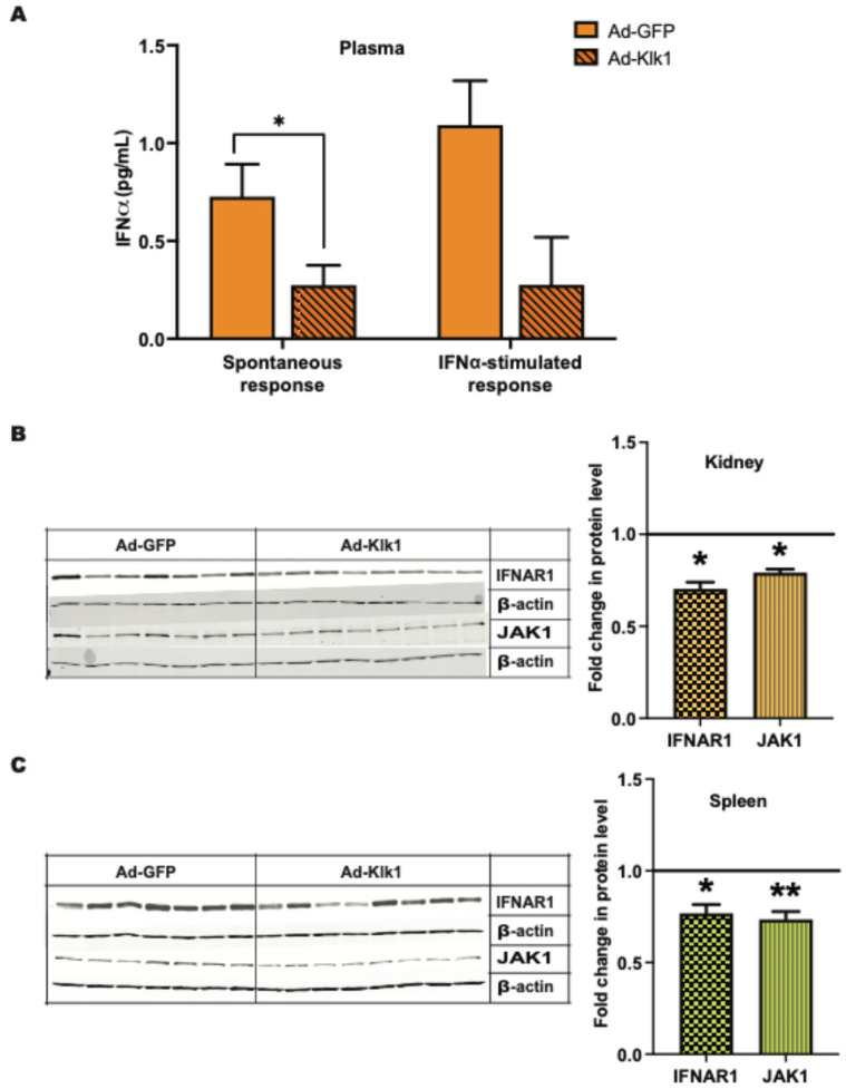
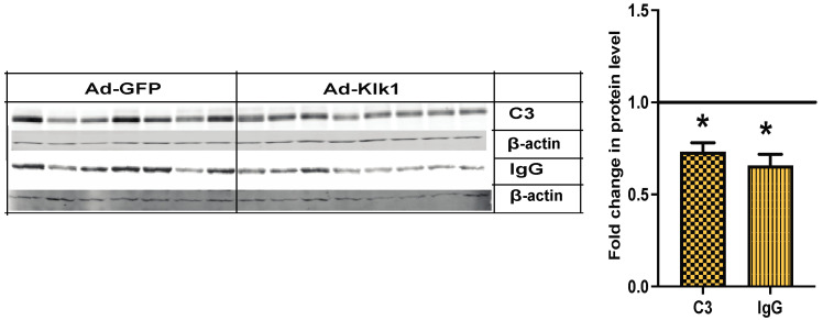
Excessive production and response to Type I interferons (IFNs) is a hallmark of systemic lupus erythematosus (SLE). Neuropsychiatric lupus (NPSLE) is a common manifestation of human SLE, with major depression as the most common presentation. Clinical studies have demonstrated that IFNα can cause depressive symptoms. We have shown that the kallikrein-kinin system (KKS) [comprised of kallikreins (Klks) and bradykinins] and angiotensin-converting enzyme inhibitors suppressed Type I IFN responses in dendritic cells from lupus-prone mice and human peripheral blood mononuclear cells. Tissue Klk genes are decreased in patients with lupus, and giving exogenous Klk1 ameliorated kidney pathology in mice. We retro-orbitally administered mouse klk1 gene-carrying adenovirus in the Murphy Roths Large lymphoproliferative (MRL/lpr) lupus-prone mice at early disease onset and analyzed immune responses and depressive-like behavior. Klk1 improved depressive-like behavior, suppressed interferon-responsive genes and neuroinflammation, and reduced plasma IFNα levels and proinflammatory cytokines. Klk1 also reduced IFNAR1 and JAK1 protein expression, important upstream molecules in Type I IFN signaling. Klk1 reduced bradykinin B1 receptor expression, which is known to induce proinflammatory response. Together, these findings suggest that Klk1 may be a potential therapeutic candidate to control IFNα production/responses and other inflammatory responses in SLE and NPSLE.

Keywords: depression; kallikrein–kinin system; neuropsychiatric lupus; tissue kallikreins; type I interferons.


Conflict of interest statement

The authors declare no conflicts of interest.

Figures

Figure 1
Figure 1
Administration of Ad-klk1 increases Klk1 protein expression (A) Western blot of Klk1 protein (MW: 28 kDa) and (B) immunohistochemical results (indicated by pink staining) of Klk1 protein expression in the kidney of the spontaneous cohort. Images were taken at 40× magnification and analyzed using ImageJ (1.54f software). The results are expressed as mean ±SEM. An unpaired t-test was used; * p ≤ 0.05 and *** p ≤ 0.001 compared to the Ad-GFP group; n = 7–8 mice/group.
Figure 2
Figure 2
Tissue Klk1 decreases IRGs and hmgb1 gene expression in the brain and periphery: (A) Spontaneous MRL/lpr mice and (B) IFNα-induced MRL/lpr mice. The data, obtained by qPCR, were analyzed using an unpaired t-test and are expressed as the mean ±SEM of the fold change in gene expression in the Ad-Klk1 group compared to the Ad-GFP group (marked as a solid black line at 1.0 on the Y axis). * p ≤ 0.05 and ** p ≤ 0.01 compared to the Ad-GFP group; n = 7–8 mice/group in the spontaneous cohort, and n = 5 mice/group in the IFNα-induced cohort.
Figure 3
Figure 3
Tissue Klk1 reduces inflammatory cytokine levels in the plasma: (A) Spontaneous MRL/lpr mice and (B) IFNα-induced MRL/lpr mice. Multiplex cytokine analysis was performed using the MSD ELISA kit. The data were analyzed using an unpaired t-test and are expressed as the mean ±SEM. * p ≤ 0.05 compared to the Ad-GFP group; n = 7–8 mice/group in the spontaneous cohort, and n = 5 mice/group in the IFNα-induced cohort.
Figure 4
Figure 4
Tissue Klk1 decreases plasma IFNα levels and proteins in Type I IFN signaling: (A) Plasma IFNα levels in the spontaneous MRL/lpr mice were measured using the high sensitivity Verikine ELISA kit and are expressed as the mean ±SEM of IFNα levels (pg/mL). Western blot analysis of IFNAR1 (MW: 64 kDa) and JAK1 (MW: 80 kDa) protein levels in the (B) kidney and (C) spleen of spontaneous MRL/lpr mice. Data were analyzed using an unpaired t-test and are expressed as the mean ±SEM of the fold change in protein levels in the Ad-Klk1 group compared to the Ad-GFP group (marked as a solid black line at 1.0 on the Y axis). * p ≤ 0.05 and ** p ≤ 0.01 compared to the Ad-GFP group; n = 7–8 mice/group.
Figure 5
Figure 5
Tissue Klk1 decreases depressive-like behavior and depression-related gene expression in the spontaneous MRL/lpr mice: (A) Tail suspension test (TST). (B) Plasma serotonin levels were estimated by using the ELISA kit. (C) Gene expression analysis of serotonin regulators in the brain; the results are expressed as the mean ±SEM of the fold change in gene expression in the Ad-Klk1 group compared to the Ad-GFP group (marked as a solid black line at 1.0 on the Y axis). (D) Correlation of the immobility time in the TST with plasma IFNα levels and serotonin-related markers (brain isg15, 5htt, and tph-2). The data were analyzed using an unpaired t-test and * p ≤ 0.05 compared to the Ad-GFP group; n = 7–8 mice/group.
Figure 6
Figure 6
Tissue Klk1 reduced microglial inflammatory state and number. Immunohistochemical staining of IBA-1 in microglia. Data were analyzed using an unpaired t-test and are expressed as the mean ±SEM of the percentage area of IBA-1 staining in the cortex region of the brain at 40× magnification (brown staining, indicated by yellow arrows) using ImageJ (1.54f software). * p ≤ 0.05 compared to Ad-GFP group; n = 7–8 mice/group.
Figure 7
Figure 7
Tissue Klk1 decreases C3 and IgG protein levels in the kidney: Western blot analysis of C3 (MW: 50 kDa) and IgG (MW: 50 kDa) protein levels in the kidney of spontaneous MRL/lpr mice. Data were analyzed using an unpaired t-test and are expressed as the mean ±SEM of the fold change in protein levels in the Ad-Klk1 group compared to the Ad-GFP group (marked as a solid black line at 1.0 on the Y axis). * p ≤ 0.05 compared to the Ad-GFP group; n = 7–8 mice/group.
Figure 8
Figure 8
Tissue Klk1 alters BK receptor expression in the spontaneous MRL/lpr mice: (A) Fold change in b1r and b2r gene expression in the brain of the Ad-Klk1 group compared to those of the Ad-GFP group (marked as a solid black line at 1.0 on the Y axis), as measured by qPCR. Fold change in B1R (MW: 40 kDa) and B2R (MW: 50 kDa) protein levels in the (B) kidneys and (C) spleen of the Ad-Klk1 group compared to those of the Ad-GFP group (marked as a solid black line at 1.0 on the Y axis), as measured by western blot. The results are expressed as mean ±SEM. An unpaired t-test was used; * p ≤ 0.05 and p ≤ 0.001 compared to the Ad-GFP group; n = 7–8 mice/group.
Figure 9
Figure 9
Scheme of the experiment: One cohort of nine-week-old MRL/lpr mice was administered with IFNα- or saline-carrying osmotic pump (IFNα-induced response) on day −2, followed by the retro-orbital administration of Ad-klk1 or Ad-gfp vector on day 0. The spontaneous cohort received Ad-klk1 or control (Ad-gfp) vector only. Behavioral studies were performed on day 15, and the mice were sacrificed on day 17 to collect the brain, spleen, kidneys, and blood.
Figure 10
Figure 10
Tissue Klk1 suppresses Type I IFN responses through IFNAR1, BRs, and PAR2 and reduces depressive-like behavior in the MRL/lpr lupus-prone mice: The present study demonstrates that tissue Klk1 administration reduces IFNα, IFNAR1, JAK1, and IRF7, consequently suppressing IRG expression. Our previous study [11] has shown a reduction in signal transducer and activator of transcription (STAT) phosphorylation (shown in blue color) following bradykinin (BK) administration, which, along with other pathway proteins (shown in grey color), will be studied in detail in the future. We observed a consequential reduction in the levels of tryptophan hydroxylase 2 (TPH2, a rate-limiting enzyme in serotonin biosynthesis), serotonin (5-HT), serotonin transporter (5-HTT), and 5-hydroxytryptamine receptor 2A (HTR2A, a serotonin receptor). We also observed depressive-like behaviors in MRL/lpr mice. Alleviation of inflammatory and IFN responses may be mediated via the alteration in BK receptors (B1R and B2R) and protease-activated receptor-2 (PAR2) expression; the mechanisms need to be studied in detail (indicated with dashed arrows). The therapeutic effects of Klk1 are shown in green notation, and SLE and NPSLE pathogenesis are represented in red notation.

References

    1. Hawro T., Krupinska-Kun M., Rabe-Jablonska J., Sysa-Jedrzejowska A., Robak E., Bogaczewicz J., Wozniacka A. Psychiatric disorders in patients with systemic lupus erythematosus: Association of anxiety disorder with shorter disease duration. Rheumatol. Int. 2011;31:1387–1391. - PMC - PubMed
    1. Morrison E., Carpentier S., Shaw E., Doucette S., Hanly J.G. Neuropsychiatric systemic lupus erythematosus: Association with global disease activity. Lupus. 2014;23:370–377. - PubMed
    1. Govoni M., Hanly J.G. The management of neuropsychiatric lupus in the 21st century: Still so many unmet needs? Rheumatology. 2020;59((Suppl. S5)):v52–v62. doi: 10.1093/rheumatology/keaa404. - DOI - PMC - PubMed
    1. Palagini L., Mosca M., Tani C., Gemignani A., Mauri M., Bombardieri S. Depression and systemic lupus erythematosus: A systematic review. Lupus. 2013;22:409–416. doi: 10.1177/0961203313477227. - DOI - PubMed
    1. Obermoser G., Pascual V. The interferon-alpha signature of systemic lupus erythematosus. Lupus. 2010;19:1012–1019. doi: 10.1177/0961203310371161. - DOI - PMC - PubMed

MeSH terms