Review
. 2024 Sep 20;25(18):10097.
doi: 10.3390/ijms251810097.

Autophagy as a Guardian of Vascular Niche Homeostasis

Affiliations
Review

Autophagy as a Guardian of Vascular Niche Homeostasis

Konstantin Dergilev et al. Int J Mol Sci. .

Abstract

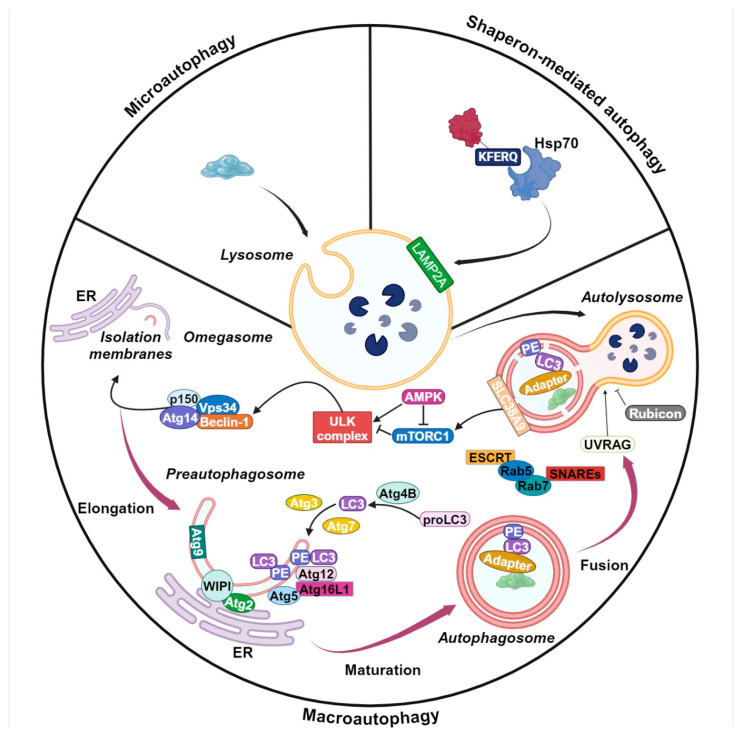
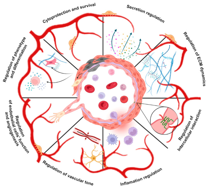
The increasing burden of vascular dysfunction on healthcare systems worldwide results in higher morbidity and mortality rates across pathologies, including cardiovascular diseases. Vasculopathy is suggested to be caused by the dysregulation of vascular niches, a microenvironment of vascular structures comprising anatomical structures, extracellular matrix components, and various cell populations. These elements work together to ensure accurate control of the vascular network. In recent years, autophagy has been recognized as a crucial regulator of the vascular microenvironment responsible for maintaining basic cell functions such as proliferation, differentiation, replicative senescence, and apoptosis. Experimental studies indicate that autophagy activation can be enhanced or inhibited in various pathologies associated with vascular dysfunction, suggesting that autophagy plays both beneficial and detrimental roles. Here, we review and assess the principles of autophagy organization and regulation in non-tumor vascular niches. Our analysis focuses on significant figures in the vascular microenvironment, highlighting the role of autophagy and summarizing evidence that supports the systemic or multiorgan nature of the autophagy effects. Finally, we discuss the critical organizational and functional aspects of the vasculogenic niche, specifically in relation to autophagy. The resulting dysregulation of the vascular microenvironment contributes to the development of vascular dysfunction.

Keywords: autophagy; vascular niche; vascular regeneration.


Conflict of interest statement

The authors declare no conflicts of interest.

Figures

Figure 1
Figure 1
Basic mechanisms of autophagy. Microautophagy involves the direct engulfment of cytoplasmic components into lysosomes. Chaperone-mediated autophagy specifically targets and degrades proteins with a KFERQ motif. These proteins are recognized by the cytoplasmic chaperone Hsp70 and subsequently transported into the lysosome via LAMP2A. Macroautophagy, more commonly referred to as autophagy, involves the sequestration of cytoplasmic components within a double-membrane structure known as the autophagosome, which subsequently fuses with the lysosome for degradation. Autophagosome formation and maturation necessitate the involvement of several key protein complexes. The activation of the ULK complex is a critical initial step involving the phosphorylation of downstream ATG proteins. Autophagy is commonly initiated by nutrient deprivation mediated by AMPK and mTORC1. AMPK activates autophagy, while mTORC1 suppresses the ULK complex through phosphorylation. Following ULK activation, the PI3K complex, comprising Vps34, p150, Atg14, and Beclin-1, is essential for omegasome formation and PI3P generation. PI3P recruits WIPI, Atg9, and Atg2 proteins, forming a structure for elongating pre-autophagosome membranes. Maturation involves two ubiquitin-like conjugation systems: the Atg5/Atg12/Atg16L1 complex and the LC3 protein complex. LC3, processed from proLC3 to LC3 I by Atg4B and then lipidated to LC3 II, integrates into the autophagosome membrane. Finally, the mature autophagosome fuses with the lysosome to form the autolysosome. This fusion is regulated by SNAREs, ESCRT, and Rab GTPases, with Vps34/p150/Beclin-1 and UVRAG promoting fusion and Rubicon inhibiting autophagy. Created with BioRender.com (https://app.biorender.com accessed on 21 August 2024).
Figure 2
Figure 2
Autophagy regulates pivotal processes associated with vasculature niche homeostasis. Created with BioRender.com (https://app.biorender.com accessed on 21 August 2024).

Similar articles

Cited by

References

    1. Ribatti D., Tamma R., Annese T. The Role of Vascular Niche and Endothelial Cells in Organogenesis and Regeneration. Exp. Cell Res. 2021;398:112398. doi: 10.1016/j.yexcr.2020.112398. - DOI - PubMed
    1. Lei Z., Hu X., Wu Y., Fu L., Lai S., Lin J., Li X., Lv Y. The Role and Mechanism of the Vascular Endothelial Niche in Diseases: A Review. Front. Physiol. 2022;13:863265. doi: 10.3389/fphys.2022.863265. - DOI - PMC - PubMed
    1. He H., Xu J., Warren C.M., Duan D., Li X., Wu L., Iruela-Arispe M.L. Endothelial Cells Provide an Instructive Niche for the Differentiation and Functional Polarization of M2-like Macrophages. Blood. 2012;120:3152–3162. doi: 10.1182/blood-2012-04-422758. - DOI - PMC - PubMed
    1. Rouget C. Memoire Sur Les Development, La Structure et La Proprietes Physiologiques Des Capillaires Sanguines et Lymphatiques. Arch. Physiol. 1873;5:603–663.
    1. Eberth C.J. Handbuch Der Lehre von Der Gewegen Des Menschen und Der Tiereitle Bd.1. Verlag von Wilhelm Engelmann; Leipzig, Germany: 1871.

LinkOut - more resources