. 2024 Sep 20;25(18):10129.
doi: 10.3390/ijms251810129.

Chromate Affects Gene Expression and DNA Methylation in Long-Term In Vitro Experiments in A549 Cells

Affiliations

Chromate Affects Gene Expression and DNA Methylation in Long-Term In Vitro Experiments in A549 Cells

Franziska Fischer et al. Int J Mol Sci. .

Abstract

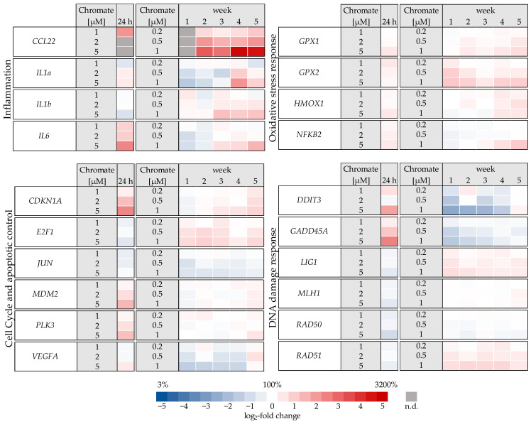
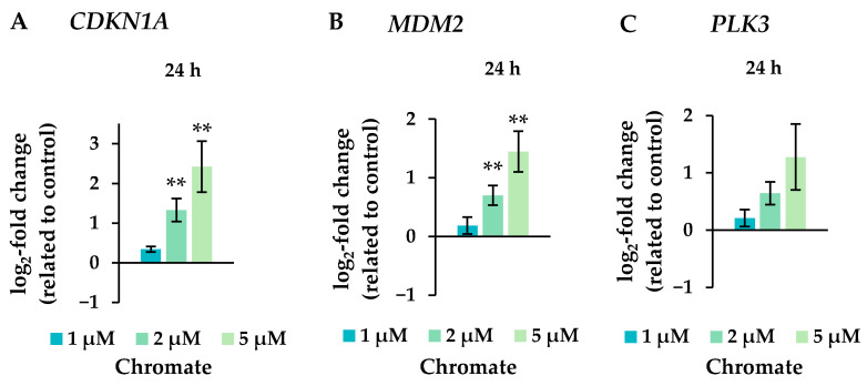
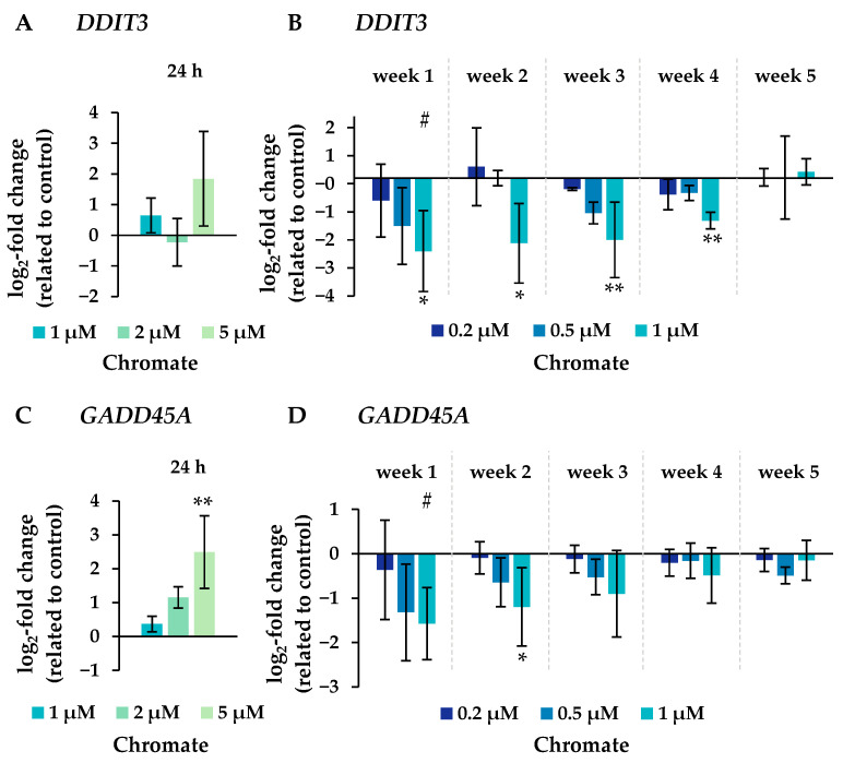
Chromate has been shown to dysregulate epigenetic mechanisms such as DNA methylation, leading to changes in gene expression and genomic instability. However, most in vitro studies are limited to short incubation periods, although chronic exposure may be more relevant for both environmental and occupational exposure. In this study, human adenocarcinoma A549 cells were treated with 1, 2 or 5 µM chromate for 24 h and compared with incubations with 0.2, 0.5 or 1 µM chromate for 1 to 5 weeks. Chromium accumulated in a pronounced time- and concentration-dependent manner after short-term treatment, whereas a plateau of intracellular chromium content was observed after long-term treatment. While short-term treatment induced a G2 arrest of the cell cycle, this effect was not observed after long-term treatment at lower concentrations. The opposite was observed for global DNA methylation: while short-term treatment showed no effect of chromate, significant dose-dependent hypomethylation was observed in the long-term experiments. Time-dependent effects were also observed in a high-throughput RT-qPCR gene expression analysis, particularly in genes related to the inflammatory response and DNA damage response. Taken together, the results suggest specific differences in toxicity profiles when comparing short-term and long-term exposure to chromate in A549 cells.

Keywords: DNA damage response; DNA methylation; chromate; epigenetic; gene expression profiles; inflammation; long-term exposure; oxidative stress.


Conflict of interest statement

The authors declare no conflicts of interest.

Figures

Figure 1
Figure 1
RCC of A549 cells after incubation for 24 h (A) or for 1–5 weeks (B) with Cr(VI). Shown are mean values of three independent experiments each performed in duplicates ± SD. Statistical analysis was performed to assess differences between treated and untreated cells within a time point using ANOVA followed by Dunnett’s T post hoc test. * p ≤ 0.05, ** p ≤ 0.01. Differences between 1 µM chromate short-term and long-term experiments were analyzed by ANOVA followed by Dunnett’s T post hoc test. ##: p ≤ 0.01.
Figure 2
Figure 2
Uptake of chromium in A549 cells after 24 h (A) or 1–5 weeks (B) of incubation with Cr(VI). The intracellular chromium content was determined via AAS, and intracellular chromium concentrations were calculated. Shown are mean values of three independent experiments, each performed in duplicates ± SD. Statistical analysis was performed to assess differences between treated and untreated cells within a time point by ANOVA followed by Dunnett’s T post hoc test. * p ≤ 0.05, ** p ≤ 0.01. Differences between 1 µM chromate after short-term and long-term treatment were determined by ANOVA followed by Dunnett’s T post hoc test. ## p ≤ 0.01.
Figure 3
Figure 3
Cell cycle phase distribution of A549 cells after 24 h Cr(VI) incubation (A) or incubation for 1–5 weeks (B). The cells were stained with DAPI and cell cycle stages were assigned via flow cytometry. Statistical analysis of the G2/M-phase was performed to assess differences between treated and untreated cells within a time point by using ANOVA followed by Dunnett’s T post hoc test. ** p ≤ 0.01.
Figure 4
Figure 4
Impact of Cr(VI) on the global 5-methyl-cytosine content in A549 cells after 24 h of treatment (A) and after incubation for 1–5 weeks (B), shown as a 5-methyl-cytosine level related to the untreated control. DNA isolated from untreated or treated cells was digested into single nucleosides and separated by HPLC on a C18 column. Cytidine was detected at 272 nm and 5-methylcytidine at 280 nm. Shown are mean values of three independent experiments performed in duplicates ± SD. Statistical analysis was performed to assess differences between treated and untreated cells within a time point using ANOVA followed by Dunnett’s T post hoc test. * p ≤ 0.05, ** p ≤ 0.01. Differences between 1 µM chromate after short-term and long-term treatment were determined by ANOVA followed by Dunnett’s T post hoc test. # p ≤ 0.05.
Figure 5
Figure 5
Overview of gene expression profiles of A549 cells treated with Cr(VI) for 24 h or 1–5 weeks using a high-throughput RT-qPCR. Genes depicted encode proteins involved in apoptotic and cell cycle control, inflammation, DNA damage response, and oxidative stress response. The log2 changes relative to the untreated control are illustrated. Blue represents a repression, and red represents an induction. Shown are mean values of at least three independent determinations performed in duplicates. n.d. = not determinable.
Figure 6
Figure 6
Changes in the expression of the inflammatory genes CCL22 after incubation for 1–5 weeks (A) and IL6 after 24 h of treatment (B) with chromate in A549 cells. Shown are mean values of at least three independent experiments performed in duplicates. Statistical analysis was performed to assess differences between treated and untreated cells within a time point followed by Dunnett’s T post hoc test. * p ≤ 0.05, ** p ≤ 0.01. Differences between the short-term (24 h) and long-term (1 to 5 weeks) experiments were determined by ANOVA followed by Dunnett’s T post hoc test. # p ≤ 0.05. n.d. = not determinable.
Figure 7
Figure 7
Expression levels of the cell cycle and apoptotic control genes CDKN1A (A), MDM2 (B), and PLK3 (C) in A549 cells after 24 h treatment with Cr(VI). The log2 changes relative to the untreated control are illustrated. Shown are mean values of at least three independent determinations performed in duplicates. Statistical analysis was performed to assess differences between treated and untreated cells by ANOVA followed by Dunnett’s T post hoc test. ** p ≤ 0.01.
Figure 8
Figure 8
Expression levels of genes related to DNA damage response DDIT3 (A,B) and GADD45A (C,D) in A549 cells after 24 h treatment (A,C) and after incubation for 1–5 weeks (B,D) with Cr(VI). The log2 changes relative to the untreated control are illustrated. Shown are mean values of at least three independent determinations performed in duplicates. Statistical analysis was performed to assess differences between treated and untreated cells within a time point using ANOVA followed by Dunnett’s T post hoc test. * p ≤ 0.05, ** p ≤ 0.01. Differences between short-term and long-term trials were determined by ANOVA followed by Dunnett’s T post hoc test. # p ≤ 0.05.

Similar articles

Cited by

References

    1. International Agency for Research on Cancer (IARC) A Review of Human Carcinogens. Volume 100C. International Agency for Research on Cancer; Lyon, France: 2012. pp. 147–167. Arsenic, Metals Fibres, and Dusts.
    1. Wetterhahn K.E., Hamilton J.W. Molecular basis of hexavalent chromium carcinogenicity: Effect on gene expression. Sci. Total Environ. 1989;86:113–129. doi: 10.1016/0048-9697(89)90199-X. - DOI - PubMed
    1. Zhitkovich A. Chromium in drinking water: Sources, metabolism, and cancer risks. Chem. Res. Toxicol. 2011;24:1617–1629. doi: 10.1021/tx200251t. - DOI - PMC - PubMed
    1. Wise S.S., Aboueissa A.E., Martino J., Wise J.P., Sr. Hexavalent Chromium-Induced Chromosome Instability Drives Permanent and Heritable Numerical and Structural Changes and a DNA Repair-Deficient Phenotype. Cancer Res. 2018;78:4203–4214. doi: 10.1158/0008-5472.CAN-18-0531. - DOI - PMC - PubMed
    1. Myers J.M., Antholine W.E., Myers C.R. The intracellular redox stress caused by hexavalent chromium is selective for proteins that have key roles in cell survival and thiol redox control. Toxicology. 2011;281:37–47. doi: 10.1016/j.tox.2011.01.001. - DOI - PMC - PubMed

MeSH terms

LinkOut - more resources