

Antioxidant Defenses, Oxidative Stress Responses, and Apoptosis Modulation in Spontaneous Abortion: An Immunohistochemistry Analysis of First-Trimester Chorionic Villi
- PMID: 39337859
- PMCID: PMC11432807
- DOI: 10.3390/life14091074
Antioxidant Defenses, Oxidative Stress Responses, and Apoptosis Modulation in Spontaneous Abortion: An Immunohistochemistry Analysis of First-Trimester Chorionic Villi
Abstract
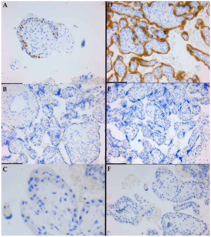
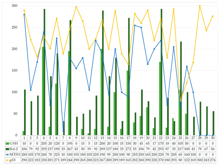
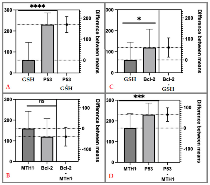
Oxidative stress (OS) and apoptosis are critical factors in placental development and function. Their interplay influences trophoblast proliferation, differentiation, and invasion, as well as vascular development. An imbalance between these processes can lead to pregnancy-related disorders such as preeclampsia, intrauterine growth restriction, and even spontaneous abortion. Our study seeks to elucidate the associations between preventive antioxidant/protective OS response factors-glutathione (GSH), MutT Homolog 1 (MTH1), and apoptotic regulation modulators-tumor protein p53 and B-cell lymphoma (Bcl-2) transcripts, in the context of spontaneous abortion (30 samples) versus elective termination of pregnancy (20 samples), using immunohistochemistry (IHC) to determine their proteomic expression in chorionic villi within abortive fetal placenta tissue samples. Herein, comparative statistical analyses revealed that both OS response factors, GSH and MTH1, were significantly under-expressed in spontaneous abortion cases as compared to elective. Conversely, for apoptotic regulators, p53 expression was significantly higher in spontaneous abortion cases, whereas Bcl-2 expression was significantly lower in spontaneous abortion cases. These findings suggest that a strong pro-apoptotic signal is prevalent within spontaneous abortion samples, alongside reduced anti-apoptotic protection, depleted antioxidant defenses and compromised oxidative DNA damage prevention/repair, as compared to elective abortion controls. Herein, our hypothesis that OS and apoptosis are closely linked processes contributing to placental dysfunction and spontaneous abortion was thus seemingly corroborated. Our results further highlight the importance of maintaining redox homeostasis and apoptotic regulation for a successful pregnancy. Understanding the mechanisms underlying this interplay is essential for developing potential therapies to manage OS, promote placentation, and avoid unwanted apoptosis, ultimately improving pregnancy outcomes. Antioxidant supplementation, modulation of p53 activity, and the enhancement of DNA repair mechanisms may represent potential approaches to mitigate OS and apoptosis in the placenta. Further research is needed to explore these strategies and their efficacy in preventing spontaneous abortion.
Keywords: B-cell lymphoma (Bcl)-2 transcript; MutT Homolog 1 (MTH1) protein; autophagy; histopathology of the placenta; immunohistochemistry; miscarriage; molecular markers; reduced glutathione (GSH); spontaneous abortion; tumor protein p53 transcript.
Conflict of interest statement
The authors declare no conflicts of interest.
Figures


References
-
- Latcu S., Novacescu D., Buciu V.-B., Dumitru C.-S., Ceauşu R., Raica M., Cut T.G., Ilina R., Malita D., Tarta C., et al. The Cavernous Nerve Injury Rat Model: A Pictorial Essay on Post-Radical Prostatectomy Erectile Dysfunction Research. Life. 2023;13:2337. doi: 10.3390/life13122337. - DOI - PMC - PubMed
-
- Vornic I., Pop O., Pascalau A., Andreescu G., Beiusan C., Manole F., Paiusan F., Pop-Crisan A., Furau C., Bodog A.D. Assessing the Role of Bcl-2 and P53 in Apoptotic Mechanisms in Spontaneous Abortions. Pharmacophore. 2024;15:1–6. doi: 10.51847/CO2qttSgIN. - DOI
-
- Pascual Z.N., Langaker M.D. StatPearls. StatPearls Publishing; Treasure Island, FL, USA: 2024. Physiology, Pregnancy. - PubMed
LinkOut - more resources
Full Text Sources
Research Materials
Miscellaneous
