. 2024 Sep 2;14(9):1103.
doi: 10.3390/life14091103.

Murine Retina Outer Plexiform Layer Development and Transcriptome Analysis of Pre-Synapses in Photoreceptors

Affiliations

Murine Retina Outer Plexiform Layer Development and Transcriptome Analysis of Pre-Synapses in Photoreceptors

Soo-Young Kim et al. Life (Basel). .

Abstract

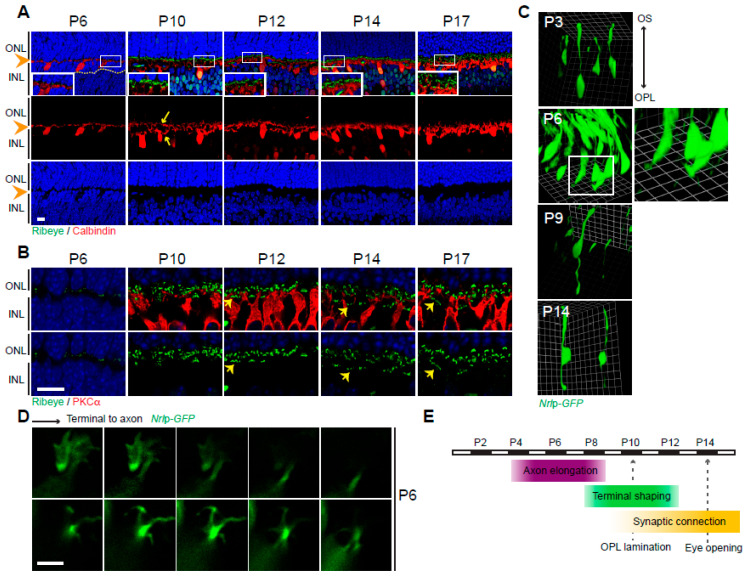
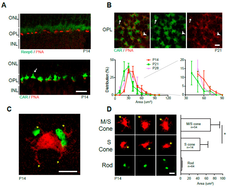
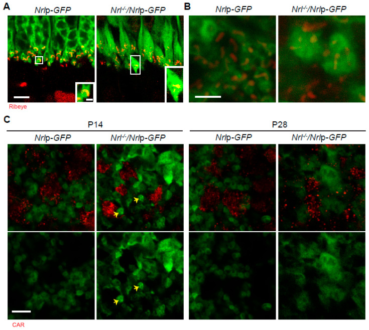
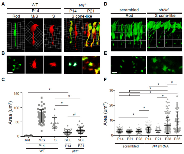
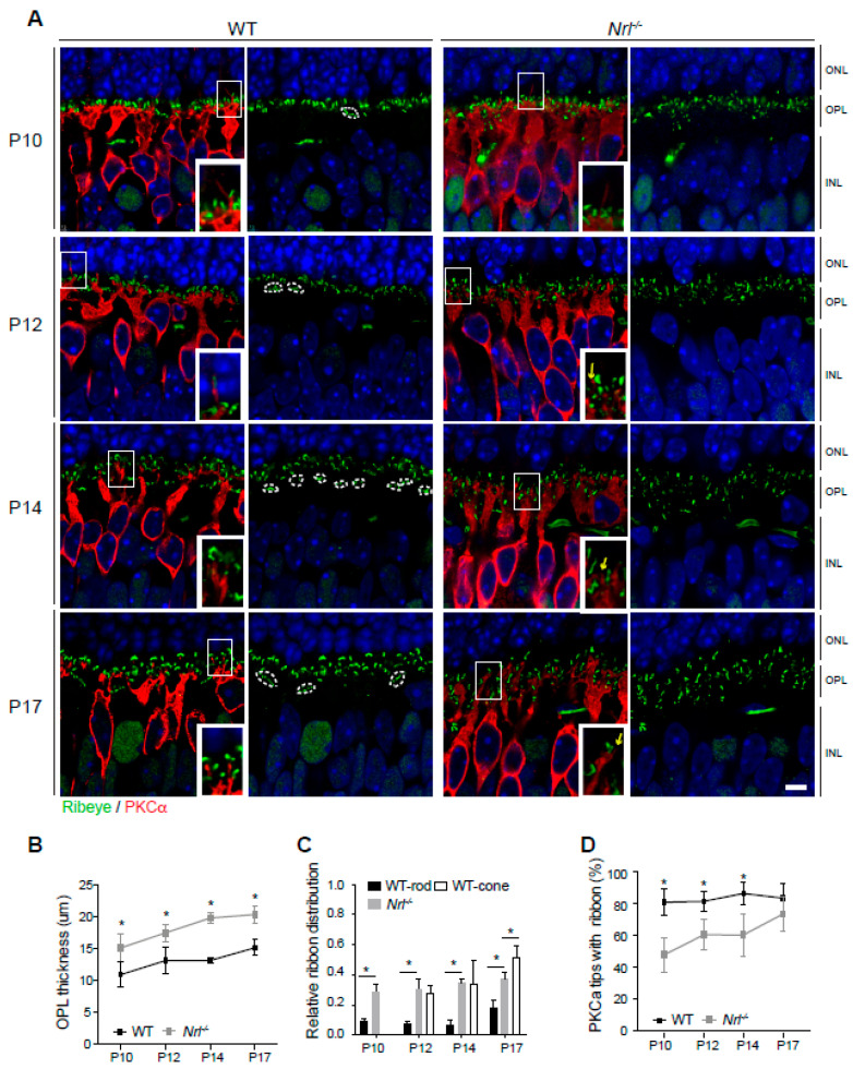
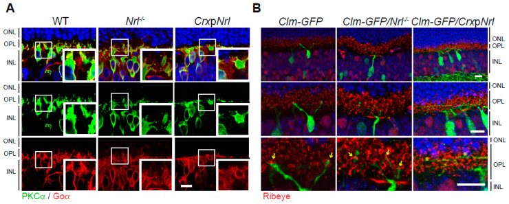
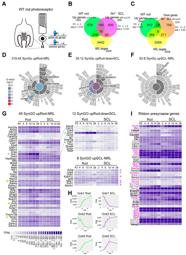
Photoreceptors in the mammalian retina convert light signals into electrical and molecular signals through phototransduction and transfer the visual inputs to second-order neurons via specialized ribbon synapses. Two kinds of photoreceptors, rods and cones, possess distinct morphology and function. Currently, we have limited knowledge about rod versus (vs.) cone synapse development and the associated genes. The transcription factor neural retina leucine zipper (NRL) determines the rod vs. cone photoreceptor cell fate and is critical for rod differentiation. Nrl knockout mice fail to form rods, generating all cone or S-cone-like (SCL) photoreceptors in the retina, whereas ectopic expression of Nrl using a cone-rod homeobox (Crx) promoter (CrxpNrl) forms all rods. Here, we examined rod and cone pre-synapse development, including axonal elongation, terminal shaping, and synaptic lamination in the outer plexiform layer (OPL) in the presence or absence of Nrl. We show that NRL loss and knockdown result in delayed OPL maturation and plasticity with aberrant dendrites of bipolar neurons. The integrated analyses of the transcriptome in developing rods and SCLs with NRL CUT&RUN and synaptic gene ontology analyses identified G protein subunit beta (Gnb) 1 and p21 (RAC1) activated kinase 5 (Pak5 or Pak7) transcripts were upregulated in developing rods and down-regulated in developing SCLs. Notably, Gnb1 and Gnb5 are rod dominant, and Gnb3 is enriched in cones. NRL binds to the genes of Gnb1, Gnb3, and Gnb5. NRL also regulates pre-synapse ribbon genes, and their expression is altered in rods and SCLs. Our study of histological and gene analyses provides new insights into the morphogenesis of photoreceptor pre-synapse development and regulation of associated genes in the developing retina.

Keywords: gene expression; neural retina leucine zipper; pedicle; photoreceptor synapse; retina outer plexiform layer; spherule; transcriptional regulation.


Conflict of interest statement

The authors declare no competing interests.

Figures

Figure 1
Figure 1
Wild-type outer plexiform layer (OPL) development. (A) Developing OPL stained by anti-Ribeye (synaptic ribbons, green) and anti-Calbindin (horizontal neurons, red). Nuclei stained with DAPI. The boxed areas are shown in insets with higher magnification. OPL (arrowheads) and separate fields of dendrites and axon branches from horizontal neurons (arrows) are shown. (B) Developing OPL stained by anti-Ribeye (green) and anti-PKCα (rod bipolar neurons, red). Clusters of pedicle ribbons (arrows) are shown. (C) Developing rod photoreceptors. Three-dimensional Volocity converted confocal images labeled by in vivo electroporation of Nrlp-EGFP plasmids. Insert at P6 is shown with higher magnification. (D) Growth cone-like structure at P6 terminals of rod photoreceptors. Confocal images displayed at z-thickness of 0.5 μm from synaptic terminal to axon stalk, labeled by in vivo electroporation of Nrlp-EGFP. (E) Schematic summary of OPL and photoreceptor synapse development. Abbreviations: ONL, outer nuclear layer; INL, inner nuclear layer; OPL, outer plexiform layer; DAPI, 4′,6-diamidino-2-phenylindole; P, postnatal day; PKCα, protein kinase C alpha. Scale bars, 10 μm in (A,B) and 5 μm in (D).
Figure 2
Figure 2
Wild-type spherule and pedicle. (A) P14 vertical retina sections stained by anti-Reep6 (spherules; green) and PNA (pedicles; red) (upper panel) or anti-CAR (M-cone, green) and PNA (S-cone, red) (lower panel). (B) Horizontal OPL images of retina whole mounts stained by anti-CAR and PNA (upper panels). Pure M-cone (arrows) and S-cone (arrowheads) pedicles are observed. The graph shows the distribution (%, average ± SEM) of CAR pedicle areas in OPL of P14, 21, and 28 whole mount retinas. Over 180 CAR positive pedicles were measured from 3 wild-type C57BL/6J retinas. The area distribution after 30 μm2 is magnified on the left side. (C) Spherules (green) and a pedicle (red) in CD1 retina whole mount labeled by in vivo electroporation of Nrlp-EGFP and S-opsinp-tdT. Telodendria (yellow arrows) are observed. (D) Representative images of M/S-, S-cone pedicles and spherules and their area size comparison. Telodendria (yellow arrows) are observed in cones. M-cone and pure S-cone pedicles are segregated by anti-M-opsin staining in the retina whole mounts labeled by S-opsinp-tdT electroporation. M/S-pedicles (n = 54), S-pedicles (n = 14), and Rod spherules (n = 64) from 3 to 5 wild-type CD1 retinas were measured. The graph displays the average ± SD of each: 70.79 ± 21.48 μm2 for M pedicles, 45.91 ± 18.97 μm2 for S pedicles, and 2.64 ± 0.81 μm2 for rod spherules. * p ≤ 0.05, two-tailed T-test. Abbreviations: CAR, cone arrestin; PNA, peanut agglutin lectin; S-opsin promoter-driven tdTomato (S-opsinp-tdT); P, postnatal day; ONL, outer nuclear layer; OPL, outer plexiform layer; INL, inner nuclear layer. Scale bars, 10 μm in (A), and 5 μm in (BD).
Figure 3
Figure 3
Photoreceptor pre-synapse terminals in Nrl-/- retina. (A) Vertical retina sections of P18 Nrlp-GFP and Nrl-/-/Nrlp-GFP mice, stained by anti-Ribeye (red). (B) Horizontal OPL of retina whole mounts in P18 Nrlp-GFP and Nrl-/-/Nrlp-GFP mice, stained by anti-Ribeye (red). (C) Horizontal OPL of retina whole mounts in P14 and P28 Nrlp-GFP and Nrl-/-/Nrlp-GFP mice, stained by anti-CAR (red). Arrows indicate pre-synaptic terminals in small size. Abbreviations: Nrl, neural retina leucine zipper; GFP, green fluorescent protein; Nrlp-GFP, Nrl promoter-driven GFP; P, postnatal day; CAR, cone arrestin. Scale bars, 1 μm in magnified box of (A), 5 μm in (A,C), and 2.5 μm in (B).
Figure 4
Figure 4
Pre-synapse comparison in wild-type rod, M/S-cone, pure S-cone, and Nrl-/- or Nrl knockdown S-cone-like (SCL) photoreceptors. (A) Representative Volocity 3D images of wild-type rod, M/S-cone, pure S-cone, and Nrl-/- SCL photoreceptors, taken from wild-type or Nrl-/- retina whole mounts labeled by Nrlp-EGFP, S-opsinp-tdT. Rods (green only), cones (red only) and SCLs (mixed green and red) were imaged. M/S- and pure S-cones were differentiated by staining with an anti-M-opsin antibody. (B) Representative confocal images of pre-synapse terminals of wild-type rod, M/S-cone, pure S-cone, and Nrl-/- SCL photoreceptors. (C) Size distribution of pre-synapses in wild-type rod (n = 64), M/S-cone (n = 54), pure S-cone (n = 14) and Nrl-/- SCL (P14, n = 25; P21, n = 38) photoreceptors. (D) Representative Volocity 3D images of P28 retina whole mounts labeled by electroporation of scrambled or Nrl shRNA plasmid (shNrl) with Nrlp-EGFP (2:1 ratio). (E) Representative confocal images of pre-synaptic terminals expressing scrambled or Nrl shRNA. (F) Size distribution of pre-synapses in control (P14, n = 248; P21, n = 64; P28, n = 98) and developing Nrl shRNA SCL photoreceptors (P14, n = 246; P21, n = 31; P28, n = 124; P35, n = 36) labeled with Nrlp-EGFP. Data of area in measurement were analyzed by one-way ANOVA (Tukey or Kriskal–Wallis test) and T-test (two-tailed) in Prism. *, statistically meaningful in one-way ANOVA and T-test; *t, statistically meaningful in T-test. Abbreviations: WT, wild-type; Nrl, neural retina leucine zipper; 3D, three-dimensional; Nrlp-EGFP, Nrl promoter-driven enhanced GFP; S-opsin promoter-driven tdTomato (S-opsinp-tdT); P, postnatal day; SCL, S-cone-like; shRNA, short hairpin ribonucleic acid. Scale bars, 5 μm in (B,E).
Figure 5
Figure 5
Outer plexiform layer development and synaptic connection in Nrl-/- retina. (A) Developing (P10 to P17) retinas of wild-type and Nrl-/- mice stained by anti-Ribeye (green) and anti-PKCα (red). Clustered pedicle ribbons (white dotted lines) and dendritic tips of rod bipolar neurons without synaptic ribbons (yellow arrows) are observed. (B) Comparison of OPL thickness in developing wild-type and Nrl-/- retinas. Measurement was quantified on five images of the middle retina (with optic nerve head) from each of three to four animals in different developing stages. Values represent mean ± SD. * p ≤ 0.05, two-tailed T-test. (C) Comparison of the ribbon distribution in OPL. Distance of ribbon location from the ONL bottom when the OPL thickness is considered 1.0. The location of individual ribbons was measured with each OPL thickness in over two images from each of three to four animals. Values represent mean ± SD. * p ≤ 0.05, two-tailed T-test. (D) Number comparison (%) of rod bipolar neuron dendritic tips with or without ribbons aligned at their tops. Dendritic tips of rod bipolar neurons were measured at P10 (WT, n = 363; Nrl-/-, n = 627), P12 (WT, n = 445; Nrl-/-, n = 953), P14 (WT, n = 433; Nrl-/-, n = 691), and P17 (WT, n = 197; Nrl-/-, n = 269). Values represent mean ± SD. * p ≤ 0.05, two-tailed T-test. Abbreviations: WT, wild-type; Nrl, neural retina leucine zipper; P, postnatal day; PKCα, Protein Kinase C alpha; ONL, outer nuclear layer; OPL, outer plexiform layer; INL, inner nuclear layer. Scale bars, 5 μm in (A).
Figure 6
Figure 6
Synaptic plasticity in Nrl transgenic retinas. (A) 1.5-month retinas of wild-type, Nrl-/- (cone-only) and CrxpNrl (rod-only) mice stained by anti-PKCα (rod bipolar neurons, green) and anti-Goα (all ON bipolar neurons, red). (B) Retinas from 1.5-month-old Clm-GFP (type 9 cone bipolar neurons, green), Clm-GFP/Nrl-/-, and Clm-GFP/CrxpNrl mice stained with anti-Ribeye (red). Abbreviations: WT, wild-type; Nrl, neural retina leucine zipper; CrxpNrl, Cone-rod homeobox promoter-driven Nrl; Clm, clomeleon; GFP, green fluorescent protein; PKCα, Protein Kinase C alpha; Goα, guanine nucleotide-binding protein alpha subunit; ONL, outer nuclear layer; OPL, outer plexiform layer; INL, inner nuclear layer. Scale bars, 10 μm in (A,B).
Figure 7
Figure 7
RNA-seq and CUT&RUN-seq analyses of photoreceptor pre-synapse genes. (A) Schematic drawing of NRL gene regulation in rod photoreceptors. NRL activates rod genes and suppresses cone genes. (B,C) Venn diagrams displaying differently expressed genes in rods, SCLs, and NRL-binding genes. (DF) SynGO visualizations of upRod-NRL, upRod-downSCL, and upSCL-NRL. (G) SynGO synapse genes, in order from P2 to P28 in transcript per million (TPM) heatmaps: SynGO upRod-NRL (left), SynGO upRod-downSCL (upper right), SynGO upSCL-NRL (down right). Genes in which transcripts are upregulated in developing rods and down-regulated in developing SCLs up to P28 are indicated with a green asterisk, and genes in which transcripts are down-regulated in rods and upregulated in developing SCLs are indicated with a purple asterisk. Known ribbon-associated genes are indicated with a yellow dot. (H) Gnb1, Gnb3, and Gnb5 expression in developing rods and SCLs measured by RNAseq dataset (TPM). The line plots (average ± SD) from all relevant transcripts in 2 to 4 bio-replicates were generated using ggplot2 from R studio. (I) Ribbon synapse genes, in order from P2 to P29 in the TPM heatmap. Gene names in magenta correspond to genes showing higher expression in SCLs than rods at P28. Green gene names correspond to genes showing higher expression in rods than SCLs at P28. Gene names in bold font indicate that NRL binds to the genes. Abbreviations: WT, wild-type; Nrl, neural retina leucine zipper; upRod-NRL, upregulated NRL binding genes in developing WT rods; upSCL-NRL, upregulated NRL binding genes in developing S-cone-like photoreceptors; downSCL, down-regulated genes in developing S-cone-like photoreceptors; adj. p, adjusted p-value; TPM, transcript per million; SynGO, synaptic Gene Ontologies and annotations.

Similar articles

References

    1. Sperry R.W. Chemoaffinity in the Orderly Growth of Nerve Fiber Patterns and Connections. Proc. Natl. Acad. Sci. USA. 1963;50:703–710. doi: 10.1073/pnas.50.4.703. - DOI - PMC - PubMed
    1. Katz L.C., Shatz C.J. Synaptic activity and the construction of cortical circuits. Science. 1996;274:1133–1138. doi: 10.1126/science.274.5290.1133. - DOI - PubMed
    1. Peichl L., Gonzalez-Soriano J. Morphological types of horizontal cell in rodent retinae: A comparison of rat, mouse, gerbil, and guinea pig. Vis. Neurosci. 1994;11:501–517. doi: 10.1017/S095252380000242X. - DOI - PubMed
    1. Ghosh K.K., Bujan S., Haverkamp S., Feigenspan A., Wassle H. Types of bipolar cells in the mouse retina. J. Comp. Neurol. 2004;469:70–82. doi: 10.1002/cne.10985. - DOI - PubMed
    1. Swaroop A., Kim D., Forrest D. Transcriptional regulation of photoreceptor development and homeostasis in the mammalian retina. Nat. Rev. Neurosci. 2010;11:563–576. doi: 10.1038/nrn2880. - DOI - PMC - PubMed

LinkOut - more resources