

Retinal Microvascular Changes in Association with Endothelial Glycocalyx Damage and Arterial Stiffness in Patients with Diabetes Mellitus Type 2: A Cross-Sectional Study in a Greek Population
- PMID: 39338249
- PMCID: PMC11433242
- DOI: 10.3390/jpm14090995
Retinal Microvascular Changes in Association with Endothelial Glycocalyx Damage and Arterial Stiffness in Patients with Diabetes Mellitus Type 2: A Cross-Sectional Study in a Greek Population
Abstract
Purpose: To evaluate the potential association between endothelial glycocalyx damage, as well as arterial stiffness, and the retinal changes on optical coherence tomography (OCT) and OCT-angiography (OCT-A) in patients with type 2 diabetes mellitus (DM).
Methods: Participants in this cross-sectional study were 65 patients with DM type 2 and 42 age- and gender-matched controls without DM. The demographic and clinical characteristics of the participants were recorded. All patients underwent a thorough ophthalmological examination and multimodal imaging, including fundus photography, OCT, and OCT-A. In addition, evaluation of the endothelial glycocalyx thickness by measuring the perfused boundary region (PBR5-25) of the sublingual microvessel, as well as of the arterial stiffness, by measuring the carotid-femoral pulse wave velocity (PWV), the central aortic pressures and the augmentation index (Aix) was performed. Univariate and multivariate logistic regression analysis was performed for the examination of the potential association between the eye imaging variables and the cardiovascular-related variables. The odds ratios (OR) with the respective 95% confidence intervals (CI) were calculated. A p-value < 0.05 was considered statistically significant.
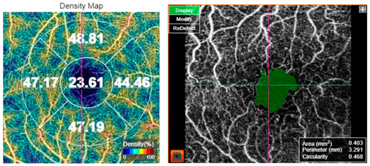
Results: Patients with DM presented significantly higher PBR5-25 compared to controls without DM (p = 0.023). At the univariate analysis, increased PBR5-25 (≥2.19 μm vs. <2.19 μm) was associated with decreased peripapillary VD at the superior quadrant (univariate OR (95% CI) = 0.34 (0.12-0.93), p = 0.037). Multivariate logistic regression analysis showed that increased PWV (≥13.7 m/s vs. <13.7 m/s) was associated with an increased foveal avascular zone (FAZ) area on OCT-A (p = 0.044) and increased FAZ perimeter (p = 0.048). Moreover, increased Aix (≥14.745% vs. <14.745%) was associated with diabetic macular edema (DME) presence (p = 0.050) and increased perifoveal and parafoveal superior and temporal thickness on OCT (p < 0.05 for all associations).
Conclusions: Markers of endothelial damage and arterial stiffness were associated with structural and microvascular retinal alterations in patients with DM, pointing out that OCT-A could be a useful biomarker for detecting potential cardiovascular risk in such patients.
Keywords: OCT-A; arterial stiffness; diabetic; endothelial dysfunction; microvascular.
Conflict of interest statement
The authors declare no conflicts of interest.
Figures
References
-
- International Diabetes Federation . IDF Diabetes Atlas. 10th ed. International Diabetes Federation; Brussels, Belgium: 2021.
LinkOut - more resources
Full Text Sources
