

Neuroregeneration Improved by Sodium-D,L-Beta-Hydroxybutyrate in Primary Neuronal Cultures
- PMID: 39338322
- PMCID: PMC11435142
- DOI: 10.3390/ph17091160
Neuroregeneration Improved by Sodium-D,L-Beta-Hydroxybutyrate in Primary Neuronal Cultures
Abstract
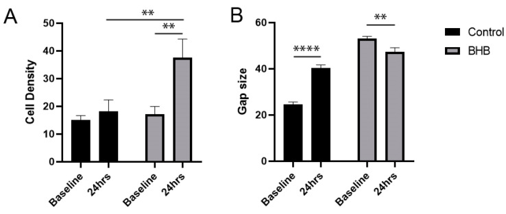
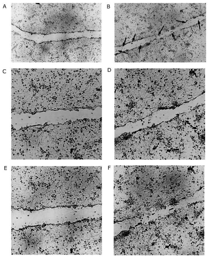
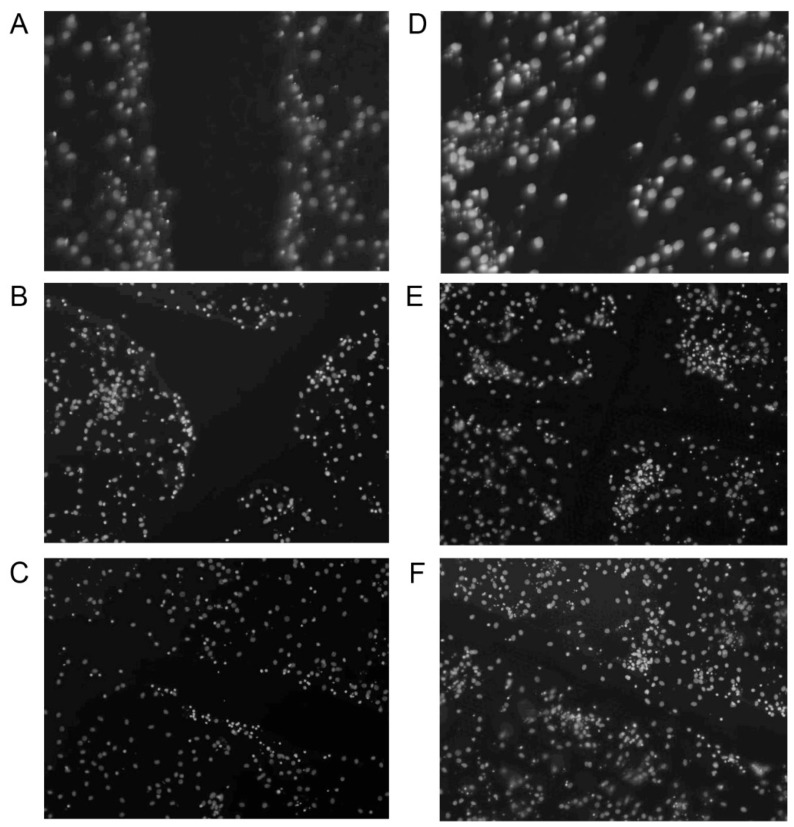
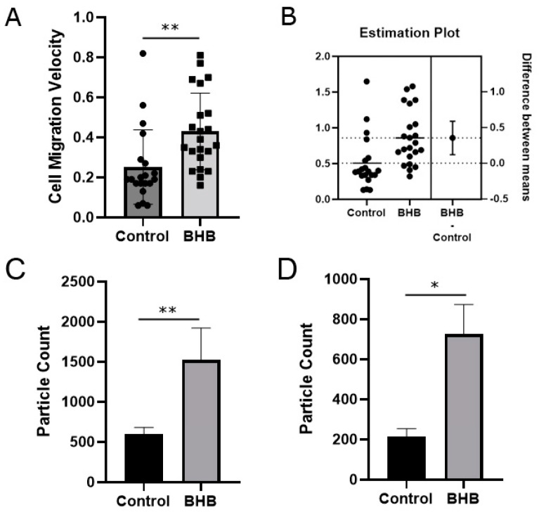
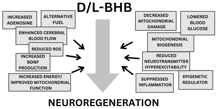
Ketone bodies are considered alternative fuels for the brain when glucose availability is limited. To determine the neuroregenerative potential of D,L-sodium-beta-hydroxybutyrate (D/L-BHB), Sprague Dawley rat primary cortical neurons were exposed to simulated central nervous system injury using a scratch assay. The neuronal cell migration, cell density and degree of regeneration in the damaged areas (gaps) in the absence (control) and presence of BHB (2 mM) were documented with automated live-cell imaging by the CytoSMART system over 24 h, which was followed by immunocytochemistry, labeling synapsin-I and β3-tubulin. The cell density was significantly higher in the gaps with BHB treatment after 24 h compared to the control. In the control, only 1.5% of the measured gap areas became narrower over 24 h, while in the BHB-treated samples 49.23% of the measured gap areas became narrower over 24 h. In the control, the gap expanded by 63.81% post-injury, while the gap size decreased by 10.83% in response to BHB treatment, compared to the baseline. The cell density increased by 97.27% and the gap size was reduced by 74.64% in response to BHB, compared to the control. The distance travelled and velocity of migrating cells were significantly higher with BHB treatment, while more synapsin-I and β3-tubulin were found in the BHB-treated samples after 24 h, compared to the control. The results demonstrate that D/L-BHB enhanced neuronal migration and molecular processes associated with neural regeneration and axonogenesis. These results may have clinical therapeutic applications in the future for nervous system injuries, such as for stroke, concussion and TBI patients.
Keywords: BHB; beta-hydroxybutyrate; cell migration; exogenous ketones; ketone salt; neural injury; neuroplasticity; scratch assay; synapsin; tubulin.
Conflict of interest statement
International Patent # PCT/US2014/031237, University of South Florida, D.P.D’Agostino, S. Kesl, Patrick Arnold, “Compositions and Methods for Producing Elevated and Sustained Ketosis”. Patent: US 10,980,764 B1, C. Ari, D.P.D’Agostino, “Exogenous ketone supplements for reducing anxiety-related behavior”; Ari, C., D’Agostino, D.P. Technology Title: “Exogenous Ketone Supplementation Improved Motor Function in Sprague–Dawley Rats”. USF Ref. No: 16A019; Ari, C., D’Agostino, D.P. Technology Title: “Lowering of Blood Glucose in Exercising and Non-Exercising Rats Following Administration of Exogenous Ketones and Ketone Formulas”. USF Ref. No: 16A049; Ari, C., D’Agostino, D.P. Technology Title: “Neuroregeneration improved by ketone”. USF Ref. No: 16B128 (provisional patent); Patent: US 10,945,975 B2: Ari, C., D’Agostino, D.P. Dean, J.B. Technology Title: “Delaying latency to seizure by combinations of ketone supplements”. Non provisional patent No. 210112–9018-US02 for AC and DPD. Technology Title: “Methods of Increasing Latency of Anesthetic Induction Using Ketone Supplementation”. US Patent Application No. 17/576,375, patent: Z. Kovacs, C. Ari, D.P.D’Agostino: “Ketone supplements evoked effect on absence epilepsy by co-administration of Uridine”, D.P. D’Agostino and C. Ari are the co-owners of the company Ketone Technologies LLC, and C. Ari is the owner of Fortis World LLC. These interests have been reviewed and managed by the University in accordance with its Institutional and Individual Conflict of Interest policies. All authors declare that there are no additional conflicts of interest.
Figures
References
-
- Lawrence A.B., Orman J.A., Miller T.R., Spicer R.S., Hendrie D. Cost of traumatic brain injuries in the United States and the return on helmet investments. In: Jallo J., Loftus C.M., editors. Neurotrauma and Critical Care of the Brain. 2nd ed. Thieme Medical Publishers, Inc.; New York, NY, USA: 2018. Chapter 32.
Grants and funding
LinkOut - more resources
Full Text Sources
Miscellaneous
