. 2024 Sep 14;17(9):1212.
doi: 10.3390/ph17091212.

Pharmacological Inhibition of Endogenous Hydrogen Sulfide Production Slows Bladder Cancer Progression in an Intravesical Murine Model

Affiliations

Pharmacological Inhibition of Endogenous Hydrogen Sulfide Production Slows Bladder Cancer Progression in an Intravesical Murine Model

Sydney Relouw et al. Pharmaceuticals (Basel). .

Abstract

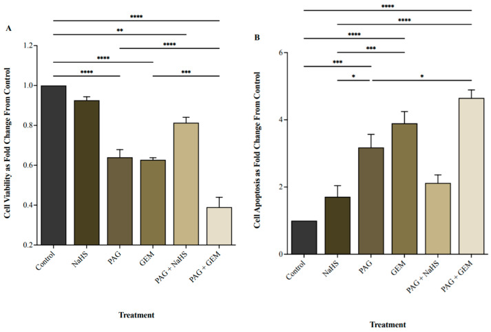
Present bladder cancer therapies have relatively limited therapeutic impact and account for one of the highest lifetime treatment costs per patient. Therefore, there is an urgent need to explore novel and optimized treatment strategies. The present study investigated the effects of inhibiting endogenous hydrogen sulfide (H2S) production on bladder cell viability and in vivo tumor progression. We targeted the H2S-producing enzyme, cystathionine γ-lyase, in 5637 cells using propargylglycine (H2S inhibitor) and performed cytofluorimetric analysis to evaluate cell viability. We then tested the efficacy of propargylglycine alone or in combination with gemcitabine (conventional chemotherapy) in an intravesical murine model of bladder cancer. Magnetic resonance imaging and immunohistochemical staining for cell proliferation, apoptosis, immune-cell infiltration, and neovascularization were performed to evaluate tumor response. Compared to control conditions or cohorts, propargylglycine administration significantly attenuated bladder cancer cell viability in vitro (p < 0.0001) and tumor growth (p < 0.002) and invasion in vivo. Furthermore, propargylglycine enhanced the anti-cancer effects of gemcitabine, resulting in tumor regression (p < 0.0001). Moreover, propargylglycine induced cleaved PARP-1-activated apoptosis (p < 0.05), as well as intratumoral CD8+ T cell (p < 0.05) and F4/80+ macrophage (p < 0.002) infiltration. Propargylglycine also reduced intratumoral neovascularization (p < 0.0001) and cell proliferation (p < 0.0002). Importantly, the pro-apoptotic and anti-neovascularization effects of gemcitabine were enhanced by propargylglycine co-administration. Our findings suggest that inhibition of endogenous H2S production can be protective against bladder cancer by enhancing the chemotherapeutic action of gemcitabine and may be a novel pharmacological target and approach for improved bladder cancer diagnosis and treatments in the future.

Keywords: apoptosis; bladder cancer (BC); cystathionine γ-lyase (CSE); gemcitabine; hydrogen sulfide (H2S); intravesical administration; magnetic resonance imaging; propargylglycine (PAG); tumor progression.


Conflict of interest statement

Melissa J. Huynh has received honoraria from Knight Therapeutics Inc. and Astellas Pharma Inc. All other authors declare no conflicts of interest.

Figures

Figure 1
Figure 1
Relative gene expression of cystathionine γ-lyase (CSE), cystathionine β-synthase (CBS), and 3-mercaptopyruvate sulftransferase (3-MST) under hypoxic conditions. Quantitative PCR (qPCR) analysis of 5637 cells for CSE, CBS, and 3-MST gene expression levels after 8 and 36 h of hypoxia. Genes were normalized against β-actin, and fold changes of gene expression were compared to cells exposed to 0 h of hypoxia and calculated using the ∆∆Ct method. Data (n = 5) are expressed as mean ± standard error of the mean (SEM). Means were compared using two-way ANOVA followed by Tukey’s post hoc test. * p < 0.05, **** p < 0.0001.
Figure 2
Figure 2
Cell viability and apoptosis following single and combination treatments with propargylglycine (PAG), sodium hydrosulfide (NaHS), and gemcitabine (GEM). (A) Cell viability and (B) apoptotic levels of 5637 cells following 8 h of hypoxia, single or combination treatments of 20 mM PAG, 100 μM NaHS, and 100 μM GEM, and an additional 24 h of hypoxia. Flow cytometry was used to quantify cell viability as the portion of cells negative for the apoptosis and necrosis markers, FITC-Annexin-V, and propidium iodide. Cell viability is represented as fold change from control cells that had undergone hypoxia without treatment. Apoptosis was quantified as the portion of cells positive for FITC-Annexin-V and negative for propidium iodide and represented as fold change from control cells that had undergone hypoxia without treatment. Data (n = 5) are expressed as mean ± SEM. Means were compared using one-way ANOVA followed by Tukey’s post hoc test. * p < 0.05, ** p < 0.002, *** p < 0.0002, **** p < 0.0001.
Figure 3
Figure 3
Tumor response as evaluated by magnetic resonance imaging (MRI) and cancer presence and degree of invasion following intravesical therapy. (A) MRI was used to assess the change in bladder wall volume, representative of tumor progression, before and after intravesical therapy of saline, PAG, NaHS, GEM, PAG + NaHS, or PAG + GEM. Changes in bladder wall volume of the N-butyl-N-(4-hydroxybutyl) nitrosamine+ (BBN+) groups were normalized to the change in bladder wall volume of the BBN group. (B) Representative images of hematoxylin- and eosin-stained bladder tumor tissue; 40× magnification. Scale bar represents 100 µm. (C) Percentage of mice within each group that had normal tissue, dysplasia, cancer with no invasion, lamina propria (LP) invasion, or muscularis propria (MP) invasion. (D) Percentage of mice with no invasion, LP invasion, or MP invasion among the mice that had bladder tumors. Data are expressed as mean ± SEM. Means were compared using one-way ANOVA followed by Tukey’s post hoc test. ** p < 0.002, *** p < 0.0002, **** p < 0.0001.
Figure 4
Figure 4
Immunohistochemical (IHC) staining of bladder tumors for cell proliferation, apoptosis, immune-cell infiltration, and neovascularization following intravesical therapy. (A) Representative images of bladder tumor samples stained for caspase-9, cleaved PARP-1, Ki67, VEGF, F4/80, CD163, CD8, and CD4 after intravesical therapy of saline, PAG, NaHS, GEM, PAG + NaHS or PAG + GEM; 40× magnification. Scale bar represents 100 µm. Arrows point to positively stained areas (BI) Corresponding digital analyses show percent area of sections positive for caspase-9, cleaved PARP-1, Ki67, VEGF, F4/80, CD163, CD8, and CD4. Data are expressed as mean ± SEM. Means were compared using one-way ANOVA followed by Tukey’s post-hoc test. * p < 0.05, ** p < 0.002, *** p < 0.0002, **** p < 0.0001.
Figure 4
Figure 4
Immunohistochemical (IHC) staining of bladder tumors for cell proliferation, apoptosis, immune-cell infiltration, and neovascularization following intravesical therapy. (A) Representative images of bladder tumor samples stained for caspase-9, cleaved PARP-1, Ki67, VEGF, F4/80, CD163, CD8, and CD4 after intravesical therapy of saline, PAG, NaHS, GEM, PAG + NaHS or PAG + GEM; 40× magnification. Scale bar represents 100 µm. Arrows point to positively stained areas (BI) Corresponding digital analyses show percent area of sections positive for caspase-9, cleaved PARP-1, Ki67, VEGF, F4/80, CD163, CD8, and CD4. Data are expressed as mean ± SEM. Means were compared using one-way ANOVA followed by Tukey’s post-hoc test. * p < 0.05, ** p < 0.002, *** p < 0.0002, **** p < 0.0001.

References

    1. Sung H., Ferlay J., Siegel R.L., Laversanne M., Soerjomataram I., Jemal A., Bray F. Global cancer statistics 2020: GLOBOCAN estimates of incidence and mortality worldwide for 36 cancers in 185 countries. CA Cancer J. Clin. 2021;71:209–249. doi: 10.3322/caac.21660. - DOI - PubMed
    1. Dugbartey G.J., Relouw S., McFarlane L., Sener A. Redox System and Oxidative Stress-Targeted Therapeutic Approaches in Bladder Cancer. Antioxidants. 2024;13:287. doi: 10.3390/antiox13030287. - DOI - PMC - PubMed
    1. Relouw S., Dugbartey G.J., Sener A. Non-Invasive Imaging Modalities in Intravesical Murine Models of Bladder Cancer. Cancers. 2023;15:2381. doi: 10.3390/cancers15082381. - DOI - PMC - PubMed
    1. Burger M., Catto J.W., Dalbagni G., Grossman H.B., Herr H., Karakiewicz P., Kassouf W., Kiemeney L.A., La Vecchia C., Shariat S., et al. Epidemiology and risk factors of urothelial bladder cancer. Eur. Urol. 2013;63:234–241. doi: 10.1016/j.eururo.2012.07.033. - DOI - PubMed
    1. Sylvester R.J., Van Der Meijden A.P., Oosterlinck W., Witjes J.A., Bouffioux C., Denis L., Newling D.W., Kurth K. Predicting recurrence and progression in individual patients with stage Ta T1 bladder cancer using EORTC risk tables: A combined analysis of 3596 patients from seven EORTC trials. Eur. Urol. 2006;49:466–744. doi: 10.1016/j.eururo.2005.12.031. - DOI - PubMed

LinkOut - more resources