. 2024 Aug 26;12(9):1766.
doi: 10.3390/microorganisms12091766.

Evidence for the Presence of Borrelia burgdorferi Biofilm in Infected Mouse Heart Tissues

Affiliations

Evidence for the Presence of Borrelia burgdorferi Biofilm in Infected Mouse Heart Tissues

Sahaja Thippani et al. Microorganisms. .

Abstract

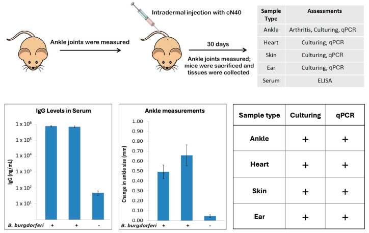
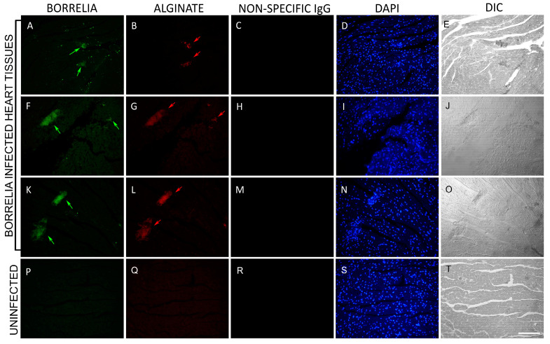
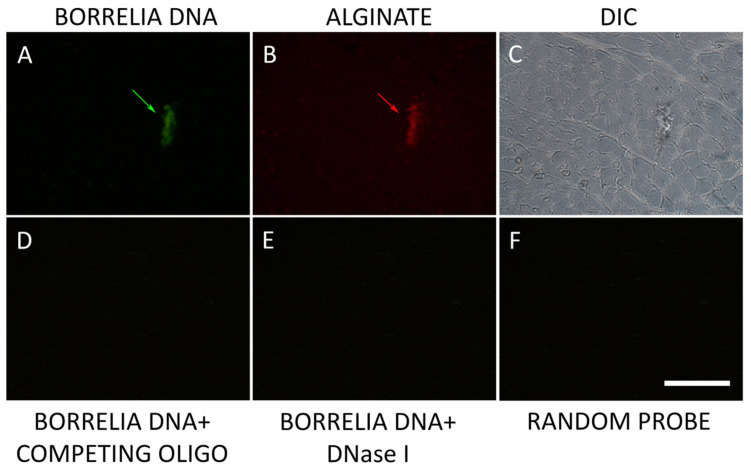
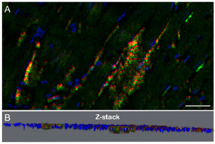
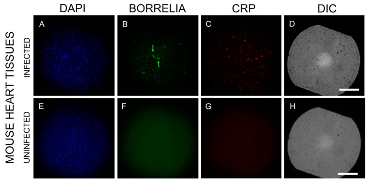
Borrelia burgdorferi, the bacterium responsible for Lyme disease, has been shown to form antimicrobial-tolerant biofilms, which protect it from unfavorable conditions. Bacterial biofilms are known to significantly contribute to severe inflammation, such as carditis, a common manifestation of Lyme disease. However, the role of B. burgdorferi biofilms in the development of Lyme carditis has not been thoroughly investigated due to the absence of an appropriate model system. In this study, we examined heart tissues from mice infected with B. burgdorferi for the presence of biofilms and inflammatory markers using immunohistochemistry (IHC), combined fluorescence in situ hybridization FISH/IHC, 3D microscopy, and atomic force microscopy techniques. Our results reveal that B. burgdorferi spirochetes form aggregates with a known biofilm marker (alginate) in mouse heart tissues. Furthermore, these biofilms induce inflammation, as indicated by elevated levels of murine C-reactive protein near the biofilms. This research provides evidence that B. burgdorferi can form biofilms in mouse heart tissue and trigger inflammatory processes, suggesting that the mouse model is a valuable tool for future studies on B. burgdorferi biofilms.

Keywords: C-reactive protein; atomic force microscopy; biofilm; carditis; immunohistochemistry; inflammation.


Conflict of interest statement

The authors declare no conflicts of interest.

Figures

Figure 1
Figure 1
Summary of the design of the experimental infection of C3H/HeN mice with B. burgdorferi and the collection of different samples (upper panel). The lower left panel shows the results of ELISA for IgG serum levels in the two infected groups (20 mice/group) and one uninfected group (4 mice). The lower middle panel shows results of the ankle measurements in the infected and uninfected groups, while the lower right panel summarizes the results of the culture and qPCR experiments on infected mice.
Figure 2
Figure 2
Immunohistochemical (IHC) detection of B. burgdorferi spirochetes and biofilm in heart tissue sections of C3H/HeN infected and uninfected mice. Panels (A,F,K,P) show IHC results using a FITC-labeled anti-borrelia antibody. Green arrows depict B. burgdorferi aggregates; green arrowheads point to small spirochetes. Panels (B,G,L) show IHC results using an anti-alginate antibody (red staining). Red arrows depict the presence of alginate on B. burgdorferi aggregates. Panels (C,H,M,R) show IHC results with non-specific IgG antibody (negative control). Panels (D,I,N,S) show DAPI stain for nuclear DNA. Panels (E,J,O,T) show the structure of the tissues by DIC microscopy. Panels (P,Q) show uninfected mouse heart sections that were stained with the same B. burgdorferi and alginate antibodies. Images were taken at 400×. Bar: 200 μm.
Figure 3
Figure 3
Representative images of B. burgdorferi and alginate-specific combined FISH/IHC on infected mouse heart tissue. Panel (A) shows B. burgdorferi stained with fluorescently labeled 16S rDNA probe (green staining with a green arrow). Panel (B) shows IHC results using an anti-alginate antibody on subsequent section (red staining with a red arrow). Panel (C) shows the structure of the tissue (DIC). Panels (DF) show negative controls for the FISH experiments: competing oligo, DNase 1, and random probes respectively as described in Methods. Images were taken at 400×. Bar: 200 μm.
Figure 4
Figure 4
Representative IHC images of B. burgdorferi infected mouse heart tissue section demonstrating the 3D spatial arrangement (Panel (A)) with the corresponding Z-stack (Panel (B)). Green staining: B. burgdorferi, red staining: alginate and blue staining: DAPI. Bar: 20 μm.
Figure 5
Figure 5
Three-dimensional analysis of Borrelia aggregates stained positive for B. burgdorferi and alginate via atomic force microscopy (AFM) in infected mouse heart tissue sections. Panel (A) shows the AFM topographical scans of B. burgdorferi biofilm (white arrowhead). Panel (B) shows results using a FITC-labeled anti-Borrelia antibody (green staining, white arrowhead). Panel (C) shows results using an anti-alginate antibody (red staining, white arrowhead). Panel (D) shows the DIC image of the infected mouse tissue section (white arrowhead), which was used for the AFM study. Scale bar: 50 μm.
Figure 6
Figure 6
Representative IHC images of B. burgdorferi and C-reactive protein (CRP) staining in infected and uninfected C3H/HeN mouse heart sections. Panels (B,F) show results using a B. burgdorferi-specific antibody (green). Panels (C,G) show results using a CRP antibody (red). Panels (A,E) show DAPI stain for nuclear DNA. Panels (F,G) show negative control, uninfected mouse heart sections that were stained with the same B. burgdorferi and CRP antibodies. Panels (D,H) show the structure of the tissues by DIC microscopy. Green arrows indicate two small B. burgdorferi aggregates. Images were taken at 200×. Bar: 200 μm.

References

    1. Perronne C. Lyme and associated tick-borne diseases: Global challenges in the context of a public health threat. Front. Cell. Infect. Microbiol. 2014;4:74. doi: 10.3389/fcimb.2014.00074. - DOI - PMC - PubMed
    1. Marques A.R., Strle F., Wormser G.P. Comparison of Lyme Disease in the United States and Europe. Emerg. Infect. Dis. 2021;27:2017–2024. doi: 10.3201/eid2708.204763. - DOI - PMC - PubMed
    1. Coburn J., Garcia B., Hu L.T., Jewett M.W., Kraiczy P., Norris S.J., Skare J. Lyme Disease Pathogenesis. Curr. Issues. Mol. Biol. 2021;42:473–518. - PMC - PubMed
    1. Liegner K.B., Shapiro J.R., Ramsay D., Halperin A.J., Hogrefe W., Kong L. Recurrent erythema migrans despite extended antibiotic treatment with minocycline in a patient with persisting Borrelia burgdorferi infection. J. Am. Acad. Dermatol. 1993;28:312–314. doi: 10.1016/0190-9622(93)70043-S. - DOI - PubMed
    1. Stricker R.B., Johnson L. Lyme disease: The next decade. Infect. Drug Resist. 2011;4:1–9. doi: 10.2147/IDR.S15653. - DOI - PMC - PubMed

LinkOut - more resources