. 2024 Aug 29;12(9):1794.
doi: 10.3390/microorganisms12091794.

Chikungunya Virus RNA Secondary Structures Impact Defective Viral Genome Production

Affiliations

Chikungunya Virus RNA Secondary Structures Impact Defective Viral Genome Production

Laura I Levi et al. Microorganisms. .

Abstract

Chikungunya virus (CHIKV) is a mosquito-borne RNA virus that poses an emerging threat to humans. In a manner similar to other RNA viruses, CHIKV encodes an error-prone RNA polymerase which, in addition to producing full-length genomes, gives rise to truncated, non-functional genomes, which have been coined defective viral genomes (DVGs). DVGs have been intensively studied in the context of therapy, as they can inhibit viral replication and dissemination in their hosts. In this work, we interrogate the influence of viral RNA secondary structures on the production of CHIKV DVGs. We experimentally map RNA secondary structures of the CHIKV genome using selective 2'-hydroxyl acylation analyzed by primer extension and mutational profiling (SHAPE-MaP), which couples chemical labelling with next-generation sequencing. We correlate the inferred secondary structure with preferred deletion sites of CHIKV DVGs. We document an increased probability of DVG generation with truncations at unpaired nucleotides within the secondary structure. We then generated a CHIKV mutant bearing synonymous changes at the nucleotide level to disrupt the existing RNA secondary structure (CHIKV-D2S). We show that CHIKV-D2S presents altered DVG generation compared to wild-type virus, correlating with the change in RNA secondary structure obtained by SHAPE-MaP. Our work thus demonstrates that RNA secondary structure impacts CHIKV DVG production during replication.

Keywords: RNA secondary structure; SHAPE-MaP; chikungunya virus; defective viral genome.


Conflict of interest statement

L.I.L., V.B, T.V. and M.V. are co-inventors of the DI 2018-21 patent. The rest of the authors declare no conflicts of interest. None of the funders described below had any influence on study design, data collection and analysis, decision to publish, or preparation of the manuscript.

Figures
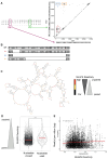
Figure 1
Figure 1
CHIKV genome secondary structures correlated with DVG generation. (A) Schematic of start and stop breakpoints of DVGs generated by high-MOI passages of CHIKV in Vero cells. Start (x axis) and stop (y axis) positions of the breakpoints of DVGs are plotted. The different clusters are called A, B, and C. (B) Schematic of WT CHIKV genome. (C) CHIKV Carib secondary structures from the SHAPE-MaP analysis [35] around the start breakpoints of DVGs A and B (Nucleotides 300 to 819). Nucleotides are depicted in black (low), red (high) or yellow (intermediate), depending on their SHAPE reactivity. (D) In first passage, nucleotides that were used as breakpoints (start and/or stop) or left unused were plotted according to their median SHAPE reactivity value around a 5-nucleotide interval. *** p < 0.001 (unpaired t-test). (E) Reads-per-million values for each nucleotide position used as a start and/or stop breakpoint (start or stop), matched with their respective rolling median SHAPE reactivity (of a 5-nucleotide window) over all passages. Values r and p denote Pearson’s correlation coefficient and its associated p-value.
Figure 2
Figure 2
Engineering a mutant with disrupted secondary structure around the breakpoints of clusters A, B and C. (A) Schematic of WT CHIKV, and the DVGs from clusters A, B and C and the D2S mutant, highlighting the 3 mutated regions and the number of silent mutations inserted. (B) Predicted secondary structures, determined using SHAPE-MaP data for WT virus (black lines) or mFold software (UNAfold, http://www.unafold.org) for D2S (blue lines), after introducing mutations around regions 1, 2 and 3. The black arrows represent CHIKV genome parts, and each line represents an interaction between 2 nucleotides. Several lines forming an arc form a hairpin. (C) Growth curves of D2S CHIKV (blue line) and WT CHIKV (black line) at MOI 0.01 (multi-step growth curve) and 1 (one-step growth curve) in Vero cells. Bars represent mean ± SD, n = 3 biological replicates; **** p < 0.0001 (two-way ANOVA with Bonferroni post-test).
Figure 3
Figure 3
D2S mutant generates a greater diversity of DVGs. (A) DVGs were mapped on their start and stop positions, with colors representing summed frequencies of the DVGs with the same start and stop bins. (B) DVG entropy of WT and D2S mutant over serial passaging. **** p < 0.001 (unpaired t-test).
Figure 4
Figure 4
DVG generation is governed by secondary structures. (A) All DVG (start, stop) positions for pairs found in WT and D2S are listed and associated with the number of DVGs supporting this pair for each virus over all replicates. We separated DVGs that had a start position before (red) or after (blue) the 5000th nucleotide. (B) Distributions of the absolute difference between DVG counts in WT vs. DS2 viruses for each (start, stop) pair. p < 0.0001 (Kolmogorov–Smirnov test and unpaired t-test). (C) Median change in SHAPE reactivity of the D2S mutant compared to WT, inside or outside modified regions (R1, R2 and R3 pooled together), *** p < 0.001 (unpaired t-test). (D) Reads-per-million values for each nucleotide position used as a start and/or stop breakpoint were matched with the SHAPE reactivity value for the nucleotide. Values r and p denote Pearson’s correlation coefficient and its associated p-value. (E), Nucleotides that were used as breakpoints (start and/or stop) or left unused by D2S mutant over all passages were plotted according to their SHAPE reactivity value (determined in D2S mutant). *** p < 0.001 (unpaired t-test).

References

    1. Borgherini G., Poubeau P., Staikowsky F., Lory M., Moullec N.L., Becquart J.P., Wengling C., Michault A., Paganin F. Outbreak of Chikungunya on Reunion Island: Early Clinical and Laboratory Features in 157 Adult Patients. Clin. Infect. Dis. 2007;44:1401–1407. doi: 10.1086/517537. - DOI - PubMed
    1. Suhrbier A., Jaffar-Bandjee M.-C., Gasque P. Arthritogenic Alphaviruses—An Overview. Nature Reviews. Rheumatology. 2012;8:420–429. doi: 10.1038/nrrheum.2012.64. - DOI - PubMed
    1. Bouquillard E., Fianu A., Bangil M., Charlette N., Ribéra A., Michault A., Favier F., Simon F., Flipo R.-M. Rheumatic Manifestations Associated with Chikungunya Virus Infection: A Study of 307 Patients with 32-Month Follow-up (RHUMATOCHIK Study) Jt. Bone Spine Rev. Rhum. 2018;85:207–210. doi: 10.1016/j.jbspin.2017.01.014. - DOI - PubMed
    1. Levi L.I., Vignuzzi M. Arthritogenic Alphaviruses: A Worldwide Emerging Threat? Microorganisms. 2019;7:133. doi: 10.3390/microorganisms7050133. - DOI - PMC - PubMed
    1. Couturier E., Guillemin F., Mura M., Léon L., Virion J.-M., Letort M.-J., De Valk H., Simon F., Vaillant V. Impaired Quality of Life after Chikungunya Virus Infection: A 2-Year Follow-up Study. Rheumatology. 2012;51:1315–1322. doi: 10.1093/rheumatology/kes015. - DOI - PubMed

LinkOut - more resources