. 2024 Aug 30;12(9):1801.
doi: 10.3390/microorganisms12091801.

Rethinking Keratoplasty for Patients with Acanthamoeba Keratitis: Early "Low Load Keratoplasty" in Contrast to Late Optical and Therapeutic Keratoplasty

Affiliations

Rethinking Keratoplasty for Patients with Acanthamoeba Keratitis: Early "Low Load Keratoplasty" in Contrast to Late Optical and Therapeutic Keratoplasty

Yaser Abu Dail et al. Microorganisms. .

Abstract

Background: Early therapeutic penetrating keratoplasty (TKP) for Acanthamoeba keratitis (AK) is thought to have a worse visual prognosis than the delayed optical penetrating keratoplasty (OKP) after successful conservative treatment of AK. This has led to a tendency to prolong conservative therapy and delay penetrating keratoplasty in patients with AK. This retrospective series presents the results of patients with AK that underwent early penetrating keratoplasty after reducing the corneal amoeba load through intensive conservative therapy, so-called "low load keratoplasty" (LLKP).

Patients and methods: The medical records of our department were screened for patients with AK, confirmed by histological examination and/or PCR and/or in vivo confocal microscopy, which underwent ab LLKP and had a follow-up time of at least one year between 2009 and 2023. Demographic data, best corrected visual acuity (BCVA) and intraocular pressure at first and last visit, secondary glaucoma (SG), and recurrence and graft survival rates were assessed.

Results: 28 eyes of 28 patients were included. The average time from initiation of therapy to penetrating keratoplasty (PKP) was 68 ± 113 days. The mean follow-up time after LLKP was 53 ± 42 months. BCVA (logMAR) improved from 1.9 ± 1 pre-operatively to 0.5 ± 0.6 at last visit (p < 0.001). A total of 14% of patients were under medical therapy for SG at the last visit, and two of them underwent glaucoma surgery. The recurrence rate was 4%. The Kaplan-Meier graft survival rate of the first graft at four years was 70%. The second graft survival rate at four years was 87.5%.

Conclusion: LLKP appears to achieve a good visual prognosis with an earlier visual and psychological habilitation, as well as low recurrence and SG rates. These results should encourage us to reconsider the optimal timing of PKP in therapy-resistant AK.

Keywords: acanthamoeba keratitis; graft survival rate; low load keratoplasty; optic keratoplasty; therapeutic keratoplasty.


Conflict of interest statement

The authors declare no conflicts of interest. All authors attest that they meet the current ICMJE criteria for authorship.

Figures

Figure 2
Figure 2
Flow chart for treatment of Acanthamoeba keratitis.
Figure 1
Figure 1
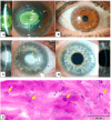
Clinical and histological findings of Acanthamoeba keratitis: (A) The left eye of a 45-year-old male patient with Acanthamoeba keratitis. Visual acuity by admission was hand motion. Please notice the ring infiltrate (grey arrows) and the central corneal infiltrate (yellow arrow). (B) Post-operative findings of the patient from (A). Visual acuity after ten years was 0.6 decimal. A penetrating excimer laser keratoplasty 7.5/7.6 mm was performed with corneal cryotherapy of the interface (“freezing-thawing-freezing”) before trephination. (C) The left eye of 21 year-old female patient with Acanthamoeba keratitis. Visual acuity by admission was hand motion. Please note the deep stromal neovascularization (grey arrows) and the central corneal infiltrate (yellow arrow). (D) Post-operative findings of the patient from (C). Visual acuity after six weeks was 0.3 decimal. A penetrating keratoplasty with mechanical trephination 7.5/7.75 mm was performed with corneal cryotherapy of the interface (“freezing-thawing-freezing”) before trephination. Please note the rapid regression of stromal neovascularization after removal of Acanthamoeba antigens with keratoplasty. (E) Several Acanthamoeba pathogens in the excised cornea of a patient with Acanthamoeba keratitis, some of them are marked with arrows (periodic acid-Schiff “PAS” coloring).
Figure 3
Figure 3
Descriptive data of the current series with a comparison of pre- and post-operative best corrected visual acuity. (A) Time from onset of symptom to keratoplasty in days. (B) Time from initiation of Acanthamoeba therapy to keratoplasty in days. (C) Best corrected visual acuity before keratoplasty and at last visit.
Figure 4
Figure 4
Kaplan–Meier charts for survival rate of the first and second corneal grafts. (A) Survival rate of the first keratoplasty in years. (B) Survival rate of the second keratoplasty in years.

References

    1. Lorenzo-Morales J., Khan N.A., Walochnik J. An update on Acanthamoeba keratitis: Diagnosis, pathogenesis and treatment. Parasite. 2015;22:10. doi: 10.1051/parasite/2015010. - DOI - PMC - PubMed
    1. Nagington J., Watson P.G., Playfair T.J., Mcgill J., Jones B.R., Steele A.D.M.G. Amoebic infection of the eye. Lancet. 1974;2:1537–1540. doi: 10.1016/S0140-6736(74)90285-2. - DOI - PubMed
    1. Ting D.S.J., Ho C.S., Deshmukh R., Said D.G., Dua H.S. Infectious keratitis: An update on epidemiology, causative microorganisms, risk factors, and antimicrobial resistance. Eye. 2021;35:1084–1101. doi: 10.1038/s41433-020-01339-3. - DOI - PMC - PubMed
    1. Nielsen S.E., Ivarsen A., Hjortdal J. Increasing incidence of Acanthamoeba keratitis in a large tertiary ophthalmology department from year 1994 to 2018. Acta Ophthalmol. 2020;98:445–448. doi: 10.1111/aos.14337. - DOI - PubMed
    1. Seal D.V., Beattie T.K., Tomlinson A., Fan D., Wong E. Acanthamoeba keratitis. Br. J. Ophthalmol. 2003;87:516. doi: 10.1136/bjo.87.4.516. - DOI - PMC - PubMed

LinkOut - more resources