. 2024 Sep 5;12(9):1833.
doi: 10.3390/microorganisms12091833.

Sulfate-Reducing Bacteria Induce Pro-Inflammatory TNF-α and iNOS via PI3K/Akt Pathway in a TLR 2-Dependent Manner

Affiliations

Sulfate-Reducing Bacteria Induce Pro-Inflammatory TNF-α and iNOS via PI3K/Akt Pathway in a TLR 2-Dependent Manner

Sudha B Singh et al. Microorganisms. .

Abstract

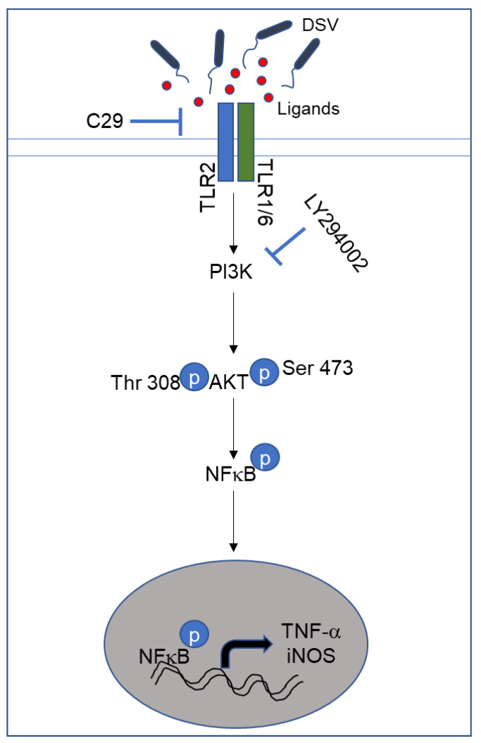
Desulfovibrio, resident gut sulfate-reducing bacteria (SRB), are found to overgrow in diseases such as inflammatory bowel disease and Parkinson's disease. They activate a pro-inflammatory response, suggesting that Desulfovibrio may play a causal role in inflammation. Class I phosphoinositide 3-kinase (PI3K)/protein kinase B (AKT) signaling pathway regulates key events in the inflammatory response to infection. Dysfunctional PI3K/Akt signaling is linked to numerous diseases. Bacterial-induced PI3K/Akt pathway may be activated downstream of toll-like receptor (TLR) signaling. Here, we tested the hypothesis that Desulfovibrio vulgaris (DSV) may induce tumor necrosis factor alpha (TNF-α) and inducible nitric oxide synthase (iNOS) expression via PI3K/Akt in a TLR 2-dependent manner. RAW 264.7 macrophages were infected with DSV, and protein expression of p-Akt, p-p70S6K, p-NF-κB, p-IkB, TNF-α, and iNOS was measured. We found that DSV induced these proteins in a time-dependent manner. Heat-killed and live DSV, but not bacterial culture supernatant or a probiotic Lactobacillus plantarum, significantly caused PI3K/AKT/TNF/iNOS activation. LY294002, a PI3K/Akt signaling inhibitor, and TL2-C29, a TLR 2 antagonist, inhibited DSV-induced PI3K/AKT pathway. Thus, DSV induces pro-inflammatory TNF-α and iNOS via PI3K/Akt pathway in a TLR 2-dependent manner. Taken together, our study identifies a novel mechanism by which SRB such as Desulfovibrio may trigger inflammation in diseases associated with SRB overgrowth.

Keywords: Desulfovibrio vulgaris (DSV); inducible nitric oxide synthase (iNOS); phosphoinositide 3-kinase (PI3K)/protein kinase B (AKT) (PI3K/AKT); toll-like receptor 2 (TLR 2); tumor necrosis factor-α (TNF-α).


Conflict of interest statement

The authors declare no conflicts of interest.

Figures

Figure 1
Figure 1
DSV induced activation of PI3K/Akt/TNF pathway in a time-dependent manner. (A) RAW 264.7 cells (8 × 10⁵) were infected with DSV at MOI 20 for 5, 15, 30, 120, or 240 min. Cells were lysed and protein lysate was prepared. Fifty μg of protein lysate was separated on SDS-PAGE and analyzed for the expression of p-Akt, total Akt, TNF-α, iNOS, p-IκB, total IκB, p-NFκB p65, and total p65 NFκB by Western blotting. Original blots were cut at different molecular weights to enable probing of as many proteins as possible in the same blot. Actin was used as a loading control. (BG) Quantification of Western blots. Blots were quantified with Fiji ImageJ by analyzing the ratio of protein of interest/Actin. For phosphorylated proteins, the ratio of phosphorylated/total protein was calculated. Data represent Mean ± SEM from at least three independent experiments. Values were normalized to control. One-way ANOVA was used to determine the statistical significance. Values were compared to control, with a post-hoc Dunnett’s test. * p < 0.05, ** p < 0.01, *** p < 0.001.
Figure 2
Figure 2
Live and heat-killed DSV, but not bacterial culture supernatant, significantly induced PI3K/Akt/TNF/iNOS pathway. (A) RAW 264.7 cells were treated with live or heat-killed (HK) DSV (MOI 20) or bacterial culture supernatant (Sup) for 240 min (4 h). Cells were lysed and protein lysate prepared. Fifty μg of protein lysate was separated on SDS-PAGE and analyzed for p-Akt, total Akt, TNF-α, and iNOS by Western blotting. Actin was used as a loading control. (BD) Quantification of Western blots. Blots were quantified with Fiji ImageJ by analyzing the ratio of protein of interest/Actin. For phosphorylated Akt, the ratio of p-Akt/total Akt was calculated. Data represent Mean ± SEM from at least three independent experiments. Values were normalized to control. One-way ANOVA was used to determine the statistical significance. Values were compared to live DSV. * p < 0.05, ns (non-significant).
Figure 3
Figure 3
Comparison of effects of DSV and Lactobacillus plantarum on PI3K/Akt/TNF/iNOS pathway. (A) RAW 264.7 cells were treated with DSV or Lacto (MOI 20) for 4 h. Cells were lysed and protein lysate prepared. Fifty μg of protein lysate was separated on SDS-PAGE and analyzed for p-Akt, total Akt, TNF-α, and iNOS by Western blotting. Lanes were spliced in the original blot and combined. Actin was used as a loading control. (BD) Quantification of Western blots. Blots were quantified with ImageJ by analyzing the ratio of protein of interest/Actin. For phosphorylated Akt, the ratio of p-Akt/total Akt was calculated. Data represent Mean ± SEM from at least three independent experiments. Values were normalized and compared to Control. One-way ANOVA was used to determine the statistical significance. * p < 0.05, *** p < 0.001, ns (non-significant).
Figure 4
Figure 4
LY294002 inhibited DSV-induced activation of PI3K/Akt/TNF pathway. (A) RAW 264.7 cells were treated with LY at either 10 or 50 μM for 2 h before infection with DSV for 4 h. Cells were lysed and protein lysate prepared. Fifty μg of protein lysate was separated on SDS-PAGE and analyzed for p-Akt, total Akt, p-P70S6K, P70S6K, TNF-α, and iNOS, by Western blotting. Actin was used as a loading control. (BE) Quantification of Western blots. (F) Post-infection, cell culture supernatant was collected and proteins were precipitated using ethanol precipitation. Proteins were resuspended in PBS and 50 μg was loaded on SDS-PAGE. Secreted TNF-α (s-TNF-α) was detected using anti-TNF-α antibody. Actin was used as a loading control. (G) Quantification of s-TNF-α. (H) Cells were pretreated with or without LY 50 μM followed by infection with DSV for 30 min and protein samples were analyzed for p-p65 NFκB and total NFκB. (I) Blots were quantified with ImageJ by analyzing the ratio of p-NFκB/NFκB. Values were normalized and compared to control. Data represent Mean ± SEM from at least three independent experiments. One-way ANOVA was used to determine the statistical significance, with a post-hoc Dunnett’s test. * p < 0.05, ** p < 0.01, *** p < 0.001, **** p < 0.0001, ns (non-significant).
Figure 5
Figure 5
Activation of PI3K/Akt pathway by DSV was dependent on TLR 2 signaling (A) RAW 264.7 cells were treated with TLR 2 signaling inhibitor C29 at 200 μM for 3 h before infection with DSV for 4 h. Cells were lysed and protein lysate prepared. Fifty μg of protein lysate was separated on SDS-PAGE and analyzed for p-Akt, total Akt, p-P70S6K, P70S6K TNF-α, and iNOS by Western blotting. Actin was used as a loading control. (BE) Quantification of Western blots. (F) Post-infection, cell culture supernatant was collected and proteins were precipitated using ethanol precipitation. Proteins were resuspended in PBS and 50 μg was loaded on SDS-PAGE. Secreted TNF-α (s-TNF-α) was detected using anti-TNF-α antibody. (G) Blots were quantified with ImageJ by analyzing the ratio of protein of interest/Actin. For phosphorylated proteins, the ratio of phosphorylated/total protein was calculated. Data represent Mean ± SEM from at least three independent experiments. Values were normalized to control. A two-tailed t-test was used to compare DSV and DSV + C29. * p < 0.05, ** p < 0.01.
Figure 6
Figure 6
Schematic representation of activation of PI3K/Akt pathway by DSV. DSV activates TLR 2 signaling which is inhibited by C29 (TL2-C29). TLR 2-mediated intracellular signaling occurs via formation of heterodimers of TLR 2 with TLR 1 or TLR 6 (TLR 1/6) that allows recognition of diverse sets of pathogen-associated patterns. TLR 2 activation further leads to the activation of PI3K/Akt as indicated by phosphorylation of Akt at Thr308 and Ser473 positions which is required for a complete activation of Akt. This step is inhibited by PI3K inhibitor LY294002. Once activated, Akt further induces transcription factor NFκB. Upon activation, NFκB induces expression of proinflammatory TNF-α and iNOS.

Similar articles

Cited by

References

    1. Gibson G.R., Macfarlane G.T., Cummings J.H. Occurrence of sulphate-reducing bacteria in human faeces and the relationship of dissimilatory sulphate reduction to methanogenesis in the large gut. J. Appl. Bacteriol. 1988;65:103–111. doi: 10.1111/j.1365-2672.1988.tb01498.x. - DOI - PubMed
    1. Singh S.B., Carroll-Portillo A., Lin H.C. Desulfovibrio in the Gut: The Enemy within? Microorganisms. 2023;11:1772. doi: 10.3390/microorganisms11071772. - DOI - PMC - PubMed
    1. Dordevic D., Jancikova S., Vitezova M., Kushkevych I. Hydrogen sulfide toxicity in the gut environment: Meta-analysis of sulfate-reducing and lactic acid bacteria in inflammatory processes. J. Adv. Res. 2021;27:55–69. doi: 10.1016/j.jare.2020.03.003. - DOI - PMC - PubMed
    1. Singh S.B., Coffman C.N., Carroll-Portillo A., Varga M.G., Lin H.C. Notch Signaling Pathway Is Activated by Sulfate Reducing Bacteria. Front. Cell. Infect. Microbiol. 2021;11:695299. doi: 10.3389/fcimb.2021.695299. - DOI - PMC - PubMed
    1. Dzierzewicz Z., Szczerba J., Lodowska J., Wolny D., Gruchlik A., Orchel A., Weglarz L. The role of Desulfovibrio desulfuricans lipopolysaccharides in modulation of periodontal inflammation through stimulation of human gingival fibroblasts. Arch. Oral Biol. 2010;55:515–522. doi: 10.1016/j.archoralbio.2010.05.001. - DOI - PubMed

LinkOut - more resources