. 2024 Sep 12;12(9):1880.
doi: 10.3390/microorganisms12091880.

Diversification of Pseudomonas aeruginosa Biofilm Populations under Repeated Phage Exposures Decreases the Efficacy of the Treatment

Affiliations

Diversification of Pseudomonas aeruginosa Biofilm Populations under Repeated Phage Exposures Decreases the Efficacy of the Treatment

Mark Grevsen Martinet et al. Microorganisms. .

Abstract

Phage therapy has been proposed as a therapeutic alternative to antibiotics for the treatment of chronic, biofilm-related P. aeruginosa infections. To gain a deeper insight into the complex biofilm-phage interactions, we investigated in the present study the effect of three successive exposures to lytic phages of biofilms formed by the reference strains PAO1 and PA14 as well as of two sequential clinical P. aeruginosa isolates from the sputum of a patient with cystic fibrosis (CF). The Calgary device was employed as a biofilm model and the efficacy of phage treatment was evaluated by measurements of the biomass stained with crystal violet (CV) and of the cell density of the biofilm bacterial population (CFU/mL) after each of the three phage exposures. The genetic alterations of P. aeruginosa isolates from biofilms exposed to phages were investigated by whole-genome sequencing. We show here that the anti-biofilm efficacy of the phage treatment decreased rapidly with repeated applications of lytic phages on P. aeruginosa strains with different genetic backgrounds. Although we observed the maintenance of a small subpopulation of sensitive cells after repeated phage treatments, a fast recruitment of mechanisms involved in the persistence of biofilms to the phage attack occurred, mainly by mutations causing alterations of the phage receptors. However, mutations causing phage-tolerant phenotypes such as alginate-hyperproducing mutants were also observed. In conclusion, a decreased anti-biofilm effect occurred after repeated exposure to lytic phages of P. aeruginosa biofilms due to the recruitment of different resistance and tolerance mechanisms.

Keywords: Pseudomonas aeruginosa; bacteriophages; biofilms; diversification of population; genomics; motility.


Conflict of interest statement

The authors declare no competing interests.

Figures

Figure 1
Figure 1
Reduction in the biofilms of P. aeruginosa PAO1 and PA14 depends on NP3 and NP1 phage concentrations. (A) The viable cells were determined as CFU/mL in four independent experiments, and the reduction was calculated as a percentage of the viable bacteria after treatment in relation to the untreated controls; means and standard error of means (SEM) are shown. (B) The reduction in biomass was determined via crystal violet staining of the phage-treated biofilms and untreated biofilms (indicated here as the horizontal lines). The measurements were performed 8 times, and means and standard deviations (SD) are shown.
Figure 2
Figure 2
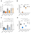
Effect of NP3 phage on PAO1 and PA14 biofilms after repeated treatments compared to the corresponding control biofilms. (A) PAO1 biomass is measured as CV absorbance. (B) Viable bacteria fraction of the resolved PAO1 biofilms as CFU/mL. (C) PA14 biomass measured as CV absorbance. (D) Viable bacteria fraction of the resolved PA14 biofilms as CFU/mL. In (A,C) mean and standard deviation (SD) and in (B,D) box blots with the 5–95%c percentile (whiskers), the 25–75% quadrille (box) with the median (line within the box) and mean (+ within the box) and outliers (grey dots) of four biological and four technical replicates are presented. Significance was assumed for p-values below or equal to 0.05, indicated as follows: * < 0.05, ** < 0.01, *** < 0.001, **** < 0.0001.
Figure 3
Figure 3
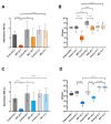
Effect of phages M32 on PAO1 and NP1 on PA14 biofilms after repeated treatments compared to the corresponding control biofilms. (A) PAO1 biomass is measured as CV absorbance. (B) Viable bacteria fraction of the resolved PAO1 biofilms as CFU/mL. (C) PA14 biomass measured as CV absorbance. (D) Viable bacteria fraction of the resolved PA14 biofilms as CFU/mL. In (A,C) mean and standard deviation (SD) and in (B,D) box blots with the 5–95%c percentile (whiskers), the 25–75% quadrille (box) with the median (line within the box) and mean (+ within the box) and outliers (grey dots) of four biological and four technical replicates are presented. Significance was assumed for p-values below or equal to 0.05, indicated as follows: * < 0.05, ** < 0.01, *** < 0.001, **** < 0.0001.
Figure 4
Figure 4
Effect of NP3 phage on biofilms of clinical isolates CF341_06 and CF341_08 after repeated treatments compared to the corresponding control biofilms. (A) CF341_06 biomass measured as CV absorbance. (B) Viable bacteria fraction of the resolved CF341_08 biofilms as CFU/mL. (C) CF341_08 biomass measured as CV absorbance. (D) Viable bacteria fraction of the resolved CF341_06 biofilms as CFU/mL. In (A,C) mean and standard deviation (SD) and in (B,D) box blots with the 5–95%c percentile (whiskers), the 25–75% quadrille (box) with the median (line within the box) and mean (+ within the box) and outliers (grey dots) of four biological and four technical replicates are presented. Significance was assumed for p-values below or equal to 0.05, indicated as follows: * < 0.05, ** < 0.01, *** < 0.001, **** < 0.0001.
Figure 5
Figure 5
Altered genes were identified in the clones of the different strains treated with the respective phages as indicated in each diagram in (AE). The colors in (AE) correspond to different pathways (see legend (F)). The number of clones with altered genes in the controls (untreated) and treated clones exhibiting phage-resistant and -sensitive or-tolerant phenotypes. The phage-resistant clones are marked in grey. WP_0031… is the coding side WP_003138482.1 corresponding to glycosyltransferase family 4 protein.

References

    1. Ciofu O., Moser C., Jensen P., Høiby N. Tolerance and resistance of microbial biofilms. Nat. Rev. Microbiol. 2022;20:621–635. doi: 10.1038/s41579-022-00682-4. - DOI - PubMed
    1. Hoiby N., Bjarnsholt T., Moser C., Bassi G.L., Coenye T., Donelli G., Hall-Stoodley L., Hola V., Imbert C., Kirketerp-Moller K., et al. ESCMID guideline for the diagnosis and treatment of biofilm infections 2014. Clin. Microbiol. Infect. 2015;21((Suppl. S1)):S1–S25. doi: 10.1016/j.cmi.2014.10.024. - DOI - PubMed
    1. Pires D.P., Melo L., Vilas B.D., Sillankorva S., Azeredo J. Phage therapy as an alternative or complementary strategy to prevent and control biofilm-related infections. Curr. Opin. Microbiol. 2017;39:48–56. doi: 10.1016/j.mib.2017.09.004. - DOI - PubMed
    1. Ng R.N., Tai A.S., Chang B.J., Stick S.M., Kicic A. Overcoming Challenges to Make Bacteriophage Therapy Standard Clinical Treatment Practice for Cystic Fibrosis. Front. Microbiol. 2020;11:593988. doi: 10.3389/fmicb.2020.593988. - DOI - PMC - PubMed
    1. Hansen M.F., Svenningsen S.L., Roder H.L., Middelboe M., Burmolle M. Big Impact of the Tiny: Bacteriophage-Bacteria Interactions in Biofilms. Trends Microbiol. 2019;27:739–752. doi: 10.1016/j.tim.2019.04.006. - DOI - PubMed

LinkOut - more resources