. 2024 Aug 23;13(9):717.
doi: 10.3390/pathogens13090717.

Modifications of Mitochondrial Network Morphology Affect the MAVS-Dependent Immune Response in L929 Murine Fibroblasts during Ectromelia Virus Infection

Affiliations

Modifications of Mitochondrial Network Morphology Affect the MAVS-Dependent Immune Response in L929 Murine Fibroblasts during Ectromelia Virus Infection

Karolina Gregorczyk-Zboroch et al. Pathogens. .

Abstract

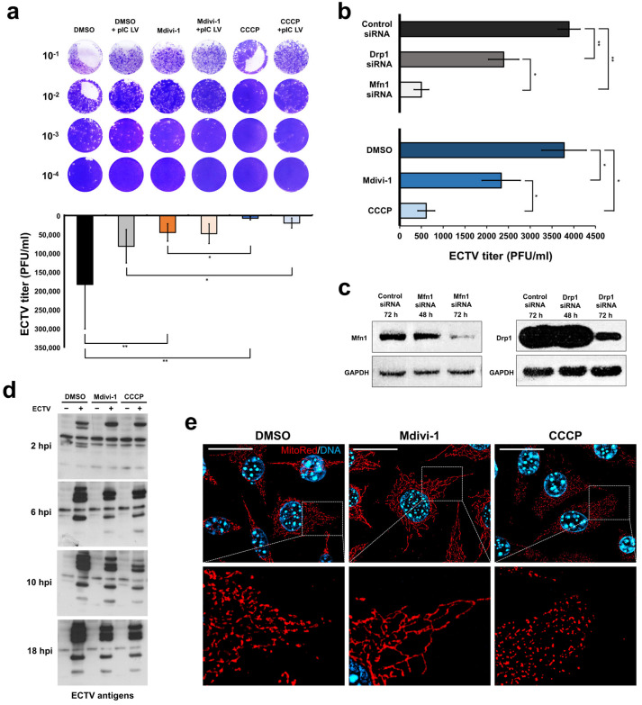
Since smallpox vaccination was discontinued in 1980, there has been a resurgence of poxvirus infections, particularly the monkeypox virus. Without a global recommendation to use the smallpox vaccine, the population is not immune, posing a severe threat to public health. Given these circumstances, it is crucial to understand the relationship between poxviruses and their hosts. Therefore, this study focuses on the ectromelia virus, the causative agent of mousepox, which serves as an excellent model for studying poxvirus pathogenesis. Additionally, we investigated the role of mitochondria in innate antiviral immunity during ECTV infection, focusing specifically on mitochondrial antiviral signaling protein. The study used a Moscow strain of ECTV and L929 mouse fibroblasts. Cells were treated with ECTV and chemical modulators of mitochondrial network: Mdivi-1 and CCCP. Our investigation revealed that an elongated mitochondrial network attenuates the suppression of MAVS-dependent immunity by ECTV and reduces ECTV replication in L929 fibroblasts compared to cells with an unaltered mitochondrial network. Conversely, a fragmented mitochondrial network reduces the number of progeny virions while increasing the inhibition of the virus-induced immune response during infection. In conclusion, our study showed that modifications of mitochondrial network morphology alter MAVS-dependent immunity in ECTV-infected mouse L929 fibroblasts.

Keywords: ECTV; MAVS; immunity; mitochondrial dynamics; mitochondrial network; poxviruses.


Conflict of interest statement

The authors declare no conflicts of interest.

Figures

Figure 1
Figure 1
Ectromelia virus (ECTV) replication in L929 murine fibroblasts with modified mitochondrial network morphology. The plaque assay determined the ECTV titer in L929 cells at two time points: (a) 18 h post infection (hpi) under various treatments, including DMSO, Mdivi-1, or CCCP, with or without transfection of poly(I:C) (MHW) LyoVec (pIC LV); and (b) 10 hpi with transfection of control siRNA, Mfn1 siRNA, or Drp1 siRNA or treatment with DMSO, Mdivi-1, or CCCP. The data from both (a,b) are presented as mean ± standard deviation (SD), with statistical significance indicated as * p ≤ 0.05 and ** p ≤ 0.01. ECTV suspension was obtained from cell lysates in post-culture media. Western blot analysis was performed to confirm Mfn1 or Drp1 gene silencing in target cells (c) and to assess ECTV antigens expression in control and infected cells treated with DMSO, Mdivi-1, or CCCP at 2, 6, 10, and 18 hpi (d). (e) Fluorescence verification of mitochondrial network morphology in L929 fibroblast after treatment with DMSO (intact network), Mdivi-1 (elongated), and CCCP (fragmented) by using MitoRed fluorochrome (red). Scale bar: 20 µm.
Figure 2
Figure 2
ECTV replication in RAW 264.7 murine macrophages with modified mitochondrial network morphology. (a) Determination of ECTV titer by plaque assay in RAW 264.7 cells at 6 hpi treated with DMSO, Mdivi-1, or CCCP. ECTV suspension was obtained from cell lysates or post-culture media. (b) Fluorescence verification of mitochondrial network morphology in RAW 264.7 murine macrophages after treatment with DMSO (intact network), Mdivi-1 (elongated), and CCCP (fragmented). Macrophages were labeled using antibodies against MAVS (green) and DNA (blue). Scale bar: 20 µm.
Figure 3
Figure 3
Distribution of MAVS protein in L929 cells at 18 hpi with ECTV. Cells were treated with DMSO, Mdivi-1, or CCCP. (a) Magnifications indicate MAVS localization in the viral factories. Arrows show the direction of fluorescence intensity measurements. Fibroblasts are labeled using antibodies against MAVS (red), HSP60 (green), or ECTV (EV; green) and DNA (blue). Scale bar: 20 µm. (b) Percentage of MAVS and HSP60 colocalization (n = 20). Different letters indicate significant differences (p < 0.05) between groups.
Figure 3
Figure 3
Distribution of MAVS protein in L929 cells at 18 hpi with ECTV. Cells were treated with DMSO, Mdivi-1, or CCCP. (a) Magnifications indicate MAVS localization in the viral factories. Arrows show the direction of fluorescence intensity measurements. Fibroblasts are labeled using antibodies against MAVS (red), HSP60 (green), or ECTV (EV; green) and DNA (blue). Scale bar: 20 µm. (b) Percentage of MAVS and HSP60 colocalization (n = 20). Different letters indicate significant differences (p < 0.05) between groups.
Figure 4
Figure 4
Flow cytometry analysis of MAVS protein level in L929 cells at 24 hpi with ECTV (EV). (a) Representative histograms indicate the fluorescence intensity of APC-labeled cells treated with DMSO, Mdivi-1, or CCCP and with or without transfection with poly(I:C)(HMW) LyoVec (pIC LV). A black line is drawn through the center of the histogram of uninfected DMSO-treated cells. (b) Dot plots of unlabeled and APC-labeled cells treated with DMSO. (c) Mean fluorescence intensity (MFI) of APC in cells treated with DMSO, Mdivi-1, or CCCP and with or without transfection with poly(I:C)(HMW) LyoVec.
Figure 5
Figure 5
Western blot analysis of proteins involved in MAVS-dependent immunity in L929 cells treated with DMSO, Mdivi-1, or CCCP during ECTV infection. (a) Representative Western blots of MDA-5, RIG-I, MAVS, pIRF3, and STING at 2, 10, 18, and 24 h post infection (hpi) with ECTV (EV). (b) Densitometry analysis of MDA-5, RIG-I, MAVS, pIRF3, and STING at 2, 10, 18, and 24 hpi with ECTV. The level of each protein was normalized to glyceraldehyde-3-phosphate dehydrogenase (GAPDH). The data ARE presented as mean ± standard deviation (SD), with statistical significance indicated as * p ≤ 0.05 AND ** p ≤ 0.01. pIC LV—cells transfected with poly(I:C)(HMW) LyoVec.
Figure 6
Figure 6
Oligomerization of MAVS protein in L929 cells transfected using poly(I:C)(HMW) LyoVec at 24 hpi with ECTV. (a,b) Uninfected control cells treated with Mdivi-1. Fibroblasts were labeled with specific antibodies against (a) MAVS (green) and pIRF3 (red) or (b) MAVS (red) and Hsp60 (green)—marker of mitochondria. (c) ECTV-infected cells treated with Mdivi-1 and labeled with specific antibodies against MAVS (red). DNA was labeled with DAPI (blue). White arrows indicate MAVS oligomers, the yellow arrow shows viral factory, and the white arrowhead indicates pIRF3 in the nucleus. Scale bar: 20 µm. (d) The bar chart presents the percentage of uninfected control cells with MAVS oligomers. Fibroblasts were treated with DMSO, Mdivi-1, or CCCP.
Figure 7
Figure 7
Extracellular level of IFN-α (a) and IFN-β (b) produced by L929 at 24 hpi with ECTV (EV). Cells were treated using DMSO, Mdivi-1, or CCCP and/or transfected with poly(I:C)(HMW) LyoVec (pIC LV). ND—non-detected. Different letters indicate significant differences (p < 0.05) between groups.
Figure 8
Figure 8
Colocalization of MAVS protein with RIG-I (a,b), MDA-5 (c,d), and STING (e,f) in L929 cells at 18 hpi with ECTV. Cells were treated using DMSO, Mdivi-1, or CCCP and/or transfected with poly(I:C)(HMW) LyoVec. Box plots (a,c,e) indicate the number of PLA dots per cell. Different letters indicate significant differences between groups (p ≤ 0.05). (b,d,f) Representative figures visualize the number of PLA dots per cell in each group. Scale bar: 20 µm.
Figure 9
Figure 9
Colocalization of MAVS with fission (a) and fusion (b) proteins in L929 at 18 hpi with ECTV. Cells were treated using DMSO, Mdivi-1, or CCCP and/or transfected with poly(I:C)(HMW) LyoVec. Box plots indicate the number of PLA dots per cell. Different letters indicate significant differences between groups (p ≤ 0.05).
Figure 10
Figure 10
Possible interaction between MAVS and selected proteins. E3—poxviral protein; ER—endoplasmic reticulum; Drp1—dynamin-related protein 1; Fis1—Fission 1 protein; IMM—inner mitochondrial membrane; MAVS—mitochondrial antiviral signaling protein; MDA5—melanoma differentiation-associated protein 5; Mdivi-1—mitochondrial division inhibitor; Mfn1/2—mitofusin 1/2; OMM—outer mitochondrial membrane; Opa1—optic atrophy 1; RIG-I—retinoic acid-inducible gene I; STING—stimulator of interferon genes.

References

    1. Lakis N.S., Li Y., Abraham J.L., Upton C., Blair D.C., Smith S., Zhao H., Damon I.K. Novel Poxvirus Infection in an Immune Suppressed Patient. Clin. Infect. Dis. 2015;61:1543–1548. doi: 10.1093/cid/civ643. - DOI - PubMed
    1. Srinivasan Rajsri K., Rao M. Poxvirus-driven human diseases and emergingtherapeutics. Ther. Adv. Infect. Dis. 2022;9:20499361221136751. doi: 10.1177/20499361221136751. - DOI - PMC - PubMed
    1. Past U.S. Cases and Outbreaks|Mpox|Poxvirus|CDC Dostępne na. [(accessed on 24 February 2024)]; Available online: https://www.cdc.gov/poxvirus/mpox/outbreak/us-outbreaks.html.
    1. Biernacka Z., Gregorczyk-Zboroch K., Lasocka I., Ostrowska A., Struzik J., Gieryńska M., Toka F.N., Szulc-Dąbrowska L. Ectromelia Virus Affects the Formation and Spatial Organization of Adhesive Structures in Murine Dendritic Cells In Vitro. Int. J. Mol. Sci. 2023;25:558. doi: 10.3390/ijms25010558. - DOI - PMC - PubMed
    1. Gieryńska M., Szulc-Dąbrowska L., Struzik J., Gregorczyk-Zboroch K.P., Mielcarska M.B., Toka F.N., Schollenberger A., Biernacka Z. Orthopoxvirus Zoonoses-Do We Still Remember and Are Ready to Fight? Pathogens. 2023;12:363. doi: 10.3390/pathogens12030363. - DOI - PMC - PubMed

Substances

LinkOut - more resources