. 2024 Sep 21;13(9):814.
doi: 10.3390/pathogens13090814.

Temperature Interference on ZIKV and CHIKV Cycles in Mosquitoes and Mammalian Cells

Affiliations

Temperature Interference on ZIKV and CHIKV Cycles in Mosquitoes and Mammalian Cells

Tiago Souza Salles et al. Pathogens. .

Abstract

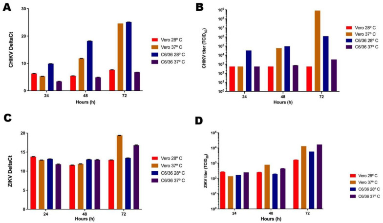
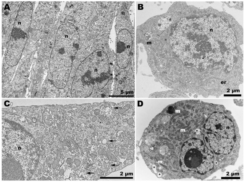
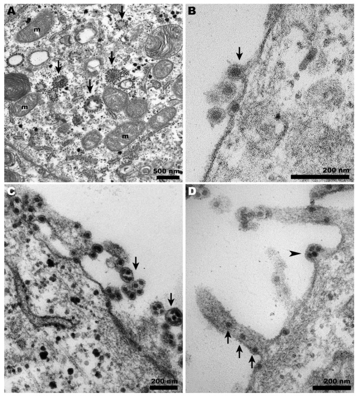
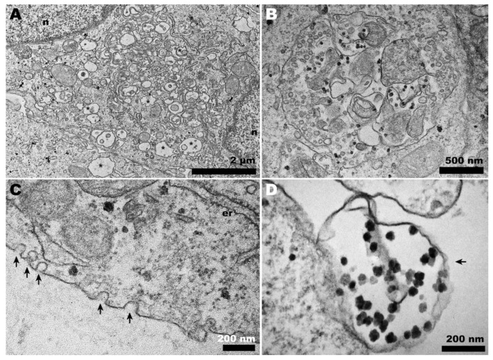
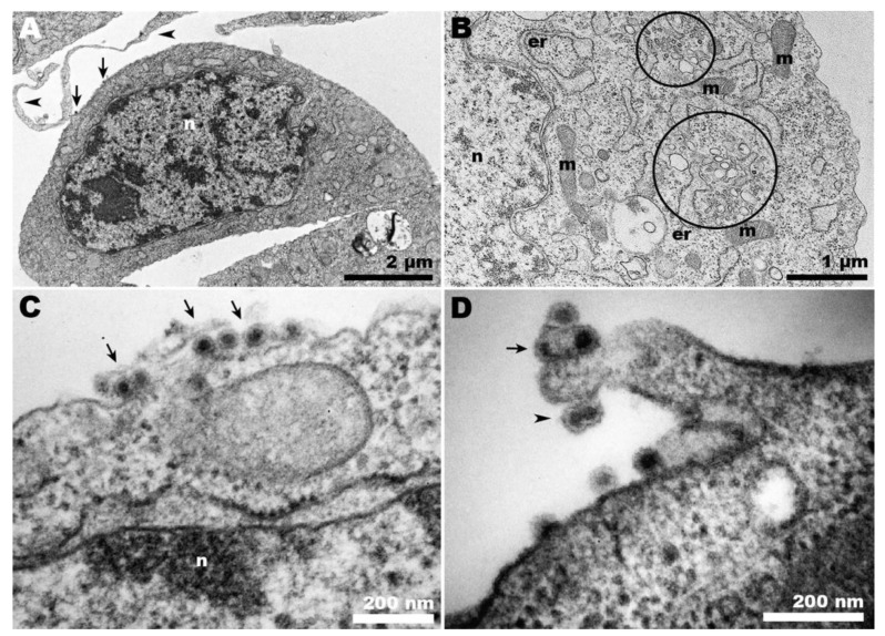
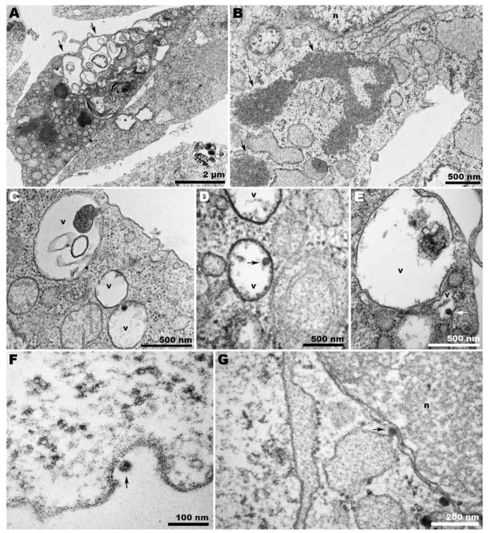
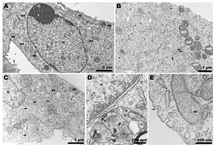
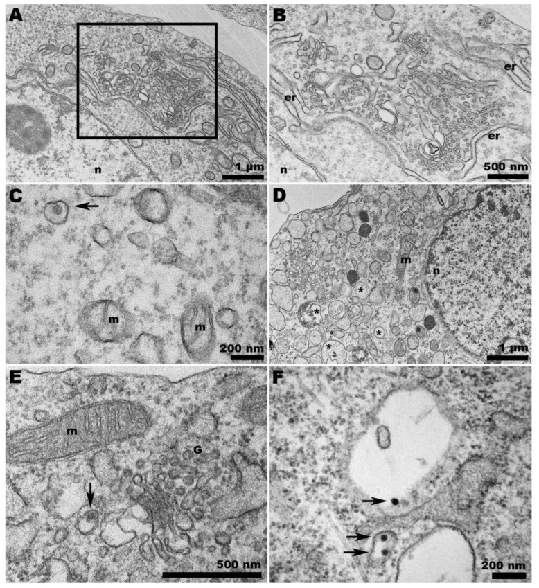
Temperature is a determining factor for the viral cycle. In this study, we investigate the effect of different temperatures on the cycles of two important arboviruses-Zika (ZIKV) and Chikungunya (CHIKV)-in Vero (mammalian) and C6/36 (mosquito) cells. We compare genome quantification to infectivity at 28 °C and 37 °C in both cell types. Virus-cell interaction was also examined by transmission electron microscopy, allowing the observation of phenomena such as virus-surfing and giant forms for CHIKV, as well as the the scarcity of ZIKV in C6/36 cells compared to its cycle in mammalian cells.

Keywords: Chikungunya virus; Zika virus; arbovírus; virus cycle.


Conflict of interest statement

The authors declare no conflicts of interests.

Figures

Figure 1
Figure 1
Quantification of viral RNA and infectious viruses. RT-PCR was performed to determine the amount of CHIKV (A) and ZIKV (B) RNA. TCID50 was performed to quantify the number of infectious particles of CHIKV (C) and ZIKV (D). Due to the small size of the error bars on the logarithmic scale, they are not visible in the graph.
Figure 2
Figure 2
Transmission electron microscopy of non-infected cells. (A) Vero cells at 37 °C were elongated and adjacent to each other; (B) C6/36 cells at 28 °C were rounded and displayed elongated endoplasmic reticulum profiles (er); (C) Vero cells at 28 °C exhibited considerable vacuolization (arrows); (D) C6/36 cells at 37 °C remained rounded, but vacuolization (asterisk) was also observed, along with an increase in the presence of mitochondria (m). (n): nucleus.
Figure 3
Figure 3
Transmission electron microscopy of CHIKV-infected Vero cells at 37 °C. (A) CHIKV-induced CPV-II (arrows) surrounded by clusters of mitochondria (m) were abundant in the cytosol; (B) characteristic budding of CHIKV at the plasma membrane (arrows); (C) giant forms adjacent to the cell surface (arrows); (D) budding of giant forms (arrowheads) and cell membrane projections carrying CHIKV (arrows).
Figure 4
Figure 4
Transmission electron microscopy of CHIKV-infected Vero cells at 28 °C. (A) Membranous rearrangement observed in the cytoplasm of Vero cells, along with vacuolization (asterisks), leading to the formation of CPV-I (B); (C) plasma membrane invaginations (arrows) that may give rise to vesicles within CPV-I; (D) giant form-like structures, with the outline of a plasma membrane bubble (arrow). (n): nucleus; (er): endoplasmic reticulum.
Figure 5
Figure 5
Transmission electron microscopy of CHIKV-infected C6/36 cells at 28 °C. (A) Panoramic view of infected cells showing condensed chromatin (n), virus adhered to the cell surface (arrows), and cell membrane projections from adjacent cells (arrowheads); (B) CHIKV-induced extensive vesiculation (circles); (C) CHIKV (arrows) adhered to the cell surface; (D) CHIKV adhered to C6/36 membrane projections (arrow and arrowhead). Some nucleocapsids appeared to be surrounded by a unique envelope (arrowhead); (n): nucleus; (m): mitochondria; (er): endoplasmic reticulum.
Figure 6
Figure 6
Transmission electron microscopy of CHIKV-infected C6/36 cells at 37 °C. (A) Panoramic view of infected cells displaying whorled contents within large vacuoles (arrows), vacuoles delimited by loose membranes (asterisk), and (B) granular content in the cytoplasm (arrows); (CE) viruses (arrows) were scarce within the vacuoles and adhered to the cell surface (F). (G) CHIKV (arrow) in the perinuclear area; (n): nucleus; (v): vacuole.
Figure 7
Figure 7
Transmission electron microscopy of ZIKV-infected Vero cells at 37 and 28 °C. (A,B) Panoramic view of Vero cell infected with ZIKV at 37 °C. (A) The cell nucleus presented condensed chromatin, and in the ultrathin sections, the virus factories were not clearly noticeable. (B) Narrow compartments enclosing viruses (arrows) were observed. (C) Panoramic view of Vero cell infected with ZIKV at 28 °C. A profusion of large vacuoles containing vesicles (arrows) was noted; additionally, at 28 °C, viral particles (arrow) were observed adjacent to the nucleus (D) and exocytosis-like events (arrow) in the cell periphery (E); (n): nucleus; (m): mitochondria; (er): endoplasmic reticulum.
Figure 8
Figure 8
Transmission electron microscopy of ZIKV-infected C6/36 cells at 28 and 37 °C. (A) Intense membrane rearrangement in the cytosol of ZIKV-infected C6/36 cells at 28 °C. The abundance of spherules is depicted in (B). (C) ZIKV confined in a vesicle (arrow) in C6/36 cells at 28 °C. (D) At 37 °C, ZIKV-infected C6/36 cells exhibited intense vacuolization (asterisks). (E) Viruses (arrow) were observed at the Golgi apparatus region but predominantly within the vacuoles (arrows) (F); (n): nucleus; (m): mitochondria; (er): endoplasmic reticulum; (G) Golgi apparatus.

References

    1. Dick G.W., Kitchen S.F., Haddow A.J. Zika virus. I. Isolations and serological specificity. Trans. R. Soc. Trop. Med. Hyg. 1952;46:509–520. doi: 10.1016/0035-9203(52)90042-4. - DOI - PubMed
    1. Gubler D.J., Vasilakis N., Musso D. History and Emergence of Zika Virus. J. Infect. Dis. 2017;216:S860–S867. doi: 10.1093/infdis/jix451. - DOI - PMC - PubMed
    1. Pathak H., Mohan M.C., Ravindran V. Chikungunya arthritis. Clin. Med. 2019;19:381–385. doi: 10.7861/clinmed.2019-0035. - DOI - PMC - PubMed
    1. Padbidri V.S., Gnaneswar T.T. Epidemiological investigations of chikungunya epidemic at Barsi, Maharashtra state, India. J. Hyg. Epidemiol. Microbiol. Immunol. 1979;23:445–451. - PubMed
    1. Chastel C. Chikungunya virus: Its recent spread to the southern Indian Ocean and Reunion Island (2005–2006) Bull. Acad. Natl. Med. 2005;189:1827–1835. - PubMed

LinkOut - more resources