. 2024 Aug 27;16(9):1131.
doi: 10.3390/pharmaceutics16091131.

Multifunctional Bioactivity Electrospinning Nanofibers Encapsulating Emodin Provide a Potential Postoperative Management Strategy for Skin Cancer

Affiliations

Multifunctional Bioactivity Electrospinning Nanofibers Encapsulating Emodin Provide a Potential Postoperative Management Strategy for Skin Cancer

Peiwen Ye et al. Pharmaceutics. .

Abstract

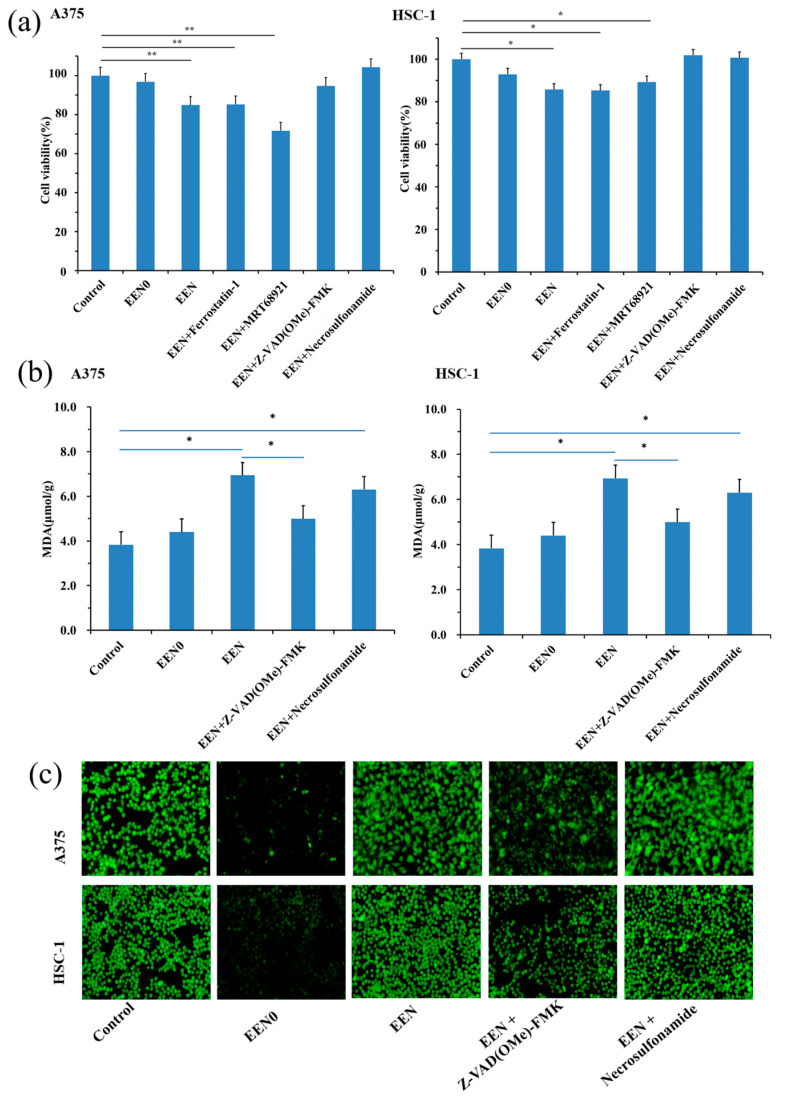
Skin cancer is threatening more and more people's health; its postoperative recurrence and wound infection are still critical challenges. Therefore, specialty wound dressings with multifunctional bioactivity are urgently desired. Emodin is a natural anthraquinone compound that has anti-cancer and anti-bacterial properties. Herein, we fabricated coaxial electrospinning nanofibers loaded with emodin to exploit a multifunctional wound dressing for skin cancer postoperative management, which encapsulated emodin in a polyvinylpyrrolidone core layer, combined with chitosan-polycaprolactone as a shell layer. The nanofibers were characterized via morphology, physicochemical nature, drug load efficiency, pH-dependent drug release profiles, and biocompatibility. Meanwhile, the anti-cancer and anti-bacterial effects were evaluated in vitro. The emodin-loaded nanofibers exhibited smooth surfaces with a relatively uniform diameter distribution and a clear shell-core structure; remarkably, emodin was evenly dispersed in the nanofibers with significantly enhanced dissolution of emodin. Furthermore, they not only display good wettability, high emodin entrapment efficiency, and biphasic release profile but also present superior biocompatibility and anti-cancer properties by increasing the levels of MDA and ROS in A-375 and HSC-1 cells via apoptosis-related pathway, and long-term anti-bacterial effects in a dose-independent manner. The findings indicate that the emodin-loaded nanofiber wound dressing can provide a potential treatment strategy for skin cancer postoperative management.

Keywords: anti-bacteria; anti-cancer; electrospinning nanofiber; emodin; postoperative management; skin cancer.


Conflict of interest statement

The authors declare no conflict of interest.

Figures

Figure 1
Figure 1
Morphology and diameter distributions of the nanofibers. (a) SEM images and (b) diameter distributions of nanofibers of 1% CS-emodin/13% PVP (A), 8% PCL/2% CS (3:2)-emodin/13% PVP (B), 16% PCL/2% CS (5:4)-emodin/13% PVP (C), 20% PCL/2% CS (3:1)-emodin/15% PVP (D), 20% PCL/2% CS (2:1)-emodin/15% PVP (E), 20% PCL/2% CS (2:1)–15% PVP without emodin (F). (c) SEM images of nanofibers after exposure to PBS solution (pH 7.4) of 8% PCL/2% CS (3:2)-emodin/13% PVP (A), 16% PCL/2% CS (5:4)-emodin/13% PVP (B), 20% PCL/2% CS (3:1)-emodin/15% PVP (C), 20% PCL/2% CS (2:1)-emodin/15% PVP (D), 20% PCL/2% CS (2:1)–15% PVP without emodin (E). (d) TEM images of 20% PCL/2% CS (2:1)-emodin/15% PVP (A), 20% PCL/2% CS (2:1)–15% PVP without emodin (B).
Figure 2
Figure 2
Characterization of nanofibers. (a) ATR–FTIR spectra, (b) XRD patterns, and (c) DSC themograms of emodin (A), CS (B), PCL (C), PVP (D), nanofibers with emodin (E), nanofibers witout emodin (F). (d) Swelling ratio of nanofibers with emodin (A) and without emodin (B) after exposure to PBS solution (pH 7.4). (e) Contact angles of nanofibers with emodin (A) and nanofibers without emodin (B) at 0, 5, 10, and 20 s, respectively.
Figure 3
Figure 3
Cumulative release profiles of emodin in different pH PBS solutions. Release profiles of emodin from the nanofibers (A) and emodin raw materials (B) in PBS solution at pH 5.0 (a), pH 8.5 (b), and pH 7.4 (c), respectively.
Figure 4
Figure 4
Effects of nanofibers on cell viabilities and migrations of HaCaT, HSF, and HUVEC cells. (a) Morphologies of HaCaT, HSF, and HUVEC cells treated with blank control, nanofibers without emodin (EEN0), nanofibers containing 0.0824 μg emodin (EEN1), 0.169 μg emodin (EEN2), 0.317 μg emodin (EEN3) for 1, 2, 3, 4, 5, 6, 7 d, respectively. Scar bars: 60 μm. (b) Cell viability statistics data. * p < 0.05 vs. control group. (c) Migration images of HaCaT, HSF, and HUVEC cells treated with blank control, nanofibers without emodin (EEN0), nanofibers containing 1.648 μg emodin (EEN1), 3.38 μg emodin (EEN2), 6.34 μg emodin (EEN3) for 24 h, respectively. Scar bars: 60 μm. (d) Migration ratio statistics data. * p < 0.05 vs. control group.
Figure 5
Figure 5
Cytotoxicity of nanofibers in A375 and HSC-1 cells. (a) Morphologies of A375 and HSC-1 cells treated for 7 d with the blank control, the nanofibers encapsulating 0 μg emodin (EEN0), 0.0824 μg emodin (EEN1), 0.169 μg emodin (EEN2) and 0.317 μg emodin (EEN3), respectively. (b) Cell viability statistics data. * p < 0.05 vs. the control group. (c) Cell migration assay of A375 and HSC-1 cells treated for 24 and 48 h with the nanofibers encapsulating 0 μg emodin (EEN0), 1.648 μg emodin (EEN1), 3.38 μg emodin (EEN2), and 6.34 μg emodin (EEN3), respectively. (d) Statistics data of A375 and HSC-1 cell migration rates, * p < 0.05 vs. the control group. (e) Cell invasion assay of A375 and HSC-1 cells treated for 48 h with the blank control, the nanofibers encapsulating 0 μg emodin (EEN0), 0.412 μg emodin (EEN1), 0.845 μg emodin (EEN2), and 1.585 μg emodin (EEN3), respectively. (f) Cell invasion statistics data of A375 and HSC-1 cells. * p < 0.05 vs. the control group.
Figure 5
Figure 5
Cytotoxicity of nanofibers in A375 and HSC-1 cells. (a) Morphologies of A375 and HSC-1 cells treated for 7 d with the blank control, the nanofibers encapsulating 0 μg emodin (EEN0), 0.0824 μg emodin (EEN1), 0.169 μg emodin (EEN2) and 0.317 μg emodin (EEN3), respectively. (b) Cell viability statistics data. * p < 0.05 vs. the control group. (c) Cell migration assay of A375 and HSC-1 cells treated for 24 and 48 h with the nanofibers encapsulating 0 μg emodin (EEN0), 1.648 μg emodin (EEN1), 3.38 μg emodin (EEN2), and 6.34 μg emodin (EEN3), respectively. (d) Statistics data of A375 and HSC-1 cell migration rates, * p < 0.05 vs. the control group. (e) Cell invasion assay of A375 and HSC-1 cells treated for 48 h with the blank control, the nanofibers encapsulating 0 μg emodin (EEN0), 0.412 μg emodin (EEN1), 0.845 μg emodin (EEN2), and 1.585 μg emodin (EEN3), respectively. (f) Cell invasion statistics data of A375 and HSC-1 cells. * p < 0.05 vs. the control group.
Figure 6
Figure 6
Anti-cancer mechanism of nanofibers. (a) Cell viabilities of A375 and HSC-1 cells, (b) MDA level in A375 and HSC-1 cells, (c) ROS level in A375 and HSC-1 cells with different treatments. * p < 0.05, ** p < 0.01 vs. the control group.
Figure 7
Figure 7
Anti-bacterial effects of nanofibers. A1–A2, B1–B2, and C1–C2 denoted the nanofibers encapsulating 210, 30, and 0 μg of emodin, respectively.

References

    1. Jin J. Screening and Prevention of Skin Cancer. JAMA-J. Am. Med. Assoc. 2023;329:1324. doi: 10.1001/jama.2023.4045. - DOI - PubMed
    1. Rojas K.D., Perez M.E., Marchetti M.A., Nichols A.J., Penedo F.J., Jaimes N. Skin cancer: Primary, secondary, and tertiary prevention. Part II. J. Am. Acad. Dermatol. 2022;87:271–288. doi: 10.1016/j.jaad.2022.01.053. - DOI - PubMed
    1. Binnewies M., Roberts E.W., Kersten K., Chan V., Fearon D.F., Merad M., Coussens L.M., Gabrilovich D.I., Ostrand-Rosenberg S., Hedrick C.C., et al. Understanding the tumor immune microenvironment (TIME) for effective therapy. Nat. Med. 2018;24:541–550. doi: 10.1038/s41591-018-0014-x. - DOI - PMC - PubMed
    1. Liao Z., Chua D., Tan N.S. Reactive oxygen species: A volatile driver of field cancerization and metastasis. Mol. Cancer. 2019;18:65. doi: 10.1186/s12943-019-0961-y. - DOI - PMC - PubMed
    1. O’Leary K. HIV and skin cancer risk. Nat. Med. 2022;24:521–530. doi: 10.1038/d41591-022-00001-7. - DOI - PubMed

LinkOut - more resources