. 2024 Aug 26;16(9):1362.
doi: 10.3390/v16091362.

Cellular N-Myristoyl Transferases Are Required for Mammarenavirus Multiplication

Affiliations

Cellular N-Myristoyl Transferases Are Required for Mammarenavirus Multiplication

Haydar Witwit et al. Viruses. .

Abstract

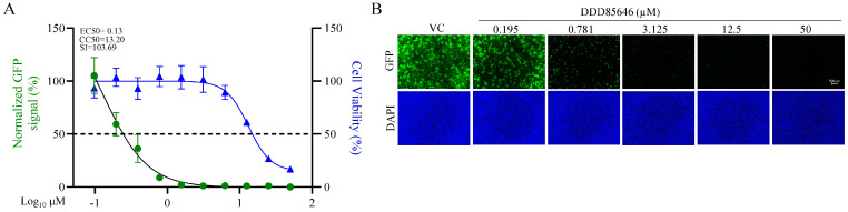
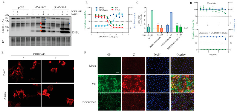
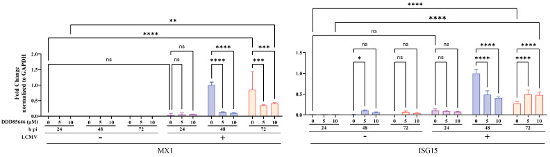
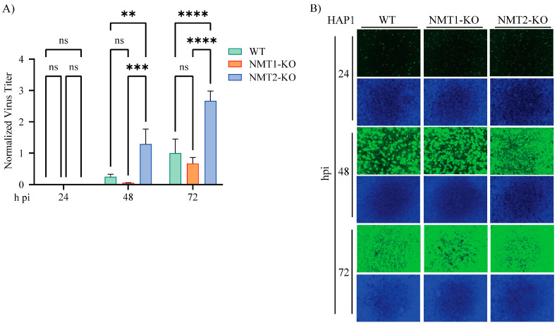
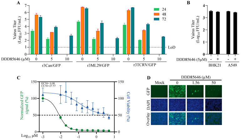
The mammarenavirus matrix Z protein plays critical roles in virus assembly and cell egress. Meanwhile, heterotrimer complexes of a stable signal peptide (SSP) together with glycoprotein subunits GP1 and GP2, generated via co-and post-translational processing of the surface glycoprotein precursor GPC, form the spikes that decorate the virion surface and mediate virus cell entry via receptor-mediated endocytosis. The Z protein and the SSP undergo N-terminal myristoylation by host cell N-myristoyltransferases (NMT1 and NMT2), and G2A mutations that prevent myristoylation of Z or SSP have been shown to affect the Z-mediated virus budding and GP2-mediated fusion activity that is required to complete the virus cell entry process. In the present work, we present evidence that the validated on-target specific pan-NMT inhibitor DDD85646 exerts a potent antiviral activity against the prototypic mammarenavirus lymphocytic choriomeningitis virus (LCMV) that correlates with reduced Z budding activity and GP2-mediated fusion activity as well as with proteasome-mediated degradation of the Z protein. The potent anti-mammarenaviral activity of DDD85646 was also observed with the hemorrhagic-fever-causing Junin (JUNV) and Lassa (LASV) mammarenaviruses. Our results support the exploration of NMT inhibition as a broad-spectrum antiviral against human pathogenic mammarenaviruses.

Keywords: LASV; LCMV; N-myristoyltransferases; antiviral; mammarenavirus; myristoylation; proteasome.


Conflict of interest statement

The authors declare that they have no conflicts of interest.

Figures

Figure 1
Figure 1
Dose-dependent effect of the NMT inhibitor DDD85646 on LCMV multiplication in A549 cells. A549 cells were seeded at 4 × 10⁴ cells/well into a 96-well plate, infected with rLCMV/WT-GFP-P2A-NP (MOI 0.05), and treated with DDD85646 at the indicated concentrations. At 72 h pi, the cells were fixed, and the number of infected cells was determined by immunofluorescence. Numbers of infected cells were normalized to those of vehicle control (VC)-infected cells and expressed as a % of infected cells (A). The total viable cell number was determined by an MTS/formazan bioreduction assay using the Cytation 5 reader. Results show the percentage of infected cells (four biological replicates). EC50 and CC50 values were calculated using a variable slope (based on four parameters) model (Prism10). Results show the mean and SD of four biological replicates. Representative immunofluorescence images of the dose-response assay of selected doses of DDD85646 are shown (B). DAPI was used to stain the DNA (nuclei). Images were taken at 4× magnification using a Keyence BZ-X710 fluorescence microscope.
Figure 2
Figure 2
Effect of the NMT inhibitor DDD85646 on LCMV multi-step growth kinetics and peak titers in A549 cells. (A) Effect of DDD85646 on the production of infectious viral progeny. A549 cells were seeded at 2 × 10⁵ cells/well in an M12-well plate, infected with rLCMV/GFP-P2A-NP (MOI 0.05), and treated with DDD85646 (5 or 10 µM) or with VC. At the indicated time points, cell culture supernatants were collected, and the titers of infectious virus were determined by the focus-forming assay (FFA) using Vero E6 cells. (B,C). Effect of DDD85646 on virus propagation. At the indicated h pi, samples from A were switched to FluoroBrite DMEM containing Hoechst dye (5 µg/mL) and stained for 15 min for live imaging fluorescence (B). GFP signals were determined using Cytation 5 and normalized to the average of the VC (72 h pi). (C,D) Effect of DDD85646 on viral RNA synthesis. Total cellular RNA was isolated from the samples described in (B), and the same RNA amount (10 ng) of each sample was analyzed by RT-qPCR using random hexamers for the RT step, followed by quantitative PCR with specific primers for LCMV NP and the host cell GAPDH. The repeated-measures analysis of variance with mixed-effect analysis and Dunnet’s correction for multiple comparisons were used to determine the statistical significance of the technical triplicates. Statistically significant values: **** p < 0.0001. (E) Effect of DDD85646 on LCMV replication and transcription. Cells were infected with rLCMV/GFP-P2A-NP (MOI 0.05), and at the indicated h pi, total cellular RNA was isolated and analyzed by Northern blotting using an LCMV NP-specific dsDNA 32P probe. Methylene blue (MB) staining was used to confirm similar transfer efficiencies for all the RNA samples.
Figure 3
Figure 3
Effect of DDD85646 on different steps of the LCMV life cycle. (A) Time-of-addition assay: Vero E6 cells were seeded into a 96-well plate at a density of 2 × 10⁴/well. The next day, the cells were infected with the single-cycle infectious rLCMV∆GPC/ZsG (MOI 0.5) and treated with DDD85646 (5 µM) or with VC, starting 1 h before (−1 h) or 2 h after (+2 h) infection. The LCMV cell-entry inhibitor F3406 (10 µM) and ribavirin (Rib) (100 µM) were used as controls. At 48 h pi, ZsG+ cells were assessed using the Synergy™ H4 Hybrid Microplate Reader from Biotek. Values were normalized to VC-treated infected cells; the data represent an average of three biological replicates. (B) LCMV cell-based MG assay: HEK293T cells were seeded into M24-well plates and transfected with plasmids expressing LCMV MG-CAT together with plasmids expressing the viral trans-acting factors NP, L polymerase, and T7 RNA polymerase and then treated with DDD85646 (5 or 10 µM); Rib- and VC-treated samples were used as controls. At 72 h post-transfection, cell lysates were prepared, and total protein was determined using a BCA protein assay. The same amount of total protein from each sample was used to determine CAT protein expression levels using a CAT ELISA kit (Roche, Sydney, Australia) and normalized by assigning the value of 100% activity to vehicle control-treated samples. (C) Z budding activity assay: HEK293T cells were seeded onto poly-L-lysine-coated M12-well plates at 1.75 × 10⁵ cells/ well. The next day, the cells were transfected with either pC-LCMV-Z-GLuc, pC-LASV-Z-Gluc, or pC-LCMV-Z-G2A-GLuc. At 5 h post-transfection, the cells were washed three times and fed with fresh medium containing the relevant drugs at the indicated concentrations. At 48 h post-transfection, cell culture supernatant (CCS) samples were collected, and whole-cell lysates (WCL) were prepared. GLuc activity in the CCS and WCL samples was determined using the SteadyGlo Luciferase Pierce: Gaussia Luciferase Glow assay kit and a Berthold Centro LB 960 luminometer (Berthold Technologies, Oak Ridge, TN, USA). The activity (relative light units) of GLuc in the CCS and WCL was used as a surrogate for Z expression and budding efficiency, defined as the ratio ZVLP/ZVLP + ZWCL. Budding efficiency values were normalized by assigning the value of 100% to vehicle control-treated samples and plotted using Prism10. (D) Virucidal assay: 105 FFU of rLCMV/GFP-P2A-NP was incubated for 30 min at RT in the presence of 0, 5, or 10 µM DDD85646 or in the presence of the validated virucidal compound sodium hypochlorite (0.5%). After treatment, the samples were diluted 1000-fold in DMEM/2%FBS, resulting in concentrations (5 nM and 10 nM) that did not have noticeable anti-LCMV activity, and the number of infectious particles was determined by the FFA.
Figure 4
Figure 4
Effect of DDD85646 on LCMV and LASV GP2-mediated fusion. HEK293T cells were seeded in a M24-well plate at a density of 2.5 × 10⁵ cells/well. The next day, the cells were transfected with pC-LCMV-GPC, pC-LASV-GPC, or pC-E (1 μg/well), all conditions received pC-GFP (50 ng/well). At 5 h post-transfection, the cells were washed once with DMEM/10% FBS and treated with DDD85646 or VC. At 24 h post-transfection, the cell monolayers were treated with acidic (pH 5.0) or neutral (pH 7.2) medium for 15 min, then returned to neutral medium (DMEM/10% FBS) and examined for fusion over time. Once fusion was observed in the VC-treated samples, the cells were fixed for IF imaging with a 20X lens using the Keyence BZ-X710 imaging system.
Figure 5
Figure 5
Effect of DDD85646 on Z expression levels and subcellular distribution. (A) HEK293T cells were seeded (4 × 10⁵ cells/M6-well) and transfected 16 h later with pCAGGS empty (pC-E), Z wild type (pC-Z-WT) or Z (G2A) (pC-Z-G2A) plasmids. At 24 h post-transfection, the cells were treated with DDD85646, MG132, or both, and at 24 h post-treatment, cell lysates were prepared and protein expression examined by Western blotting. Red arrows indicate Z protein oligomers; blue circles indicate Z protein-specific associated bands. (B) A549 cells were plated on 96-well clear-bottom plates (4.0 × 10⁴ cells/well). Sixteen hours later, the cells were infected with rLCMV/Z-HA (MOI 1), and serial dilutions (3-fold) of DDD85646 were added to cells, starting at 10 μM. At 72 h after the drug treatment, the cells were fixed with 4% PFA and stained with fluorescent antibodies for signal quantification; each point represents the mean of four biological replicates. (C) A549 cells were infected (MOI = 0.1) with rLCMV/GFP-P2A-NP. After a 90 min adsorption, the inoculum was removed, the cells were washed once, and medium containing either MG132 (20 µM), DD85646 (5 µM), or MG132 + DDD85646 was added to the cells. Cell culture supernatants were collected at 0 (after adsorption) and 24 h post-infection, and virus titers were determined by the FFA. (D) A549 cells were seeded at 4 × 10⁴ cells/well into a 96-well plate, infected with rLCMV/GFP-P2A-NP (MOI 0.05), and treated with incremental doses of clemizole or with clemizole in the presence of 5μM DDD85646. At 72 h pi, the cells were fixed, and the number of infected cells was determined by IF. (E) HEK293T cells were plated on poly-L-Lysine 96-well clear-bottom optically treated plates (2.0 × 10⁴ cells/well). The next day, the cells were transfected with LCMV Z-HA or the Z-G2A-HA mutant. At 24 h post-transfection (h pt), the media was replaced with DDD85646 (5 μM)-containing media, and at 48 h after drug treatment, the cells were fixed with 4% PFA. Images were taken using a Keyence BZX-710 microscope at 20× magnification, which were then zoomed in digitally to 1.5× magnification. The images were subjected to the Point–Spread Function (PSF) and then deconvolved using DeconvolutionLab2 (Fiji ImageJ, v154) with the following setting: Iterative Constraint Tikhonov–Miller algorithm at 100 iteration N, 1 step γ, and low reg. λ. (F) A549 cells were plated on 96-well clear-bottom optically treated plates (2.0 × 10⁴ cells/well). The next day, the cells were infected with rLCMV/Z-HA (MOI 0.05) and treated with DDD85646 (5 μM)-containing media 90 min later. At 72 h after drug treatment, the cells were fixed with 4% PFA and co-labeled with anti-HA (Z) and anti-NP antibodies and with DAPI for DNA; the Z protein is red, NP is green, and DNA is blue. The images have been zoomed in digitally to 3× magnification.
Figure 6
Figure 6
Effect of DDD85646 on the expression of ISGs. A549 cells were seeded at 2 × 10⁵ cells/well in an M12-well plate, infected with LCMV/GFP-P2A-NP (MOI 0.05), and treated with DDD85646 (5 or 10 µM) or with VC. At the indicated time points, total cellular RNA was isolated and analyzed by RT-qPCR (10 ng/sample; three technical replicates). The RT step was performed using random hexamers, followed by quantitative PCR with specific primers for ISGs MX1, or ISG15 and the cell housekeeping gene GAPDH. MX1 and ISG15 fold changes were normalized to GAPDH. The repeated-measures analysis of variance with mixed-effect analysis and Dunnet’s correction for multiple comparisons were used. Statistically significant values: ns p > 0.05, * p ≤ 0.05, ** p < 0.01, *** p < 0.001, **** p < 0.0001.
Figure 7
Figure 7
Contribution of NMT1 and NMT2 isozymes to LCMV multiplication. HAP1 NMT WT, NMT-1-KO, or NMT-2-KO cells were seeded at 2 × 10⁵ cells/well in an M12-well plate and infected with LCMV/GFP-P2A-NP (MOI 0.05). At the indicated time points, CCS was collected for virus titration. Virus titers were normalized to titers at 72 h pi in the VC-treated sample, which was assigned the value of 1 (A), and the cells were fixed for immunofluorescence imaging (B). The repeated-measures analysis of variance with mixed-effect analysis and Tukey’s correction for multiple comparisons were used. Statistically significant values: ns p > 0.05, * p ≤ 0.05, ** p < 0.01, *** p < 0.001, **** p < 0.0001.
Figure 8
Figure 8
Effect of DDD85646 on the multiplication of the mammarenaviruses JUNV, TCRV, ML29, and LASV, and the flavivirus ZIKV. (A) A549 cells were infected with the indicated mammarenavirus and treated with DDD85646 (5 or 10 µM) or VC. At the indicated h pi, titers of infectious virus in the cell culture supernatants were determined by the FFA. (B) A549 and BHK21 cells were seeded into M24-well plates at a density of 1 × 10⁵ cells/well and infected with ZIKV (MOI 0.1) for 1 h, then treated with DDD85646 (5 µM). At 24 h pi, supernatants were collected, and virus titers were determined by the FFA. Titers of technical duplicates were log-transformed and plotted as the mean ± SD (error bars). (C) A549 cells were seeded at 4 × 10⁴ cells/well into a 96-well plate, infected with r3LASV/GFP (MOI 0.05), and treated with DDD85646 at the indicated concentrations. At 72 h pi, the cells were fixed, and the numbers of infected cells were determined by immunofluorescence. Numbers of infected cells were normalized to those of vehicle control (VC)-infected cells and expressed as a % of infected cells (viral infection). Cell viability was estimated based on the signal of DAPI staining. Results show the percentage of infected cells (four biological replicates). EC50 and CC50 values were calculated using a variable slope (based on four parameters) model (Prism10). Results show the mean and SD of four replicates. Representative immunofluorescence images of the dose-response assay of selected doses of DDD85646 are shown (D).

Update of

References

    1. Radoshitzky S.R., Buchmeier M.J., de la Torre J.C. Fields Virology: Emerging Viruses: Arenaviridae. In: Knipe D.M., Howley P.M., Whelan S., editors. Fields Virology. 7th ed. Lippincott & Wilkins; Philadelphia, PA, USA: 2022. Vol I: Emerging Viruses.
    1. Lenz O., ter Meulen J., Klenk H.-D., Seidah N.G., Garten W. The Lassa Virus Glycoprotein Precursor GP-C Is Proteolytically Processed by Subtilase SKI-1/S1P. Proc. Natl. Acad. Sci. USA. 2001;98:12701–12705. doi: 10.1073/pnas.221447598. - DOI - PMC - PubMed
    1. Beyer W.R., Pöpplau D., Garten W., von Laer D., Lenz O. Endoproteolytic Processing of the Lymphocytic Choriomeningitis Virus Glycoprotein by the Subtilase SKI-1/S1P. J. Virol. 2003;77:2866–2872. doi: 10.1128/JVI.77.5.2866-2872.2003. - DOI - PMC - PubMed
    1. Kunz S., Edelmann K.H., de la Torre J.-C., Gorney R., Oldstone M.B.A. Mechanisms for Lymphocytic Choriomeningitis Virus Glycoprotein Cleavage, Transport, and Incorporation into Virions. Virology. 2003;314:168–178. doi: 10.1016/S0042-6822(03)00421-5. - DOI - PubMed
    1. Rojek J.M., Lee A.M., Nguyen N., Spiropoulou C.F., Kunz S. Site 1 Protease Is Required for Proteolytic Processing of the Glycoproteins of the South American Hemorrhagic Fever Viruses Junin, Machupo, and Guanarito. J. Virol. 2008;82:6045–6051. doi: 10.1128/JVI.02392-07. - DOI - PMC - PubMed

Publication types

Substances

LinkOut - more resources