. 2024 Aug 29;16(9):1379.
doi: 10.3390/v16091379.

Presence of CD80 and Absence of LAT in Modulating Cellular Infiltration and HSV-1 Latency

Affiliations

Presence of CD80 and Absence of LAT in Modulating Cellular Infiltration and HSV-1 Latency

Ujjaldeep Jaggi et al. Viruses. .

Abstract

CD80 is the best-known costimulatory molecule for effective T cell functions. Many different reports have summarized the role of CD80 in HSV-1 and its functions in maintaining adaptive immunity, which is the main player in causing herpes stromal keratitis (HSK). To determine the effects of absence or overexpression of CD80 in HSV-1 infection, we infected CD80-/- and WT mice with a recombinant HSV-1 expressing murine CD80 (HSV-CD80) in place of the latency associated transcript (LAT). Parental dLAT2903 virus lacking LAT was used as a control. After infection, critical components of infection like virus replication, eye disease, early cellular infiltrates into the corneas and trigeminal ganglia (TG), latency-reactivation in the infected mice were determined. Our findings reveal that the absence of CD80 in the CD80-/- mice infected with both viruses did not affect the viral titers in the mice eyes or eye disease, but it played a significant role in critical components of HSV-induced immunopathology. The WT mice infected with dLAT2903 virus had significantly higher levels of latency compared with the CD80-/- mice infected with dLAT2903 virus, while levels of latency as determined by gB DNA expression were similar between the WT and CD80-/- mice infected with HSV-CD80 virus. In contrast to the differences in the levels of latency between the infected groups, the absence of CD80 expression in the CD80-/- mice or its overexpression by HSV-CD80 virus did not have any effect on the time of reactivation. Furthermore, the absence of CD80 expression contributed to more inflammation in the CD80-/--infected mice. Overall, this study suggests that in the absence of CD80, inflammation increases, latency is reduced, but reactivation is not affected. Altogether, our study suggests that reduced latency correlated with reduced levels of inflammatory molecules and blocking or reducing expression of CD80 could be used to mitigate the immune responses, therefore controlling HSV-induced infection.

Keywords: CD8; PD-1; corneal scarring; latency; reactivation; virus replication.


Conflict of interest statement

The authors declare no conflict of interest.

Figures

Figure 1
Figure 1
Viral titers in WT and CD80-/- mice eyes following ocular infection with dLAT2903 and HSV-CD80 viruses. WT and CD80-/- mice were infected with 2 × 10⁵ PFU/eye of dLAT2903 and HSV-CD80 viruses. The presence of infectious virus in the eyes of infected mice was monitored daily for 7 days by collecting tear films and quantifying the virus using standard plaque assays as described (see Section 4). Each point represents the mean ± SEM from 24 eyes for all infected mouse groups; no differences in viral titers were seen among the four groups. The experiment was repeated twice. The levels of virus shedding were not significantly different in the respective groups.
Figure 2
Figure 2
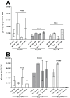
Quantification of the gB copy numbers in the corneas and TG of the infected WT and CD80-/- mice. (A) The gB copy numbers in the infected corneas. WT and CD80-/- mice were infected with 2 × 10⁵ PFU/eye of dLAT2903 and HSV-CD80 viruses, and the corneas and TG from the WT- and CD80-/--infected mice (3 mice/group) were harvested on days 3, 5, and 7 PI. The total RNA was isolated from each cornea or TG, and GAPDH expression was used to normalize the expression of the gB transcripts in the corneas or TG of the infected mice, and the gB copy numbers were determined by qRT-PCR as described in Section 2. No differences were observed in the infected corneas between the four infected mice groups (p > 0.05). Each bar represents the mean expression ± SEM in the six corneas from all the infected mouse groups; and (B) the gB copy numbers in the infected TG. The TG were harvested on days 3, 5, and 7 PI from the above infected mice. The total RNA was isolated from each TG, and GAPDH expression was used to normalize the expression of each transcript in the TG of ocularly infected mice, and the gB copy numbers were determined as above. No differences in the infected TG were observed between the four infected mice groups (p > 0.05). Each bar represents the mean expression ± SEM in the six TG from all the infected mouse groups. Only the differences that are statistically significant are shown for each gene.
Figure 3
Figure 3
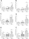
Quantification of CD4, CD8α, and F4/80 RNA transcripts in the corneas and TG of the infected WT and CD80-/- mice. (A,B) Expressions of CD4 and CD8α in the infected corneas. WT and CD80-/- mice were infected as described in Figure 2 above. The total RNA was isolated from each cornea, and GAPDH expression was used to normalize the expressions of CD4 and CD8α transcripts in the corneas of ocularly infected mice. CD4 and CD8α in the infected corneas displayed no significant differences on days 3, 5 and 7 PI among all the infected mice groups (p > 0.05); (C,D) expression of CD4 and CD8α in the infected TG. The total RNA was isolated from each TG, and GAPDH expression was used to normalize the expressions of CD4 and CD8α transcripts in the TG of ocularly infected mice. CD4 T cells transcript levels on day 3 PI in the infected TG in the WT mice infected with dLAT2903 virus displayed higher expression levels in comparison to CD4 transcript levels in CD80-/- mice infected with HSV-CD80 virus (p < 0.0031). CD8α T cells in the TG were not significantly different in any of the infected mice groups (p > 0.05); and (E,F) expression levels of F4/80 RNA transcripts in the corneas and TG. The total RNA was isolated from each cornea or TG, and GAPDH expression was used to normalize the expression levels of F4/80 transcripts in the corneas and TG of ocularly infected mice. F4/80 expression levels in the infected corneas displayed no significant differences on days 3, 5 and 7 PI among all the infected groups (p > 0.05). F4/80 expression levels in the infected TG were higher on day 5 PI in the WT mice infected with HSV-CD80 virus as compared with the WT mice infected with dLAT2903 virus (p = 0.0067). Each bar represents the mean expression ± SEM in the 6 Corneas and TG from all the infected mouse groups.
Figure 4
Figure 4
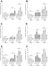
Quantification of CD11c, NK1.1, and CD45 RNA transcripts in the corneas and TG of the infected WT and CD80-/- mice. (A,B) Expression levels of CD11c in the infected corneas and TG. WT and CD80-/- mice were infected as described in Figure 2. The total RNA was isolated from each cornea and TG, and GAPDH expression was used to normalize expression of the CD11c transcript in the corneas and TG of the ocularly infected mice. No significant expression level differences were detected for CD11c in the infected mouse groups; (C,D) expression levels of NK1.1 in the infected corneas and TG. The total RNA was isolated from each cornea and TG, and GAPDH expression was used to normalize expression of the NK1.1 transcript in the ocularly infected mice. No significant expression level differences were detected for NK1.1 in the infected mouse groups; and (E,F) expression levels of CD45 in the infected corneas and TG. The total RNA was isolated from each cornea and TG, and GAPDH expression was used to normalize expression of the CD45 transcript of the ocularly infected mice. No significant expression level differences were detected for CD45 in the infected mouse groups. Each bar represents the mean expression ± SEM in the six corneas and TG for all the infected mouse groups. Only the differences that are statistically significant are shown for each gene.
Figure 5
Figure 5
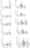
Quantification of IFN-γ, IL-6, IL-1α, IL-1β, and TNF-α RNA transcripts in the corneas and TG of the infected WT and CD80-/- mice. (A,B) Expression levels of IFN-γ in the infected corneas and TG. WT and CD80-/- mice were infected as described in Figure 2. The total RNA was isolated from each cornea and TG. GAPDH expression was used to normalize expression levels of IFN-γ transcripts in the corneas and TG of the ocularly infected mice. The corneas from the CD80-/- mice infected with dLAT2903 virus showed significantly higher expression levels of IFN-γ transcripts on day 7 PI in comparison to all three other infected mice groups (p < 0.05), whereas in the TG, IFN-γ transcript levels had no significant differences at any point of the day among the four groups (p > 0.05); (C,D) expression levels of IL-6 in the infected corneas and TG. WT and CD80-/- mice were infected as described in Figure 2. The total RNA was isolated from each cornea and TG. GAPDH expression was used to normalize expression levels of the IL-6 transcript in the corneas and TG of the ocularly infected mice. The infected WT and CD80-/- mice had no significant differences in IL-6 expression levels in the corneas or TG (p > 0.05); (E,F) expression levels of IL-1α in the infected corneas and TG. WT and CD80-/- mice were infected as described in Figure 2. The total RNA was isolated from each cornea and TG, and GAPDH expression was used to normalize expression levels of the IL-1α transcript in the corneas and TG of the ocularly infected mice. The infected WT and CD80-/- mice had no significant differences in IL-1α transcript expression levels in the corneas or TG (p > 0.05); (G,H) expression levels of IL-1β in the infected corneas and TG. WT and CD80-/- mice were infected as described in Figure 2. The total RNA was isolated from each cornea and TG. GAPDH expression was used to normalize expression levels of the IL-1β transcript in the corneas and TG of the ocularly infected mice. The infected WT and CD80-/- mice had no significant differences in IL-1β transcript expression levels in the corneas or TG (p > 0.05); and (I,J) expression levels of TNF-α in the infected corneas and TG. WT and CD80-/- mice were infected as described in Figure 2. The total RNA was isolated from each cornea and TG. GAPDH expression was used to normalize expression levels of the TNF-α transcript in the corneas and TG of the ocularly infected mice. The infected WT and CD80-/- mice had no significant differences in TNF-α transcript expression levels in the corneas or TG (p > 0.05). Each bar represents mean expression ± SEM in the six corneas and TG from all the infected mouse groups. Only the differences that are statistically significant are shown for each gene. Panels: (A,B) Expression levels of IFN-γ in the corneas and TG. (C,D) Expression levels of IL-6 in the infected corneas and TG. (E,F) Expression levels of IL-1α RNA transcripts in the corneas and TG. (G,H) Expression levels of IL-1β RNA transcripts in the infected corneas and TG. (I,J) Expression levels of TNF-α in the infected corneas and TG.
Figure 6
Figure 6
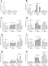
CD80, CD86, PD-L1, and CTLA4 expressions in the corneas and TG of the infected WT and CD80-/- mice. (A,B) Expression levels of CD80 in the infected corneas and TG. WT and CD80-/- mice were infected as described in Figure 2. The total RNA was isolated from each cornea and TG. GAPDH expression was used to normalize expression levels of CD80 transcripts in the corneas and TG of the ocularly infected mice. CD80 transcript levels had no significant differences at any point of the day (p > 0.05); (C,D) expression levels of CD86 in the infected corneas and TG. WT and CD80-/- mice were infected as described in Figure 2. The total RNA was isolated from each cornea and TG. GAPDH expression was used to normalize expression levels of CD86 transcripts in the corneas and TG of the ocularly infected mice. CD86 transcript levels had no significant differences at any point of the day or in any infected mice groups (p > 0.05); (E,F) expression levels of PD-L1 in the infected corneas and TG. WT and CD80-/- mice were infected as described in Figure 2. The total RNA was isolated from each cornea and TG. GAPDH expression was used to normalize expression levels of PD-L1 transcripts in the corneas and TG of the ocularly infected mice. PD-L1 transcript levels had no significant differences at any point of the day or in any infected mice groups (p > 0.05); and (G,H) expression levels of CTLA4 in the infected corneas and TG. WT and CD80-/- mice were infected as described in Figure 2. The total RNA was isolated from each cornea and TG. GAPDH expression was used to normalize expression levels of CTLA4 transcripts in the corneas and TG of the ocularly infected mice. CTLA4 transcript levels in the corneas had no significant differences at any point of the day or in any infected mice groups (p > 0.05), but in the TG on day 7 PI, CTLA4 in the WT (dLAT2903 virus-infected) mice was higher as compared with the CD80-/- (HSV-CD80 virus-infected) mice. Each bar represents mean expression ± SEM in the six corneas and TG from all the infected mouse groups. Only the differences that are statistically significant are shown for each gene.
Figure 7
Figure 7
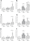
GzmA, GzmB, and perforin expressions in the corneas and TG of the infected WT and CD80-/- mice. (A,B) Expression levels of GzmA in the infected corneas and TG. WT and CD80-/- mice were infected as described in Figure 2. The total RNA was isolated from each cornea and TG. GAPDH expression was used to normalize expression levels of GzmA transcripts in the corneas and TG of the ocularly infected mice. Where GzmA transcript levels in the corneas had no significant differences, GzmA in the TG on day 7 PI was higher in the WT (HSV-CD80 virus-infected) mice as compared to the WT (dLAT2903 virus-infected) mice; (C,D) expression levels of GzmB in the infected corneas and TG. WT and CD80-/- mice were infected as described in Figure 2. The total RNA was isolated from each cornea and TG. GAPDH expression was used to normalize expression levels of GzmB transcripts in the corneas and TG of the ocularly infected mice. GzmB transcript levels in the corneas and TG had no significant differences in all the infected mice groups (p > 0.05); and (E,F) expression levels of Perforin in the infected corneas and TG. WT and CD80-/- mice were infected as described in Figure 2. The total RNA was isolated from each cornea and TG. GAPDH expression was used to normalize expression levels of perforin transcripts in the corneas and TG of the ocularly infected mice. Perforin transcript levels had no significant differences at any point of the day (p > 0.05).
Figure 8
Figure 8
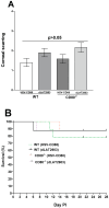
Absence of CD80 or LAT does not affect survival and eye disease. (A) Eye disease. Thirty-two eyes from and WT (HSV-CD80 virus-infected), CD80-/- (HSV-CD80 virus-infected), and CD80-/- (dLAT2903 virus-infected) mice and twenty-six eyes from WT (dLAT2903 virus-infected) mice were used to measure corneal scarring (CS). The severity of CS in mouse corneas was examined in all groups by slit lamp biomicroscopy. The CS severity was scored on day 28 PI from three independent experiments. All the infected mice groups were not significantly different from one another (p > 0.05). CS is based on 48 eyes for the WT mice infected with HSV-CD80 virus and the CD80-/- mice infected with HSV-CD80 and dLAT2903 viruses, while CS in the WT mice infected with dLAT2903 virus is based on 46 eyes; and (B) mice survival. The mice were ocularly infected as described above. Survival of the WT and CD80-/- mice was monitored over a 28-day period after infection. There were no differences in survival among all the infected mice groups (p > 0.05). Survival is based on three independent experiments.
Figure 9
Figure 9
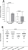
The latent gB DNA and duration of explant reactivation following ocular infection of WT and CD80-/- mice. (A) gB DNA copy numbers in the latent TG. Forty TG from each infected mice group were isolated on day 28 PI. Expressions of gB DNA were determined using qPCR, and gB copy numbers were measured as described in Section 2. gB DNA copy numbers were higher in the WT mice infected with dLAT2903 virus compared with the CD80-/- mice infected with dLAT2903 virus (p = 0.03). No differences were detected in the WT and CD80-/- mice infected with HSV-CD80 virus (p > 0.05); and (B) explant reactivation in the latent TG. On day 28 PI, the TG from the infected WT and CD80-/- mice were isolated and incubated in 1.5 mL of tissue culture media at 37 °C, and the presence of infectious virus was monitored as described in Section 2. The results are shown as the number of the TG that reactivated daily. Each point represents mean reactivated TG ± SEM of 20 TG from each mice group infected and from two independent experiments. There were no differences in reactivation (p > 0.05).

References

    1. Liesegang T.J., Melton L.J., 3rd, Daly P.J., Ilstrup D.M. Epidemiology of ocular herpes simplex. Incidence in Rochester, Minn, 1950 through 1982. Arch. Ophthalmol. 1989;107:1155–1159. doi: 10.1001/archopht.1989.01070020221029. - DOI - PubMed
    1. Young R.C., Hodge D.O., Liesegang T.J., Baratz K.H. Incidence, recurrence, and outcomes of herpes simplex virus eye disease in Olmsted County, Minnesota, 1976–2007: The effect of oral antiviral prophylaxis. Arch. Ophthalmol. 2010;128:1178–1183. doi: 10.1001/archophthalmol.2010.187. - DOI - PMC - PubMed
    1. Brandt C.R. The role of viral and host genes in corneal infection with herpes simplex virus type 1. Exp. Eye Res. 2005;80:607–621. doi: 10.1016/j.exer.2004.09.007. - DOI - PubMed
    1. Thomas J., Rouse B.T. Immunopathogenesis of herpetic ocular disease. Immunol. Res. 1997;16:375–386. doi: 10.1007/BF02786400. - DOI - PubMed
    1. Rowe A.M., St Leger A.J., Jeon S., Dhaliwal D.K., Knickelbein J.E., Hendricks R.L. Herpes keratitis. Prog. Retin. Eye Res. 2013;32:88–101. doi: 10.1016/j.preteyeres.2012.08.002. - DOI - PMC - PubMed

Publication types

MeSH terms

Substances

LinkOut - more resources