Review
. 2024 Sep 7;16(9):1427.
doi: 10.3390/v16091427.

Cytomegalovirus Retinitis: Clinical Manifestations, Diagnosis and Treatment

Affiliations
Review

Cytomegalovirus Retinitis: Clinical Manifestations, Diagnosis and Treatment

Jing Zhang et al. Viruses. .

Abstract

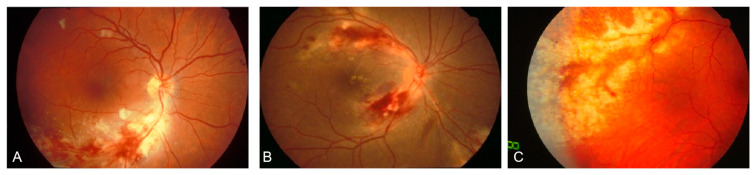
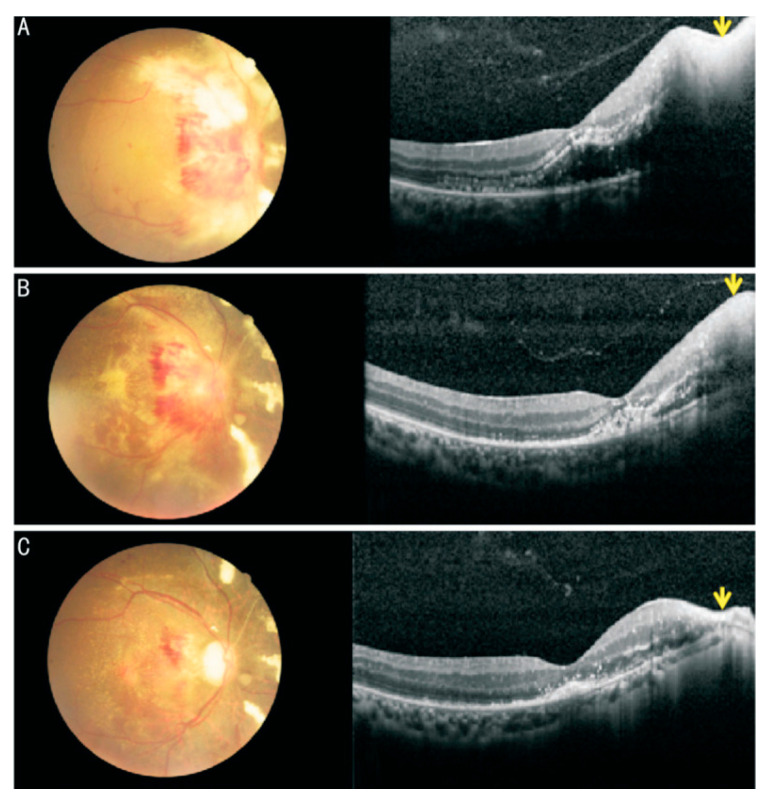
Cytomegalovirus (CMV) retinitis is the most common eye disease associated with CMV infection in immunocompromised individuals. The CMVR may initially be asymptomatic; however, relatively mild vitreous inflammation at the onset may be an important differential point from other diseases in HIV patients. Fundus photography, CD4 T-cell count, and telemedicine could be used to screen and monitor the high-risk population, particularly in resource-limited regions. Retinitis generally starts in the peripheral retina and advances toward the posterior pole, which could develop to the characteristic "pizza pie" appearance marked by central retinal necrosis and intraretinal hemorrhage. CMVR causes vision loss if left untreated, and early antiviral therapy significantly reduces the risk of vision loss. Alongside traditional antiviral treatments, immunotherapies including CMV-specific adoptive T-cell therapy and CMV immunoglobulin (CMVIG) are emerging as promising treatment options due to their favorable tolerability and reduced mortality. This review comprehensively examines CMV retinitis, encompassing the clinical features, differential diagnosis, laboratory tests, and updated treatment strategies to inform clinical management.

Keywords: antiviral treatment; cytomegalovirus; differential diagnosis; immunodeficiency; retinitis.


Conflict of interest statement

The authors declare no conflicts of interest.

Figures

Figure 1
Figure 1
Fundus photograph of cytomegalovirus retinitis: (A) wedge-shaped appearance with the apex “pointing” toward the optic disc. (B) a “hemorrhagic” appearance involving the posterior pole, characterized by retinal necrosis and edema, intraretinal hemorrhage and “satellite lesions” at the border. (C) cytomegalovirus retinitis involving the periphery, characterized by a “granular” appearance and without hemorrhage. (Modified from Standardization of Uveitis Nomenclature (SUN) Working Group, American journal of ophthalmology, 2021, 228: 245–254 [43], under a Creative Commons licence CC BY).
Figure 2
Figure 2
Cytomegalovirus (CMV) retinitis zones: Zone 1 encompasses the area within 1500 µm of the optic nerve or 3000 µm of the fovea. Zone 2 extends from the outer boundary of Zone 1 to the equator, as determined by the vortex veins. Zone 3 covers the peripheral retina from the equator to the ora serrata (Photo by Kwon H J, et al., Microorganisms, 2021 [48], under a Creative Commons licence CC BY 4.0).
Figure 3
Figure 3
Fundus photographs and OCT images of CMVR and cytomegalovirus papillitis: (A) First visit: fundus photography showed yellowish white retinal necrosis and retinal hemorrhage around the disc; OCT showed exudative retinal detachment in macular area and significant thickening and hyperreflective in temporal retina of optic disc with full-thickness disruption of retinal architecture (yellow arrow). (B) Two weeks: after 2 wk of anti-cytomegalovirus therapy, retinal necrotic lesion has disappeared on fundus photograph, OCT showed subretinal fluid absorption and the edema of the necrotic lesion relief (yellow arrow). (C) Six weeks: fundus photography showed optic atrophy and the retinal necrosis and most of the retinal hemorrhage were absorbed; OCT showed complete absorption of subretinal fluid and retinal thinning in temporal retina of optic disc (yellow arrow). (Photo by Sheng, Yan, et al. International Journal of Ophthalmology, 2020, 13(11): 1800 [77], under a Creative Commons licence CC BY 4.0).

Similar articles

Cited by

References

    1. Martinez-Gomez E., Perez-Carpena P., Flook M., Lopez-Escamez J.A. A systematic review on the association of acquired human cytomegalovirus infection with hearing loss. J. Clin. Med. 2020;9:4011. doi: 10.3390/jcm9124011. - DOI - PMC - PubMed
    1. Alonso-Álvarez S., Colado E., Moro-García M.A., Alonso-Arias R. Cytomegalovirus in haematological tumours. Front. Immunol. 2021;12:703256. doi: 10.3389/fimmu.2021.703256. - DOI - PMC - PubMed
    1. Xu J.X., Liu X.L., Zhang X.Y., Marshall B., Dong Z., Liu Y.T., Espinosa-Heidmann D.G., Zhang M. Ocular cytomegalovirus latency exacerbates the development of choroidal neovascularization. J. Pathol. 2020;251:200–212. doi: 10.1002/path.5447. - DOI - PMC - PubMed
    1. Zhang J., Kamoi K., Zong Y., Yang M., Ohno-Matsui K. Cytomegalovirus anterior uveitis: Clinical manifestations, diagnosis, treatment, and immunological mechanisms. Viruses. 2023;15:185. doi: 10.3390/v15010185. - DOI - PMC - PubMed
    1. Shukla R., Mishra A.K., Verma A., Garg P., Shrinkhal L., Shukla R., Mishra A.K., Verma A., Garg Sr P. A Rare Case of Cytomegalovirus Retinitis in a Young Immunocompetent Patient. Cureus. 2023;15:e44948. doi: 10.7759/cureus.44948. - DOI - PMC - PubMed

Publication types

MeSH terms

Substances

LinkOut - more resources