

Retrospective Analyses of Porcine Circovirus Type 3 (PCV-3) in Switzerland
- PMID: 39339907
- PMCID: PMC11437478
- DOI: 10.3390/v16091431
Retrospective Analyses of Porcine Circovirus Type 3 (PCV-3) in Switzerland
Abstract
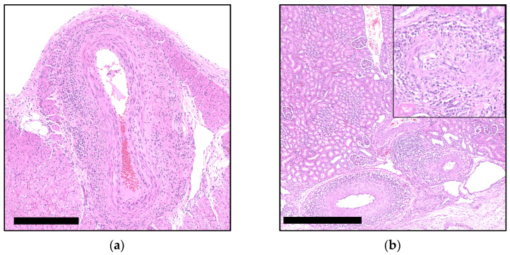
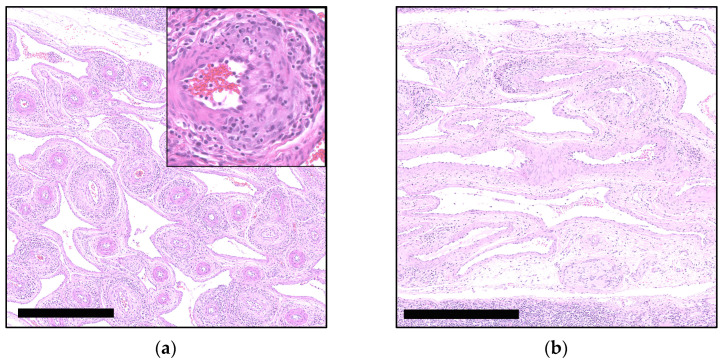
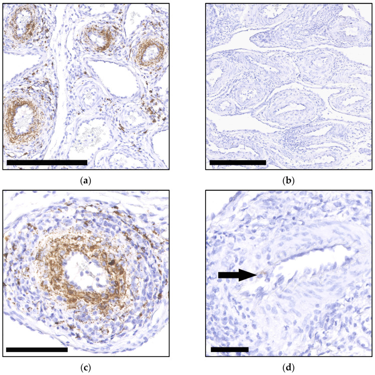
Porcine circovirus 3 (PCV-3) has emerged as a significant pathogen affecting global swine populations, yet its epidemiology and clinical implications remain incompletely understood. This retrospective study aimed to investigate the prevalence and histopathological features of PCV-3 infection in pigs from Switzerland, focusing on archival cases of suckling and weaner piglets presenting with suggestive lesions. An in-house qPCR assay was developed for detecting PCV-3 in frozen and formalin-fixed paraffin-embedded tissues, enhancing the national diagnostic capabilities. Histopathological reassessment identified PCV-3 systemic disease (PCV-3-SD) compatible lesions in 19 (6%) of archival cases, with 47% testing positive by qPCR across various organs. Notably, vascular lesions predominated, particularly in mesenteric arteries, heart, and kidneys. The study confirms the presence of PCV-3 in Switzerland since at least 2020, marking the first documented cases within the Swiss swine population. Despite challenges in in situ hybridization validation due to prolonged formalin fixation, the findings indicate viral systemic dissemination. These results contribute to the understanding of PCV-3 epidemiology in Swiss pigs, emphasizing the need for continued surveillance and further research on its clinical implications and interaction with host factors. Our study underscores the utility and limitations of molecular techniques in confirming PCV-3 infections.
Keywords: PCV-3 systemic disease (PCV-3-SD); diagnostic; histopathology; in situ hybridization (ISH); porcine circovirus 3 (PCV-3); real-time quantitative polymerase chain reaction (qPCR); tissue microarray (TMA); vascular lesions.
Conflict of interest statement
The authors declare no conflicts of interest. The funders had no role in the design of the study; in the collection, analyses, or interpretation of data; in the writing of the manuscript; or in the decision to publish the results.
Figures
Similar articles
-
Porcine Circovirus 3 Detection in Aborted Fetuses and Stillborn Piglets from Swine Reproductive Failure Cases.Viruses. 2021 Feb 9;13(2):264. doi: 10.3390/v13020264. Viruses. 2021. PMID: 33572209 Free PMC article.
-
Lack of detection of Porcine circovirus 3 (PCV-3) in formalin-fixed, paraffin- embedded tissues from porcine abortions in Switzerland.Schweiz Arch Tierheilkd. 2024 Sep;166(9):460-464. doi: 10.17236/sat00431. Schweiz Arch Tierheilkd. 2024. PMID: 39225507 English.
-
Retrospective investigation of porcine circoviruses in cases of porcine dermatitis and nephropathy syndrome.Vet Res. 2024 Nov 9;55(1):146. doi: 10.1186/s13567-024-01405-8. Vet Res. 2024. PMID: 39521988 Free PMC article.
-
Porcine circovirus 3 (PCV-3) as a causal agent of disease in swine and a proposal of PCV-3 associated disease case definition.Transbound Emerg Dis. 2021 Nov;68(6):2936-2948. doi: 10.1111/tbed.14204. Epub 2021 Jul 8. Transbound Emerg Dis. 2021. PMID: 34184834 Free PMC article. Review.
-
Porcine circovirus type 2 in China: an update on and insights to its prevalence and control.Virol J. 2014 May 14;11:88. doi: 10.1186/1743-422X-11-88. Virol J. 2014. PMID: 24885983 Free PMC article. Review.
References
-
- Palinski R., Piñeyro P., Shang P., Yuan F., Guo R., Fang Y., Byers E., Hause B.M. A Novel Porcine Circovirus Distantly Related to Known Circoviruses Is Associated with Porcine Dermatitis and Nephropathy Syndrome and Reproductive Failure. J. Virol. 2017;91:1–13. doi: 10.1128/JVI.01879-16. - DOI - PMC - PubMed
-
- Franzo G., Legnardi M., Hjulsager C.K., Klaumann F., Larsen L.E., Segales J., Drigo M. Full-Genome Sequencing of Porcine Circovirus 3 Field Strains from Denmark, Italy and Spain Demonstrates a High within-Europe Genetic Heterogeneity. Transbound. Emerg. Dis. 2018;65:602–606. doi: 10.1111/tbed.12836. - DOI - PubMed
Publication types
MeSH terms
Grants and funding
LinkOut - more resources
Full Text Sources
