. 2024 Sep 11;16(9):1448.
doi: 10.3390/v16091448.

N6-Methyladenosine Positively Regulates Coxsackievirus B3 Replication

Affiliations

N6-Methyladenosine Positively Regulates Coxsackievirus B3 Replication

Hainian Zhao et al. Viruses. .

Abstract

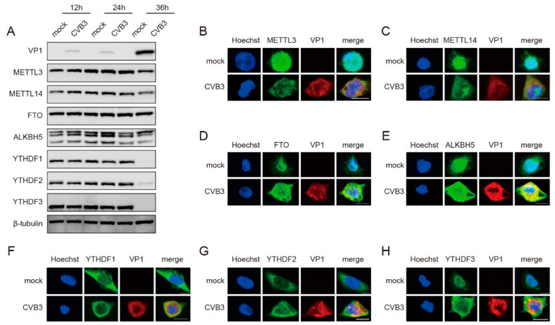
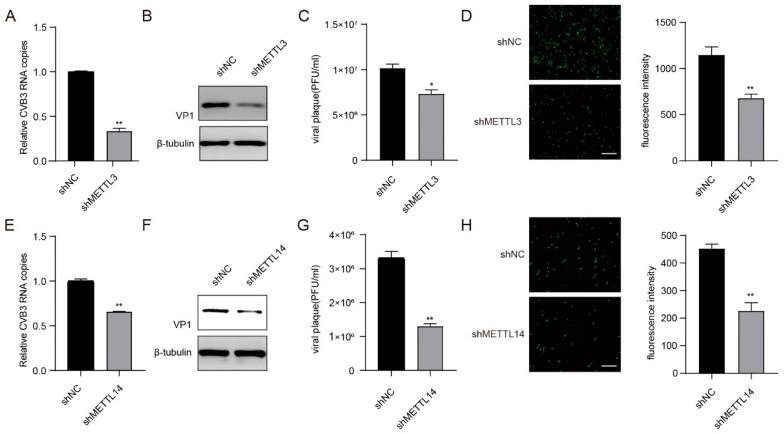
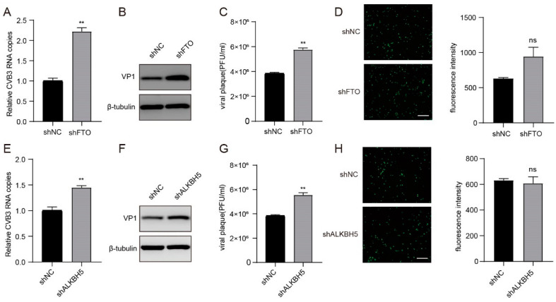
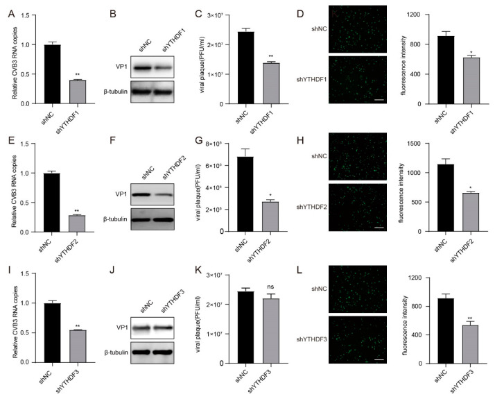
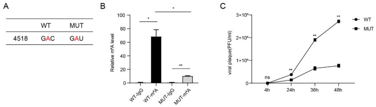
Enteroviruses such as coxsackievirus B3 are identified as a common cause of viral myocarditis, but the potential mechanism of its replication and pathogenesis are largely unknown. The genomes of a variety of viruses contain N6-methyladenosine (m6A), which plays important roles in virus replication. Here, by using the online bioinformatics tools SRAMP and indirect immunofluorescence assay (IFA), we predict that the CVB3 genome contains m6A sites and found that CVB3 infection could alter the expression and cellular localization of m6A-related proteins. Moreover, we found that 3-deazaadenosine (3-DAA), an m6A modification inhibitor, significantly decreased CVB3 replication. We also observed that the m6A methyltransferases methyltransferase-like protein 3 (METTL3) and METTL14 play positive roles in CVB3 replication, whereas m6A demethylases fat mass and obesity-associated protein (FTO) or AlkB homolog 5 (ALKBH5) have opposite effects. Knockdown of the m6A binding proteins YTH domain family protein 1 (YTHDF1), YTHDF2 and YTHDF3 strikingly decreased CVB3 replication. Finally, the m6A site mutation in the CVB3 genome decreased the replication of CVB3 compared with that in the CVB3 wild-type (WT) strain. Taken together, our results demonstrated that CVB3 could exploit m6A modification to promote viral replication, which provides new insights into the mechanism of the interaction between CVB3 and the host.

Keywords: N6-methyladenosine; coxsackievirus B3; m6A-related proteins; replication.


Conflict of interest statement

The authors declare no conflicts of interest.

Figures

Figure 1
Figure 1
m6A modification regulates CVB3 replication. (A) Prediction of m6A modification sites on CVB3 genomic RNA via the online bioinformatics tool SRAMP. (B) Total RNA was extracted at the indicated times from CVB3-infected HeLa cells, in which HeLa cells were pretreated with the indicated concentrations of 3-DAA for 24 h. CVB3 RNA was quantified via qRT–PCR, with HS-ACTB used as a control. * p < 0.05, ** p < 0.01. (C) HeLa cells were pretreated with the indicated concentrations of 3-DAA for 24 h before infection with CVB3. Western blotting was carried out to determine the expression of VP1. (D) HeLa cells were pretreated with the indicated concentrations of 3-DAA for 24 h before infection with CVB3, and the supernatants were collected at the indicated times post-infection to measure virus titres as plaque-forming units (PFUs). ** p < 0.01. (E) Fluorescence microscopy images of CVB3-infected HeLa cells. GFP expression was monitored at the indicated times via fluorescence microscopy. Micrographs at ×10 magnification (scale bar of 100 μm) are shown. The fluorescence intensity was quantified with ImageJ software. * p < 0.05.
Figure 2
Figure 2
CVB3 infection alters the expression patterns of m6A-related proteins. (A) HeLa cells infected with CVB3 (MOI = 0.01) were harvested at 12 hpi, 24 hpi and 36 hpi. The expression of m6A-related proteins was detected by Western blotting. (BH) Confocal microscopy images of CVB3- or mock-infected HeLa cells at 24 h post-infection (hpi). The nucleus (blue) and virus protein (red) were labelled with Hoechst and a VP1-specific antibody, respectively. The m6A-related proteins (green) were stained with antibodies as indicated. Scale bars, 5 μm.
Figure 3
Figure 3
METTL3 and METTL14 enhance CVB3 replication in HeLa Cells. (A,E) Total RNA was extracted from CVB3-infected HeLa cells in which METTL3 or METTL14 was knocked down at the indicated times. CVB3 RNA was quantified via qRT–PCR, with HS-ACTB used as a control. ** p < 0.01. (B,F) VP1 protein expression when METTL3 and METTL14 were knocked down in HeLa cells. (C,G) HeLa cells in which METTL3 or METTL14 was knocked down were infected with CVB3, and the supernatants were collected at the indicated times post-infection to measure virus titres as PFU. * p < 0.05, ** p < 0.01. (D,H) Fluorescence microscopy images of CVB3-infected HeLa cells. GFP expression was monitored at the indicated times via fluorescence microscopy. Micrographs at ×10 magnification (scale bar of 100 μm) are shown. The fluorescence intensity was quantified with ImageJ software. ** p < 0.01.
Figure 4
Figure 4
FTO and ALKBH5 inhibit CVB3 replication in HeLa cells. (A,E) Total RNA was extracted from CVB3-infected HeLa cells in which FTO or ALKBH5 was knocked down at the indicated times. CVB3 RNA was quantified via qRT–PCR, with HS-ACTB used as a control. ** p < 0.01. (B,F) FTO and ALKBH5 were knocked down in HeLa cells, followed by CVB3 infection, and the expression of VP1 was assessed by Western blotting. (C,G) HeLa cells in which FTO or ALKBH5 was knocked down or not knocked down were infected with CVB3, and the supernatants were collected at the indicated times post-infection to measure virus titres as PFU. ** p < 0.01. (D,H) Fluorescence microscopy images of CVB3-infected HeLa cells. GFP expression was monitored at the indicated times via fluorescence microscopy. Micrographs at ×10 magnification (scale bar of 100 μm) are shown. The fluorescence intensity was quantified with ImageJ software.
Figure 5
Figure 5
YTHDF1, YTHDF2 and YTHDF3 interfere with the expression of CVB3 RNA. (A,E,I) Total RNA was extracted at the indicated times from CVB3-infected HeLa cells in which YTHDF1, YTHDF2 or YTHDF3 was knocked down. CVB3 RNA was quantified via qRT–PCR, with HS-ACTB used as a control. ** p < 0.01. (B,F,J) YTHDF1, YTHDF2 and YTHDF3 were knocked down in HeLa cells, and Western blotting was carried out to determine the expression of VP1 after CVB3 infection. (C,G,K) HeLa cells in which YTHDF1, YTHDF2 or YTHDF3 was knocked down were infected with CVB3, and the supernatants were collected at the indicated times post-infection to measure virus titres as PFU. * p < 0.05, ** p < 0.01. (D,H,L) Fluorescence microscopy images of CVB3-infected HeLa cells. GFP expression was monitored at the indicated times via fluorescence microscopy. Micrographs at ×10 magnification (scale bar of 100 μm) are shown. The fluorescence intensity was quantified with ImageJ software. * p < 0.05, ** p < 0.01.
Figure 6
Figure 6
m6A-abrogating CVB3 mutants have defects in replication. (A) Diagram of wild-type (WT) and m6A mutant CVB3. The sequences of the WT and mutant strains are presented. (B) MeRIP-qPCR was used to assess the RNA m6A modification of wild-type (WT) and m6A mutant CVB3. The enrichment of m6A in each group was calculated by m6A IP/input and IgG IP/input. * p < 0.05, ** p < 0.01. (C) Viral titres (PFU/mL) at 4, 24, 36 and 48 hpi. HeLa cells were infected with CVB3 wild-type (WT) or m6A mutant strains, and the supernatants were collected at the indicated times post-infection to measure virus titres as PFU. * p < 0.05, ** p < 0.01.

Similar articles

Cited by

References

    1. Garmaroudi F.S., Marchant D., Hendry R., Luo H., Yang D., Ye X., Shi J., McManus B.M. Coxsackievirus B3 replication and pathogenesis. Future Microbiol. 2015;10:629–653. doi: 10.2217/fmb.15.5. - DOI - PubMed
    1. Yip F., Lai B., Yang D. Role of Coxsackievirus B3-Induced Immune Responses in the Transition from Myocarditis to Dilated Cardiomyopathy and Heart Failure. Int. J. Mol. Sci. 2023;24:7717. doi: 10.3390/ijms24097717. - DOI - PMC - PubMed
    1. Peischard S., Ho H.T., Theiss C., Strutz-Seebohm N., Seebohm G. A Kidnapping Story: How Coxsackievirus B3 and Its Host Cell Interact. Cell. Physiol. Biochem. 2019;53:121–140. - PubMed
    1. Zaccara S., Ries R., Jaffrey S. Reading, writing and erasing mRNA methylation. Nat. Rev. Mol. Cell Biol. 2019;20:608–624. doi: 10.1038/s41580-019-0168-5. - DOI - PubMed
    1. Xiao W., Adhikari S., Dahal U., Chen Y., Hao Y., Sun B., Sun H., Li A., Ping X., Lai W., et al. Nuclear m(6)A Reader YTHDC1 Regulates mRNA Splicing. Mol. Cell. 2016;61:507–519. doi: 10.1016/j.molcel.2016.01.012. - DOI - PubMed

Publication types

MeSH terms

LinkOut - more resources