. 2024 Sep 11;16(9):1447.
doi: 10.3390/v16091447.

Structural Heterogeneity of the Rabies Virus Virion

Affiliations

Structural Heterogeneity of the Rabies Virus Virion

Xiaoying Cai et al. Viruses. .

Abstract

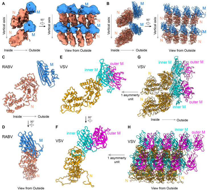
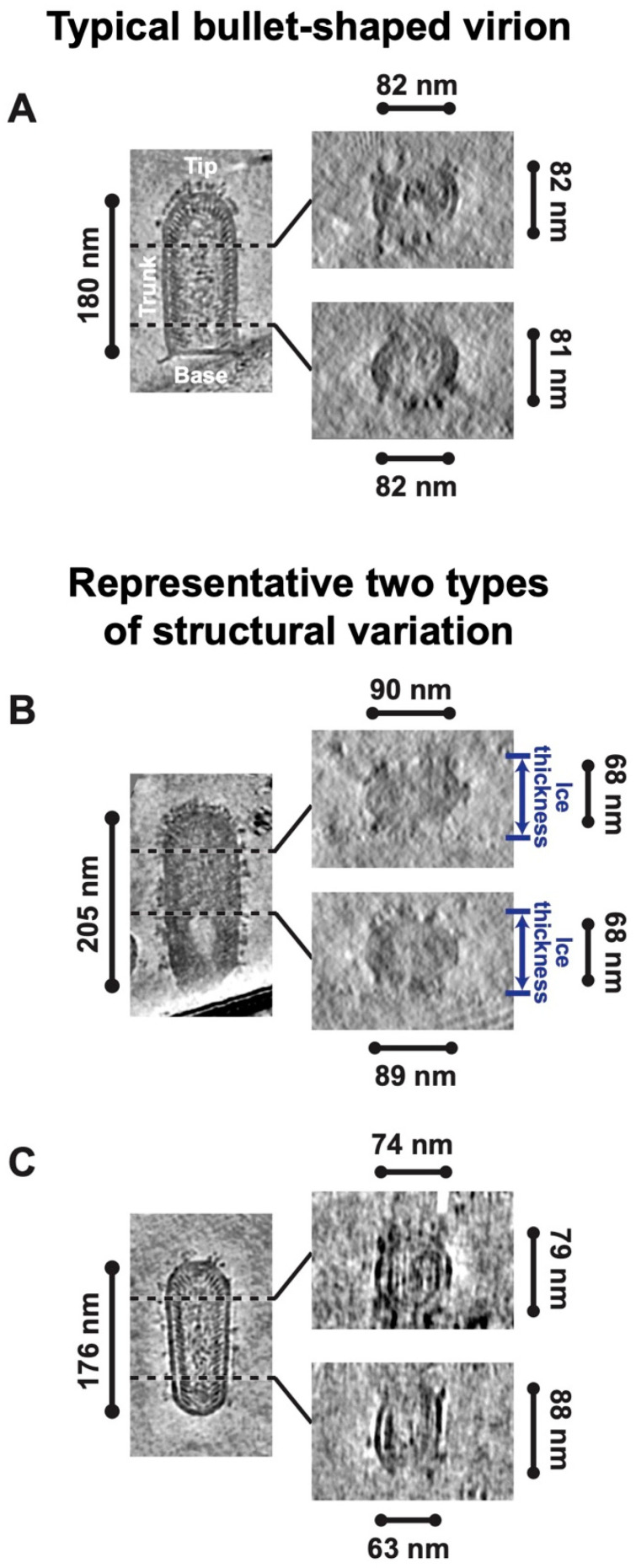
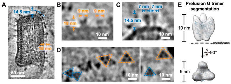
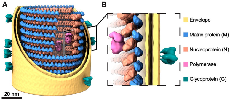
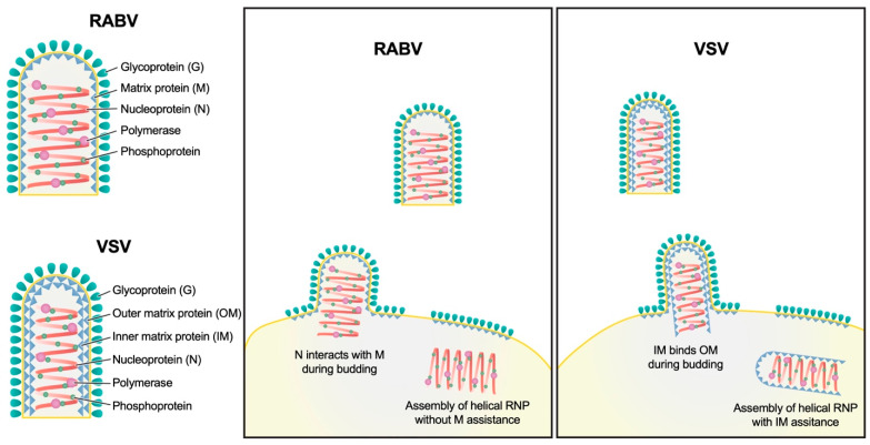
Rabies virus (RABV) is among the first recognized viruses of public health concern and has historically contributed to the development of viral vaccines. Despite these significances, the three-dimensional structure of the RABV virion remains unknown due to the challenges in isolating structurally homogenous virion samples in sufficient quantities needed for structural investigation. Here, by combining the capabilities of cryogenic electron tomography (cryoET) and microscopy (cryoEM), we determined the three-dimensional structure of the wild-type RABV virion. Tomograms of RABV virions reveal a high level of structural heterogeneity among the bullet-shaped virion particles encompassing the glycoprotein (G) trimer-decorated envelope and the nucleocapsid composed of RNA, nucleoprotein (N), and matrix protein (M). The structure of the trunk region of the virion was determined by cryoEM helical reconstruction, revealing a one-start N-RNA helix bound by a single layer of M proteins at an N:M ratio of 1. The N-M interaction differs from that in fellow rhabdovirus vesicular stomatitis virus (VSV), which features two layers of M stabilizing the N-RNA helix at an M:N ratio of 2. These differences in both M-N stoichiometry and binding allow RABV to flex its N-RNA helix more freely and point to different mechanisms of viral assembly between these two bullet-shaped rhabdoviruses.

Keywords: cryogenic electron microscopy; cryogenic electron tomography; dynamics; flexibility; rabies virus; rhabdoviruses; wild type.


Conflict of interest statement

The authors declare no conflicts of interest.

Figures

Figure 3
Figure 3
Sub-particle reconstruction of RABV trunk by cryoEM. (A) CryoEM density maps of partial RABV nucleocapsid shown in two orthogonal views. An 8 Å low-pass filter was applied to the cryoEM map for better visualization. Only intact N and intact M are colored for clarity, other densities are hidden. N subunits are colored in orange, and M are colored in blue. (B) Two orthogonal views of the fitted N protein models (PDB: 2GTT) [11] and AlphaFold2-predicted M protein models [36] corresponding to the cryoEM density maps in (A). (C,D) Two orthogonal views of the one asymmetry unit from RABV. RNA was hidden for clarity. (E,F) Two orthogonal views of the one asymmetry unit from VSV. N subunits are colored in yellow, inner M in cyan, and outer M in magenta. (G,H) Two orthogonal views of 8-asymmetry unit from VSV. A cross-sectional view is provided in (G) to more clearly display the models’ details.
Figure 1
Figure 1
Typical bullet-shaped virion and its structural variations of rabies virus (RABV). (A) Depiction of 7 Å thick density slices from a reconstructed tomogram displaying a typical bullet-shaped RABV virion. The longitudinal central section of the virion is shown on the left, and the perpendicular sections at different heights along the virion trunk are presented on the right. (B,C) Depiction of 7 Å thick density slices from reconstructed tomograms displaying two types of structural variations of RABV virions: an overall flatten virion (B) and a virion with gradual alterations in trunk morphology (C). The longitudinal central sections of the virions are shown on the left, and the perpendicular sections at different heights along the virion trunk are presented on the right.
Figure 2
Figure 2
Architecture analysis of the helical RABV nucleocapsid by cryoET. (A) A slice through a reconstructed tomogram showing a section of a representative RABV virion. The scale bar corresponds to 50 nm. (B,C) The horizontal and vertical histograms show the distribution of measured length (B) and diameter (C), respectively, from thirty-five RABV virions. The solid black lines are normal distribution fits to the histograms with means and standard deviations of 178 ± 15 nm for the length and 85 ± 4 nm for the diameter. (D,E) RNP helix model of RABV and vesicular stomatitis virus (VSV). One turn of RNP helix is colored in yellow. (F) The box plots of the pitches measured from 35 reconstructed tomograms (left) and from placed-back M-N subtomograms in 10 reconstructed tomograms (right). Boxes and whiskers show interquartile range and maximum and minimum values of data; center lines in boxes represent the median. The means and standard deviations of the pitches measured from the reconstructed tomograms and the placed-back M-N subtomograms are both 6.3 ± 0.4 nm. (G) Three types of RABV tips are shown in slices from reconstructed tomograms in the order of conical, dome-like, and flat-topped tips. The turn layers of the helical RNP in the tips are annotated. The scale bar corresponds to 30 nm. (H) Side (left) and top views (right) of the subtomogram-averaged structure of RABV M-N. The dashed box in (A) shows a sample subtomogram extraction. The arrow on the left indicates the directionality of the virion. The pitch of RNP helix and the distance between subsequent units in one turn are indicated. (I) Placed-back M-N subtomograms in tomogram of (A). The place-back is guided by the coordinates and orientations of the aligned subtomogram in Figure S3B. The scale bar corresponds to 50 nm. (J) The histogram displays the distribution of pitch measurements obtained from the placed-back M-N subtomograms within (I).
Figure 4
Figure 4
G trimers in both prefusion and postfusion conformations on RABV virion envelope. (A) A 7 Å thick density slice from a reconstructed tomogram showing a RABV virion with both prefusion and postfusion G trimers on viral envelope. The average height of 10 nm for glycoproteins indicates the prefusion conformation, whereas the average height of 14.5 nm corresponds to the postfusion conformation. The scale bar corresponds to 50 nm. (B,C) The side views of representative G trimers in prefusion (B) and postfusion conformations (C). Length and width of the G trimers are annotated. (D) The top views of G trimers. Trimers in prefusion conformation are framed by yellow dashed lines, while those in postfusion conformation are framed by blue dashed lines. The scale bar corresponds to 10 nm. (E) Prefusion G trimer segmented from a reconstructed tomogram. The atomic model of G trimer (PDB: 7U9G, ribbon diagrams colored in light blue) was docked into the segmented density map.
Figure 5
Figure 5
A model of the trunk of an RABV virion. (A) The trunk model of an RABV virion, with partial removal of the M and N proteins and the envelope membrane bilayer, enhances visual clarity and shows the interior structure. Two nested left-handed helices, consisting of an outer layer M (colored in blue) and an inner layer N (colored in orange), along with the envelope membrane bilayer (colored in yellow), were modeled based on the cryoEM density map of the trunk. The published cryoEM reconstructions of the RABV polymerase (EMD-20753, colored in magenta) and glycoprotein (EMD-26397, colored in dark cyan) were low-pass filtered to 20 Å and integrated into the model according to their positions within the virion density segmented from a reconstructed tomogram. The scale bar corresponds to 20 nm. (B) A zoomed-in view of a section of the RABV structural model.
Figure 6
Figure 6
Schematic model illustrating the process of RABV assembly and budding and the differences with VSV. N of the helical RNP of VSV interact with the inner matrix protein (IM). Because the IMs in neighboring turns of the helix interact with each other, the pitch is fixed during the assembly of the VSV RNP helix. Subsequently, the outer matrix protein (OM), distributed on the cytoplasmic side of the envelope membrane lipid, binds to IM-RNP helix during virion budding. The helical RNP of RABV, on the contrary, assembles without assistance of M; thus, the pitch of the RNP helix is variable. M distributed on the cytoplasmic side of the envelope interacts with the RNP helix during the budding without securing neighboring turns, resulting in a variable pitch in the RNP helix.

Similar articles

References

    1. Dubos R.J. Louis Pasteur: Freelance of Science. Little, Brown and Company; Boston, MA, USA: 1950. pp. 335–336.
    1. Matsumoto S. Electron microscopy of nerve cells infected with street rabies virus. Virology. 1962;17:198–202. doi: 10.1016/0042-6822(62)90099-5. - DOI - PubMed
    1. Hummeler K., Koprowski H., Wiktor T.J. Structure and development of rabies virus in tissue culture. J. Virol. 1967;1:152–170. doi: 10.1128/jvi.1.1.152-170.1967. - DOI - PMC - PubMed
    1. Guichard P., Krell T., Chevalier M., Vaysse C., Adam O., Ronzon F., Marco S. Three dimensional morphology of rabies virus studied by cryo-electron tomography. J. Struct. Biol. 2011;176:32–40. doi: 10.1016/j.jsb.2011.07.003. - DOI - PubMed
    1. Ng A.K., Zhang H., Tan K., Li Z., Liu J.H., Chan P.K., Li S.M., Chan W.Y., Au S.W., Joachimiak A., et al. Structure of the influenza virus A H5N1 nucleoprotein: Implications for RNA binding, oligomerization, and vaccine design. FASEB J. 2008;22:3638–3647. doi: 10.1096/fj.08-112110. - DOI - PMC - PubMed

Publication types