. 2024 Sep 13;16(9):1456.
doi: 10.3390/v16091456.

Coxsackievirus B3 Activates Macrophages Independently of CAR-Mediated Viral Entry

Affiliations

Coxsackievirus B3 Activates Macrophages Independently of CAR-Mediated Viral Entry

Yasir Mohamud et al. Viruses. .

Abstract

Enteroviruses are a genus of small RNA viruses that are responsible for approximately one billion global infections annually. These infections range in severity from the common cold and flu-like symptoms to more severe diseases, such as viral myocarditis, pancreatitis, and neurological disorders, that continue to pose a global health challenge with limited therapeutic strategies currently available. In the current study, we sought to understand the interaction between coxsackievirus B3 (CVB3), which is a model enterovirus, and macrophage cells, as there is limited understanding of how this virus interacts with macrophage innate immune cells. Our study demonstrated that CVB3 can robustly activate macrophages without apparent viral replication in these cells. We also showed that myeloid cells lacked the viral entry receptor coxsackievirus and adenovirus receptor (CAR). However, the expression of exogenous CAR in RAW264.7 macrophages was unable to overcome the viral replication deficit. Interestingly, the CAR expression was associated with altered inflammatory responses during prolonged infection. Additionally, we identified the autophagy protein LC3 as a novel stimulus for macrophage activation. These findings provide new insights into the mechanisms of CVB3-induced macrophage activation and its implications for viral pathogenesis.

Keywords: LC3; coxsackievirus B3 (CVB3); coxsackievirus and adenovirus receptor (CAR); innate immunity; macrophages.


Conflict of interest statement

The authors declare no conflicts of interest.

Figures

Figure 1
Figure 1
CVB3 induced pro-inflammatory gene expression in macrophages independent of the viral replication. (A) HL1 cardiomyocytes or RAW264.7 macrophage cells were subjected to CVB3 infection (MOI = 100) for the indicated timepoints. Lysates were subjected to Western analysis for the viral capsid protein (VP1) and normalized to the loading control ACTB. Densitometric analysis results are shown in the adjacent bar plot. (B) RAW264.7 murine macrophages and (C) THP1-derived human macrophages were infected with CVB3 (MOI = 10; 4 h). RNA was harvested and subjected to qPCR analysis for inflammatory gene markers IFNB, TNFA, IL-1B, and IL-6. Viral gene marker 2A was utilized as a control to assess the viral treatment. The results are presented as the relative gene fold change expression between sham- and CVB3-infected groups (mean ± S.D., n = 3), where they were statistically evaluated via an unpaired Student’s t-test.
Figure 2
Figure 2
CVB3-induced macrophage activation was independent of the cGAS-STING pathway. (A) RAW264.7 murine macrophages were subjected to a dose ramp of the STING agonist 2′3′-cGAMP (10 and 20 μg/mL, 4 h) and subjected to Western analysis for STING and p-TBK1 protein expression; ACTB was used as the loading control. (B) RAW264.7 macrophages were treated with STING agonists 2′3′-cGAMP (20 ug/mL) and diABZI (10 μM) or cGAS agonists herring testis (HT)-DNA (3 μg/mL) or poly-dAdT (3 μg/mL). cGAS-STING pathway activation was assessed via Western analysis of the STING and p-TBK1 protein expressions. (C) STING was not activated following CVB3 infection. RAW264.7 cells were infected with CVB3 (MOI = 10; 4 h) as above, and lysates were harvested for Western analysis of the STING activation, p-TBK1, and ACTB loading control. The RAW264.7 cells were subjected to a timecourse STING agonist treatment with diABZI (10 μM), and cGAMP (32 μM) was the positive control. (D) RAW264.7 macrophages were subjected to a time-course infection with CVB3 (MOI = 10), and cell lysates were analyzed by Western blot for macrophage activation marker COX2 and cGAS-STING pathway activation (anti-STING, anti-p-STING). ACTB was used as the loading control. (E) RAW264.7 macrophages were infected with CVB3 (MOI = 10; 5 h) and the cells were fixed for confocal microscopy imaging of the dsDNA/mtDNA release. Scale bars: 20 µm. (F) RAW264.7 macrophages were sham or CVB3 infected (MOI = 10; 8 h) in the presence of cGAS and STING inhibitors RU.521 (10 μM) and H151 (10 μM), respectively. Cell lysates were subjected to Western analysis for macrophage activation (COX2), STING activation (anti-STING, anti-p-STING, anti p-TBK1), and viral replication (VP1). (G) RAW264.7 macrophages were treated with cGAS and STING inhibitors RU.521 (10 μM) and H-151 (10 μM) as above, and the drug efficacy was evaluated with a qPCR assessment of the relative IFNB gene expression (mean ± S.D., n = 3).
Figure 3
Figure 3
CVB3-induced macrophage activation was dependent on NF-κB (A) RAW 264.7 macrophages were infected with CVB3 (MOI = 10; 5 h), and the cells were fixed for confocal microscopy imaging of NFkB activation and COX2 expression. Scale bars: 20 µm. White arrows denote the nuclear-localized p65 present in CVB3- but not sham-infected cells. (B) RAW264.7 macrophages were infected with CVB3 (MOI = 10; 3 h) and subjected to specific inhibitors of TLR1/2, CU-CPT22 (500 nM), TLR3-inhibitor, CU-CPT4A (27 μM), TLR4-inhibitor C34 (10 μM), or NFkB inhibitor peptide (50 ug/mL). Cell lysates were subjected to Western analysis for the macrophage activation marker (COX2). (C) RAW264.7 macrophages were infected as in (A) and subjected to a dose ramp of the NFkB peptide inhibitor. Lysates were subjected to Western analysis for the macrophage activation marker COX2.
Figure 4
Figure 4
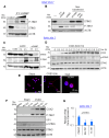
CVB3-induced macrophage activation was independent of the viral replication, CAR expression, and phagocytosis. (A) RAW264.7 macrophages were sham or CVB3 infected (MOI = 10; 8 h) in the presence of the phagocytosis inhibitor cytochalasin D (1 μM) or a DMSO control. Cell lysates were assessed by Western blotting for the macrophage activation marker COX2. Densitometry results of the COX2 activation are presented in the right-hand panel (mean ± S.D., n = 3). (B) The SIM-A9 murine microglia cell line was assessed for myeloid activation through Western analysis with a COX2 marker following sham or CVB3 infection as above. (C) CVB3 replication and virus-induced autophagy was assessed in the NSC-34 murine motor neuron cells and SIM-A9 macrophages (MOI = 10; 8 h). Lysates were subjected to Western analysis for the autophagy marker protein LC3 and viral replication marker VP1. (D) The specificity of the COX2 myeloid expression marker was assessed in the NSC-34 motor neuron cells and SIM-A9 microglia cell line through Western analysis in the presence or absence of CVB3 infection (MOI = 10; 8 h). ACTB served as the loading control. (E) Virus-induced COX2 expression in SIM-A9 microglia was assessed with a control CVB3 virus and UV-inactivated CVB3 (MOI = 10; 8 h). UV inactivation of CVB3 was verified in the CVB3-susceptible cell line HEK293 through Western analysis for the viral replication marker VP1. (F) Expression of the coxsackievirus entry receptor CAR was assessed in HL-1, NSC-34, SIM-A9, HeLa, and RAW264.7 cells through Western analysis with the anti-CAR antibody. ACTB served as a protein loading control. (G) Schematic illustration of CVB3-susceptible cells and CVB3-resistant cells. * Denotes NSC-34 as the only CVB3-susceptible cell line that did not have detectable CAR expression.
Figure 5
Figure 5
CAR expression regulated the late, but not early, response to the CVB3 infection. (A) RAW264.7 cells were transduced with either a control GFP- or hCAR-expressing lentivirus for 72 h, followed by CVB3 infection (MOI = 10) for 24 h. Cell lysates were harvested and subjected to Western analysis for anti-VP1 and anti-CAR antibody. Ponceau Red was used as a total protein loading stain. (B) Densitometry of VP1 expression from (A) was quantified and normalized to the total protein and presented in the bar plot (mean ± S.D., n = 3). (C) RAW264.7 cells were transduced as above and subjected to confocal analysis for GFP (green) and CAR (red) expression. Nuclei were stained with DAPI (blue). Scale bar: 20 µm. (D,E) RAW264.7 cells transduced with a GFP or hCAR lentivirus as above were subsequently infected with CVB3 (MOI = 10) for 2 h (D) or 24 h (E). RNA was harvested and subjected to qPCR analysis for viral RNA (2A) replication or inflammatory markers IL-1β, IL-6, or TNFA and presented as a relative quantitation where the first group was set to 1 (mean ± S.D., n = 3).
Figure 6
Figure 6
Viral-induced LC3 stimulated macrophage activation. (A) Recombinant LC3 (rLC3) activated the SIMA-A9 myeloid cells. SIM-A9 were incubated with increasing doses of rLC3 for 4 h. Lysates were harvested and subjected to Western analysis for COX2 expression. ACTB was used as a loading control. (B) rLC3 potentiated CVB3-induced activation of the SIM-A9. SIM-A9 cells were co-incubated with CVB3 (MOI = 10) and increasing doses of rLC3 for 4 h. Lysates were harvested as above and subjected to Western analysis (C) SIM-A9 cells were infected with CVB3 (MOI = 10; 24 h). Lysates were harvested and subjected to Western analysis for autophagy marker LC3 and normalized to ACTB. (D) Densitometry of panel (C) where LC3-I and LC3-II were quantified and normalized to ACTB and presented as the mean ± S.D., n = 3.

Similar articles

Cited by

References

    1. Newland J.G., Romero J.R. Enteroviruses. In: Heggenhougen H.K., editor. International Encyclopedia of Public Health. Academic Press; Oxford, UK: 2008. pp. 347–352.
    1. Baggen J., Thibaut H.J., Strating J.R.P.M., van Kuppeveld F.J.M. The life cycle of non-polio enteroviruses and how to target it. Nat. Rev. Microbiol. 2018;16:368–381. doi: 10.1038/s41579-018-0005-4. - DOI - PubMed
    1. Mohamud Y., Luo H.L. The Intertwined Life Cycles of Enterovirus and Autophagy. Virulence. 2019;10:470–480. doi: 10.1080/21505594.2018.1551010. - DOI - PMC - PubMed
    1. Garmaroudi F.S., Marchant D., Hendry R., Luo H., Yang D., Ye X., Shi J., McManus B.M. Coxsackievirus B3 replication and pathogenesis. Future Microbiol. 2015;10:629–653. doi: 10.2217/fmb.15.5. - DOI - PubMed
    1. Jagdeo J.M., Dufour A., Klein T., Solis N., Kleifeld O., Kizhakkedathu J., Luo H., Overall C.M., Jan E. N-Terminomics TAILS Identifies Host Cell Substrates of Poliovirus and Coxsackievirus B3 3C Proteinases That Modulate Virus Infection. J. Virol. 2018;92:10-1128. doi: 10.1128/JVI.02211-17. - DOI - PMC - PubMed

Publication types

MeSH terms

Substances

LinkOut - more resources