. 2024 Sep 17;16(9):1475.
doi: 10.3390/v16091475.

Tuning VSV-G Expression Improves Baculovirus Integrity, Stability and Mammalian Cell Transduction Efficiency

Affiliations

Tuning VSV-G Expression Improves Baculovirus Integrity, Stability and Mammalian Cell Transduction Efficiency

Martina Mattioli et al. Viruses. .

Abstract

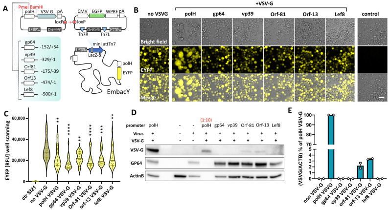
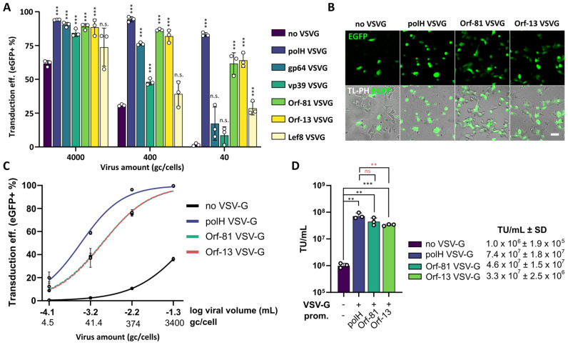
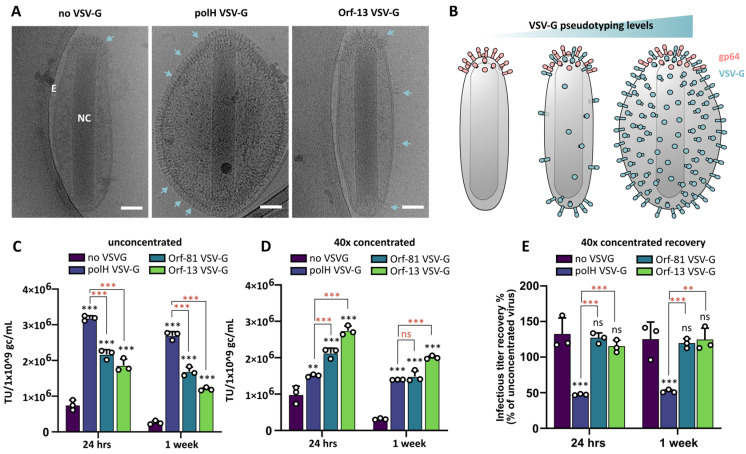
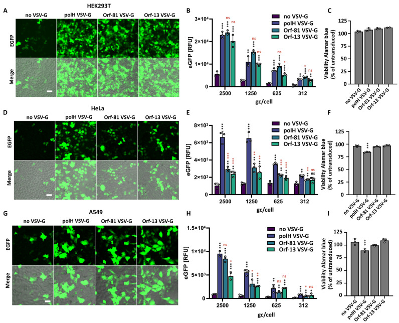
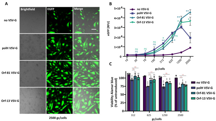
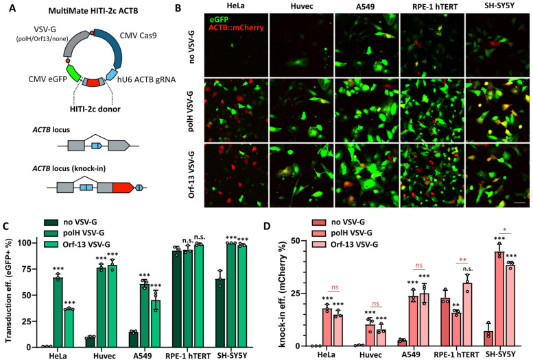
Baculoviral vectors (BVs) derived from Autographa californica multiple nucleopolyhedrovirus (AcMNPV) are an attractive tool for multigene delivery in mammalian cells, which is particularly relevant for CRISPR technologies. Most applications in mammalian cells rely on BVs that are pseudotyped with vesicular stomatitis virus G-protein (VSV-G) to promote efficient endosomal release. VSV-G expression typically occurs under the control of the hyperactive polH promoter. In this study, we demonstrate that polH-driven VSV-G expression results in BVs characterised by reduced stability, impaired morphology, and VSV-G induced toxicity at high multiplicities of transduction (MOTs) in target mammalian cells. To overcome these drawbacks, we explored five alternative viral promoters with the aim of optimising VSV-G levels displayed on the pseudotyped BVs. We report that Orf-13 and Orf-81 promoters reduce VSV-G expression to less than 5% of polH, rescuing BV morphology and stability. In a panel of human cell lines, we elucidate that BVs with reduced VSV-G support efficient gene delivery and CRISPR-mediated gene editing, at levels comparable to those obtained previously with polH VSV-G-pseudotyped BVs (polH VSV-G BV). These results demonstrate that VSV-G hyperexpression is not required for efficient transduction of mammalian cells. By contrast, reduced VSV-G expression confers similar transduction dynamics while substantially improving BV integrity, structure, and stability.

Keywords: AcMNPV; VSV-G; baculovirus; gene delivery; pseudotyping; viral vector.


Conflict of interest statement

The authors declare conflicts of interest. I.B. is shareholder in Geneva Biotech SARL, related to this correspondence. Geneva Biotech owns patents and trademarks in the field of baculovirus vector technology. All the other authors declare no conflicts of interest.

Figures

Figure 1
Figure 1
Baculovirus VSV-G pseudotyping using alternative viral promoters. (A) Schematic representation of the cloning strategy implemented to generate BVs encoding CMV EGFP (mammalian cells transduction marker), polH EYFP (viral amplification and protein expression marker in insect cells), and VSV-G expression under alternative viral promoters (pseudotyping module). (BE) Characterisation of Sf21 insect cells at 72 h post inoculation with titre-normalised viral supernatants. (B) Live-cell imaging, scalebar = 50 µm. (C) polH EYFP expression readout; violin plot of plate reading measurement (plate scanning, 25 readings). Adj. p-value (** ≤ 0.01, *** ≤ 0.001, **** ≤ 0.0001); all samples were compared against non-VSV-G BVs; ANOVA test, n = 25 measurements. (D) Representative western blot of VSV-G and GP64 in insect cells. Actin B was used as loading control. VSV-G levels, protein extracts of cells inoculated with polH VSV-G BVs (lane 1) were additionally loaded 1:10 (lane 4). (E) Quantification of VSV-G expression levels, relative to (D). Averages + S.D. of 2 independent experiments.
Figure 2
Figure 2
VSV-G hyperexpression is not required for efficient transduction of mammalian cells. (A) HEK293T transduction rates 24 h after transduction with control BVs (non-VSV-G) or BVs pseudotyped with different levels of VSV-G using alternative viral promoters. CMV EGFP expression was used as transduction marker. Mean + S.D. of 3 independent replicates. p-value(*** ≤ 0.001); Student’s t-test, all samples compared to non-VSV-G virus. (B) HEK293T transduced with the indicated BVs at 4000 gc/cell, scalebar = 50 µm. (C,D) Fluorescent titration assay in HEK293T at 24 h post-transduction with the indicated BVs. (C) Flow cytometry data of n = 3 independent replicates and transducing units per mL (D). p-value (** ≤ 0.01, *** ≤ 0.001), Student’s t-test, all samples compared to non-VSV-G virus (black symbols), Orf-13, and Orf-81 compared to polH VSV-G virus (red symbols).
Figure 3
Figure 3
VSV-G hyperexpression affects BV morphology and stability. (A) Nanostructure of control baculoviruses (non-VSV-G) and VSV-G-pseudotyped BVs with standard (polH) or reduced (Orf-13) expression levels. Cyro-EM imaging, scalebar = 40 nm. E = envelope; NC = nucleocapsid. Arrows indicate position of glycoproteins (GP64 or VSV-G) embedded in the lipid bilayer envelope. (B) Model of BVs nanostructures with increasing VSV-G incorporation levels. (CE) Transducing viral titres upon concentration and resuspension at 1 (C) or 1:40 (D) of the original volume assessed on HEK293T via flow cytometry; viral stocks were tested after 24 h or 1 week storage at 4 °C. (D) Transducing titres recovery of 40× concentrated viral stocks compared to their unconcentrated controls. Mean + S.D. of 3 independent replicates (CE). p-value (** ≤ 0.01, *** ≤ 0.001), Student’s t-test, all samples compared to non-VSV-G virus (black symbols), Orf-81 and Orf-13 compared to polH VSV-G virus (red symbols).
Figure 4
Figure 4
Analysis of transgene expression and viability in immortalised human cell lines. HEK293T (AC), HeLa (DF), and A549 (GI). (A,D,G) Live brightfield imaging of cells transduced with the indicated BVs at 2500 gc/cell. Scalebar = 50 μm. (B,E,H) Transgene (EGFP) expression levels in cells transduced with serial dilutions of polH, Orf-81, and Orf-13 VSV-G BVs or with a non-pseudotyped control virus (non-VSV-G). Plate reader, EGFP relative fluorescence units (RFUs), mean ± S.D. n = 3 independent replicates, p-value (* ≤ 0.05, ** ≤ 0.01, *** ≤ 0.001), Student’s t-test, all samples compared to non-VSV-G virus (black symbols), Orf-81 and Orf-13 compared to polH VSV-G virus (red symbols). (C,F,I) Viability of transduced cells measured via alamarBlue staining at 48 h post-transduction (2500 gc/cell). alamarBlue RFUs are presented as % of the untransduced control. Mean + S.D of n = 3 independent replicates. p-value (*** ≤ 0.001), Student’s t-test, all samples compared to non-VSV-G virus.
Figure 5
Figure 5
Analysis of transgene expression and viability in primary HUVEC. (A) Live brightfield imaging of HUVEC transduced with the indicated BVs. Scalebar = 50 μm. (B) Transgene (EGFP) expression dynamics in HUVEC transduced with serial dilutions of polH, Orf-81, and Orf-13 VSV-G BVs or with a non-pseudotyped control virus (no VSV). Plate reader, EGFP relative fluorescence units (RFUs), mean ± S.D. n = 3 independent replicates. p-value (* ≤ 0.05, ** ≤ 0.01), Student’s t-test, Orf-13 (light green symbols) and Orf-81 (dark green symbols) samples compared to polH VSV-G virus. (C) Viability of transduced cells measured via alamarBlue staining. alamarBlue RFUs are presented as % of the untransduced control. Mean + S.D of n = 3 independent replicates. p-value (* ≤ 0.05, ** ≤ 0.01, *** ≤ 0.001), Student’s t-test, all samples compared to non-VSV-G virus (black symbols), Orf-81 and Orf-13 compared to polH VSV-G virus (red symbols).
Figure 6
Figure 6
Assessment of the impact of reduced VSV-G pseudotyping levels on gene editing efficiencies of all-in-one CRISPR BVs. (A) Schematic representation of MultiMate HITI-2c ACTB all-in-one vector encoding Cas9, gRNA, and HITI-2c donor. To generate VSV-G-pseudotyped BVs, pIDC vectors were incorporated into MultiMate HITI-2c ACTB via in vitro CRE-mediated recombination. Upon successful knock-in, endogenous ACTB is fused to a C-terminal mCherry fluorescence marker. (BD) Analysis of EGFP (transduction efficiency) and mCherry (knock-in) efficiency in the indicated cell lines at 48 h post-transduction with control (non-VSV-G), polH VSV-G, and Orf-13 VSV-G BVs encoding the MultiMate HITI-2c ACTB construct depicted in (A). (B) Confocal live-cell imaging, scalebar = 100 μm; (C) transduction and (D) knock-in efficiencies at 48 h post-transduction. Histogram of flow cytometry data, mean + S.D of n = 3 independent replicates. p-value (* ≤ 0.05, ** ≤ 0.01, *** ≤ 0.001), Student’s t-test, all samples compared to non-VSV-G virus (black symbols), Orf-13 compared to polH VSV-G virus (red symbols).

Similar articles

Cited by

References

    1. Condreay J.P., Witherspoon S.M., Clay W.C., Kost T.A. Transient and stable gene expression in mammalian cells transduced with a recombinant baculovirus vector. Proc. Natl. Acad. Sci. USA. 1999;96:127–132. doi: 10.1073/pnas.96.1.127. - DOI - PMC - PubMed
    1. Sung L.-Y., Chen C.-L., Lin S.-Y., Li K.-C., Yeh C.-L., Chen G.-Y., Lin C.-Y., Hu Y.-C. Efficient gene delivery into cell lines and stem cells using baculovirus. Nat. Protoc. 2014;9:1882–1899. doi: 10.1038/nprot.2014.130. - DOI - PubMed
    1. Mansouri M., Bellon-Echeverria I., Rizk A., Ehsaei Z., Cianciolo Cosentino C., Silva C.S., Xie Y., Boyce F.M., Davis M.W., Neuhauss S.C.F., et al. Highly efficient baculovirus-mediated multigene delivery in primary cells. Nat. Commun. 2016;7:11529. doi: 10.1038/ncomms11529. - DOI - PMC - PubMed
    1. Maghodia A.B., Jarvis D.L., Geisler C. Complete Genome Sequence of the Autographa californica Multiple Nucleopolyhedrovirus Strain E2. Genome Announc. 2014;2 doi: 10.1128/genomeA.01202-14. - DOI - PMC - PubMed
    1. Rohrmann G.F. The AcMNPV Genome: Gene Content, Conservation, and Function. National Center for Biotechnology Information (US); 2019. [(accessed on 27 August 2024)]. Available online: https://www.ncbi.nlm.nih.gov/books/NBK543457.

Publication types

Substances

Supplementary concepts

LinkOut - more resources