Review
. 2024 Sep 20;16(9):1491.
doi: 10.3390/v16091491.

Is Autophagy a Friend or Foe in SARS-CoV-2 Infection?

Affiliations
Review

Is Autophagy a Friend or Foe in SARS-CoV-2 Infection?

Asifa Khan et al. Viruses. .

Abstract

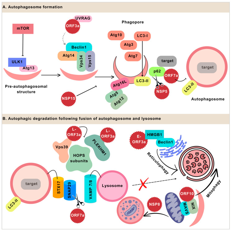
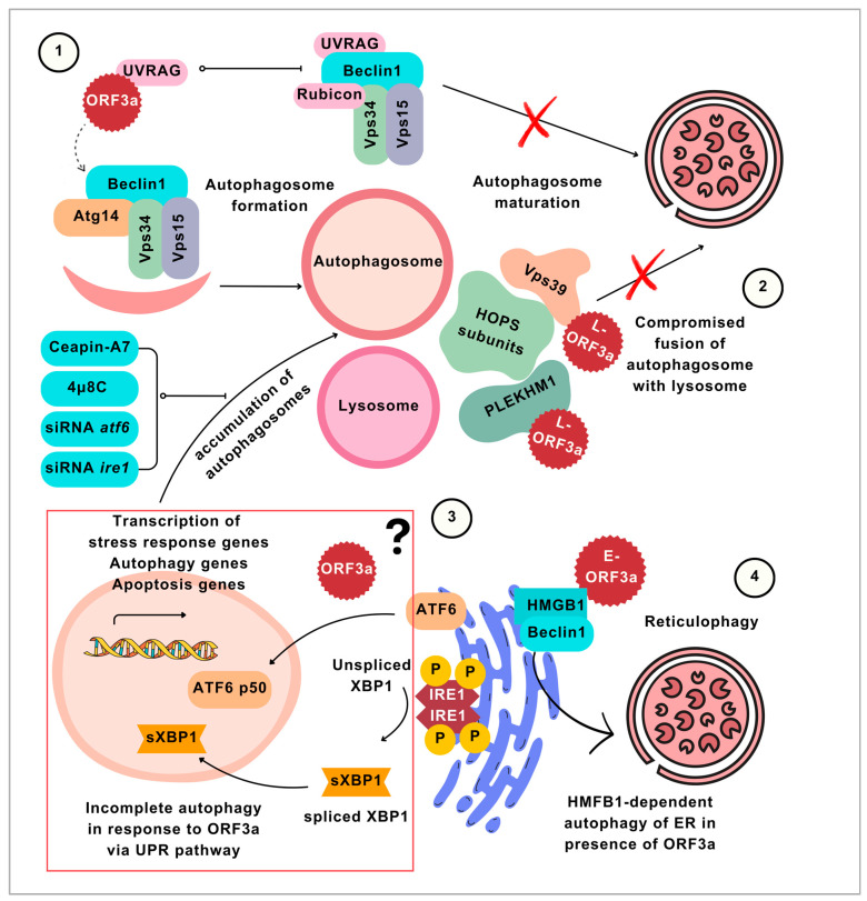
As obligate parasites, viruses need to hijack resources from infected cells to complete their lifecycle. The interaction between the virus and host determines the viral infection process, including viral propagation and the disease's outcome. Understanding the interaction between the virus and host factors is a basis for unraveling the intricate biological processes in the infected cells and thereby developing more efficient and targeted antivirals. Among the various fundamental virus-host interactions, autophagy plays vital and also complicated roles by directly engaging in the viral lifecycle and functioning as an anti- and/or pro-viral factor. Autophagy thus becomes a promising target against virus infection. Since the COVID-19 pandemic, there has been an accumulation of studies aiming to investigate the roles of autophagy in SARS-CoV-2 infection by using different models and from distinct angles, providing valuable information for systematically and comprehensively dissecting the interplay between autophagy and SARS-CoV-2. In this review, we summarize the advancements in the studies of the interaction between SARS-CoV-2 and autophagy, as well as detailed molecular mechanisms. We also update the current knowledge on the pharmacological strategies used to suppress SARS-CoV-2 replication through remodeling autophagy. These extensive studies on SARS-CoV-2 and autophagy can advance our understanding of virus-autophagy interaction and provide insights into developing efficient antiviral therapeutics by regulating autophagy.

Keywords: SARS-CoV-2; antivirals; autophagy; autophagy modulator; incomplete autophagy; virophagy.


Conflict of interest statement

The authors declare no conflicts of interest.

Figures

Figure 1
Figure 1
SARS-CoV-2 proteins hijacking autophagic machinery to affect the formation and maturation of autophagosomes. (A). Autophagy vesicle formation initiates ULK1 and Atg13 autophagy-related proteins. mTOR is an inhibitor of the initiation step. The vesicle nucleation involves the recruitment of other proteins including Beclin1, which forms a complex with Atg14, Vps34, and Vps15. The interaction of the SARS-CoV-2 protein ORF3a with UVRAG may drive the process towards autophagosome formation and prevent the autophagosome’s maturation by hindering the interaction of UVRAG with Beclin1. Other autophagy-related proteins such as Atg10, Atg7, and Atg3 catalyze the two conjugation reactions to form the Atg5–Atg12–Atg16L complex and LC3-II, which leads to the enclosure of autophagosome vesicles. The SARS-CoV-2 protein NSP15 inhibits autophagosome formation; the mechanism of this process and the phase of vesicles at which it is involved are not yet determined. The adaptor protein, p62, facilitates the interaction of the target molecules with LC3-II, which brings the target closer to the autophagosome for engulfment. SARS-CoV-2 NSP5 cleaves p62, inhibiting autophagic degradation. ORF7a is also known to interact with p62; this interaction of ORF7a prevents the fusion of the autophagosome with the lysosome. (B). ORF7a prevents the fusion by degrading SNAP29 through the caspase pathway. The degradation of SNAP29 compromises the interaction with the receptor on the lysosome, vesicle-associated membrane protein 7/8 (VAMP7/8). The ORF3a localized to the lysosome (L-ORF3) binds to Vps39, a subunit of the HOPS complex. The interaction with ORF3a prevents the HOPS formation which is required for mediating the tethering of the autophagosome on the lysosome. ORF3a also disrupts HOPS formation by binding to another HOPS subunit, PLEKHM1. While autophagic flux is reduced due to these viral proteins, the selective degradation of the host ER and mitochondrial proteins is facilitated by some SARS-CoV-2 proteins. The ER-localized ORF3a (E-ORF3a) promotes reticulophagy. ORF3a interacts with high mobility group box 1 protein (HMGB1), which is a damage-associated molecular pattern. By increasing the interaction of HMGB1 with Beclin1, ORF3a promotes reticulophagy of the ER. ORF10 of SARS-CoV-2 targets mitochondrial antiviral signaling (MAVS) for autophagy through autophagy receptor NIX. The NSP8 protein of SARS-CoV-2 also targets mitochondria, but the autophagy is incomplete, which leads to the accumulation of autophagosomes.
Figure 2
Figure 2
Three mechanisms (1–3) of incomplete autophagy stimulated by ORF3a and ORF3a-mediated reticulophagy (4). (1) ORF3a binds to UVRAG, which shifts the balance between two mutually exclusive Beclin1 complexes towards Beclin1-Atg14-Vps34-Vps16 (Beclin1–Atg14 complex) formation. The Beclin1–Atg14 complex stimulates the autophagosome formation. Meanwhile, the other complex of Beclin1 (Beclin1-UVRAG-Vps34-Vps15-Rubicon) is limited by the sequestering of UVRAG by ORF3a. The Beclin1–UVRAG complex is responsible for deriving the maturation of autophagosomes. Since the Beclin1–UVRAG complex is inhibited and more Beclin1 is available for forming the Beclin1–Atg14 complex, the process leads to more autophagosome formation and less autophagosome maturation; hence, incomplete autophagy is caused by ORF3a. (2) ORF3a inhibits HOPS complex formation by binding to HOPS complex subunits, thereby preventing the fusion of autophagosomes with lysosomes, leading to the accumulation of autophagosomes and incomplete autophagy. (3) ORF3a initiates incomplete autophagy through ATF6 and IRE1-mediated unfolded protein response (UPR). The crosstalk of autophagy and the UPR pathway has long been known; however, the mechanism by which SARS-CoV-2-ORF3a stimulates the pathway is not yet determined. The illustration of the ATF6 and IRE1 pathways and their relation to expressing autophagy genes is inspired by Estébanez et al. [62]. Downstream ATF6 and IRE1 pathways are ATF6 p50 and spliced XBP1, which translocate to the nucleus and may stimulate the autophagy genes in response to ER stress. To date, SARS-CoV-2 studies have not yet indicated which of the components of these pathways are directly influenced by ORF3a. However, it was determined that inhibiting the ATF6 and IRE1 through chemical inhibitors, Chapin-A7 and 4µ8C, and the gene knockdown of AFT6 or IRE1 reduce the autophagosome accumulation in ORF3a-transfected cells. (4) While the three mechanisms show the involvement of ORF3a in incomplete autophagy, ORF3a is also known to selectively stimulate the complete autophagy of the ER through HMGB1.

Similar articles

Cited by

References

    1. Carabelli A.M., Peacock T.P., Thorne L.G., Harvey W.T., Hughes J., de Silva T.I., Peacock S.J., Barclay W.S., de Silva T.I., Towers G.J., et al. SARS-CoV-2 Variant Biology: Immune Escape, Transmission and Fitness. Nat. Rev. Microbiol. 2023;21:162–177. doi: 10.1038/s41579-022-00841-7. - DOI - PMC - PubMed
    1. Coronavirus Disease (COVID-19) [(accessed on 24 February 2024)]. Available online: https://www.who.int/news-room/fact-sheets/detail/coronavirus-disease-(co...
    1. Wise J. COVID-19: WHO Declares End of Global Health Emergency. BMJ. 2023;381:p1041. doi: 10.1136/bmj.p1041. - DOI - PubMed
    1. Mizushima N., Klionsky D.J. Protein Turnover Via Autophagy: Implications for Metabolism. Annu. Rev. Nutr. 2007;27:19–40. doi: 10.1146/annurev.nutr.27.061406.093749. - DOI - PubMed
    1. Murrow L., Debnath J. Autophagy As A Stress Response And Quality Control Mechanism—Implications for Cell Injury and Human Disease. Annu. Rev. Pathol. 2013;8:105–137. doi: 10.1146/annurev-pathol-020712-163918. - DOI - PMC - PubMed

Publication types

Substances