. 2024 Sep 21;16(9):1497.
doi: 10.3390/v16091497.

A Quadruple Gene-Deleted Live BoHV-1 Subunit RVFV Vaccine Vector Reactivates from Latency and Replicates in the TG Neurons of Calves but Is Not Transported to and Shed from Nasal Mucosa

Affiliations

A Quadruple Gene-Deleted Live BoHV-1 Subunit RVFV Vaccine Vector Reactivates from Latency and Replicates in the TG Neurons of Calves but Is Not Transported to and Shed from Nasal Mucosa

Selvaraj Pavulraj et al. Viruses. .

Abstract

Bovine herpesvirus type 1 (BoHV-1) establishes lifelong latency in trigeminal ganglionic (TG) neurons following intranasal and ocular infection in cattle. Periodically, the latent virus reactivates in the TG due to stress and is transported anterogradely to nerve endings in the nasal epithelium, where the virus replicates and sheds. Consequently, BoHV-1 is transmitted to susceptible animals and maintained in the cattle population. Modified live BoHV-1 vaccine strains (BoHV-1 MLV) also have a similar latency reactivation. Therefore, they circulate and are maintained in cattle herds. Additionally, they can regain virulence and cause vaccine outbreaks because they mutate and recombine with other circulating field wild-type (wt) strains. Recently, we constructed a BoHV-1 quadruple mutant virus (BoHV-1qmv) that lacks immune evasive properties due to UL49.5 and glycoprotein G (gG) deletions. In addition, it also lacks the gE cytoplasmic tail (gE CT) and Us9 gene sequences designed to make it safe, increase its vaccine efficacy against BoHV-1, and restrict its anterograde neuronal transport noted above. Further, we engineered the BoHV-1qmv-vector to serve as a subunit vaccine against the Rift Valley fever virus (BoHV-1qmv Sub-RVFV) (doi: 10.3390/v15112183). In this study, we determined the latency reactivation and nasal virus shedding properties of BoHV-1qmv (vector) and BoHV-1qmv-vectored subunit RVFV (BoHV-1qmv sub-RVFV) vaccine virus in calves in comparison to the BoHV-1 wild-type (wt) following intranasal inoculation. The real-time PCR results showed that BoHV-1 wt- but not the BoHV-1qmv vector- and BoHV-1qmv Sub-RVFV-inoculated calves shed virus in the nose following dexamethasone-induced latency reactivation; however, like the BoHV-1 wt, both the BoHV-1qmv vector and BoHV-1qmv Sub-RVFV viruses established latency, were reactivated, and replicated in the TG neurons. These results are consistent with the anterograde neurotransport function of the gE CT and Us9 sequences, which are deleted in the BoHV-1qmv and BoHV-1qmv Sub-RVFV.

Keywords: BoHV-1; BoHV-1qmv vector; DIVA; bovine herpesvirus; cattle; glycoprotein E; latency reactivation; quadruple mutant virus; trigeminal ganglion; vectored vaccine.


Conflict of interest statement

The authors declare no conflicts of interest.

Figures

Figure 1
Figure 1
Infection, sample collection, dexamethasone-induced latency reaction, and euthanasia scheme for the animal experiment. PFUs—plaque-forming units; Subcut.—subcutaneous injection; Dex.—dexamethasone; Days post-Dex.—Days post-dexamethasone treatment.
Figure 2
Figure 2
The clinical score in calves following bovine herpesvirus type 1 (BoHV-1) infection and latency reactivation. Criteria: rectal temperatures were scored 0–4 (<39.0 °C, 39.5 °C, 40.0 °C, 40.5 °C, and >40.9 °C), nasal discharges were scored 0–4 (normal, serous, mild, and severe mucopurulent), and nasal lesions were scored 0–3 (normal, hyperemia, pustules and ulcers). There was a significantly higher clinical score in the BoHV-1 wild-type group compared to the BoHV-1qmv vector and BoHV-1qmv Sub-RVFV vaccine group on days 5 and 7 post infection. We used two-way ANOVA followed by Bonferroni post tests to compare replicate means by row; *** p < 0.001. dpi—days post infection; dp-Dex—days post dexamethasone injection.
Figure 3
Figure 3
Nasal virus shedding following primary infection and after dexamethasone-induced latency re-activation determined by the virus plaque assays on MDBK cells. The Shapiro–Wilk test was performed to determine the normal distribution of the data. Normally distributed data were analyzed by two-way ANOVA followed by Bonferroni post tests to compare replicate means by row; * p < 0.05; ** p < 0.01.
Figure 4
Figure 4
Nasal virus shedding following primary infection, during latency and upon dexamethasone-induced latency reactivation as determined by sensitive qPCR test using BoHV-1 major capsid protein (MCP)-specific probes. The detection limit was determined to be 10 copies per reaction, as evidenced by the ability to reliably detect the target at this concentration with a consistent amplification efficiency of 95% [25]. The assay demonstrated high reproducibility with a coefficient of variation of less than 5% across three independent runs. The Shapiro–Wilk test was performed to determine the normal distribution of the data. Normally distributed data were analyzed by two-way ANOVA followed by Bonferroni post tests to compare replicate means by row; ** p < 0.01.
Figure 5
Figure 5
Quantifying BoHV-1 genome copies in the trigeminal ganglion (TG) of infected calves by BoHV-1 major capsid protein (MCP) gene-specific qPCR. Calves were infected with BoHV-1 wild-type (wt) or immunized with BoHV-1qmv vector or BoHV-1qmv Sub-RVFV vaccine. At 28 days post infection, latently infected calves were reactivated by dexamethasone treatment. At five days post reactivation, calves were euthanized, TGs were collected, and DNA was isolated from 25 mg of TG tissues. The qPCR was performed on isolated DNA samples targeting the BoHV-1-MCP gene. The BoHV-1 gene copies were calculated by normalization of the MCP-specific CT values against the standard curve generated based on the CT values obtained from the same samples for a cellular housekeeping GAPDH gene. The mean copy numbers of the BoHV-1-MCP gene copies per one million cells are shown. Two independent qPCR tests were performed for each TG sample. The bar graph represents the individual values in each group. The Shapiro–Wilk test was performed to determine the normal distribution of the data. Normally distributed data were analyzed by two-way ANOVA followed by Bonferroni post tests to compare replicate means by row; ** p < 0.01; *** p < 0.001. BoHV-1wt—BoHV-1 wt-infected calves (n = 5; 2—latency and 3—reactivated); BoHV-1qmv—BoHV-1qmv vector-infected calves (n = 3; 1—latency and 2—reactivated); BoHV-1qmv Sub-RVFV—BoHV-1qmv Sub-RVFV vaccine-infected calves (n = 8; 4—latency and 4—reactivated); Mock—TG from mock-infected calf; LAT—latency; RE—reactivated.
Figure 6
Figure 6
Quantifying BoHV-1-specific latency-related (LR), ICP0 and glycoprotein C gene transcript copies in infected calves’ trigeminal ganglion (TG). Calves were infected with BoHV-1 wt or immunized with BoHV-1qmv vector or BoHV-1qmv Sub-RVFV vaccine. At 28 days post infection, latently infected calves were reactivated by dexamethasone treatment. At five days post reactivation, calves were euthanized, and TGs were collected, and the total RNA was isolated from 25 mg of TG tissues. DNase-treated RNA was used for cDNA synthesis, and qPCR was performed targeting BoHV-1 (A) latency-related transcripts, (B) immediate early gene transcript ICP0, and (C) late protein transcript glycoprotein C. DNase-treated RNA without cDNA synthesis was included as a control to determine the efficacy of DNase treatment. Targeted transcript copies were calculated according to CT values of the standard curve. Calculated transcript copies were normalized and expressed per ng of RNA. Two independent qPCR analyses were performed for each calf. The bar graph represents the individual values in each group. The Shapiro–Wilk test was performed to determine the normal distribution of the data. Normally distributed data were analyzed by two-way ANOVA followed by Bonferroni post tests to compare replicate means by row; *** p < 0.001 between BoHV-1 wild-type LAT and BoHV-1 wild-type LAT-RE. BoHV-1 wt—BoHV-1 wt-infected calves (n = 5; 2—latency and 3—reactivated); BoHV-1qmv—BoHV-1qmv vector-infected calves (n = 3; 1—latency and 2—reactivated); BoHV-1qmv Sub-RVFV—BoHV-1qmv Sub-RVFV vaccine-immunized calves (n = 8; 4—latency and 4—reactivated); Mock—TG from mock-infected calves; LAT—latency; RE—reactivated.
Figure 7
Figure 7
Bovine herpesvirus type 1 (BoHV-1) glycoprotein C expression in the trigeminal ganglion (TG) of the (A) BoHV-1- wild-type- (wt; af), (B) BoHV-1qmv vector- (ad) and BoHV-1qmv Sub-RVFV- (eh) infected calves determined by indirect immunofluorescence assay (IFA). Calves were infected with the respective viruses, and at 28 days post infection, latently infected calves were reactivated by dexamethasone (Dex) treatment. At five days post reactivation, calves were euthanized, TGs were collected, and formalin-fixed, and paraffin-embedded tissue sections were prepared. BoHV-1 gC-specific IIFA was performed in TG tissue sections collected from infected calves during latency or after reactivation. Bright apple-green fluorescent signals indicated positive signals.
Figure 8
Figure 8
Histological analysis of trigeminal ganglion (TG) tissues collected from calves. Calves were infected with BoHV-1 wild-type (wt) or immunized with BoHV-1qmv vector/BoHV-1qmv Sub-RVFV vaccine virus. At 28 days post infection, latently infected calves were reactivated by dexamethasone injection. At five days post reactivation, calves were euthanized, TGs were collected, and formalin-fixed, and paraffin-embedded tissue sections were prepared and stained for histological analysis. (A) TG collected from the healthy calf revealed a normal morphology. A moderately large inflammatory focus was observed in the TG of calves infected with BoHV-1 wild-type following (B) latency and (C) latency reactivation. In contrast, no apparent inflammation was seen in both (D,E) BoHV-1qmv vector-immunized and (F,G) BoHV-1qmv Sub-RVFV-immunized calves during (D and F, respectively) latency and (E and G, respectively) after reactivation. Lower panel figures are enlarged sections of corresponding figures.
Figure 9
Figure 9
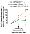
Bovine herpesvirus type 1 (BoHV-1)-specific serum neutralizing antibody (SN) titers developed in calves following primary infection, during latency, and after dexamethasone included latency reactivation. The data represent the mean ± standard deviation. The Shapiro–Wilk test was performed to determine the normal distribution of the data. Normally distributed data were analyzed by two-way ANOVA followed by Bonferroni post tests to compare replicate means by row; *** p < 0.001 between BoHV-1 wild-type LAT and BoHV-1 wild-type LAT-RE. (n = 2 for BoHV-1 wild-type latency group, n = 3 for BoHV-1 wild-type latency reactivation group, n = 4 for BoHV-1qmv Sub-RVFV latency group, and n = 4 for BoHV-1qmv Sub-RVFV latency reactivation group) LAT—latency; RE—reactivation.

Similar articles

Cited by

References

    1. Ellis J.A. Update on viral pathogenesis in BRD. Anim. Health Res. Rev. 2009;10:149–153. doi: 10.1017/S146625230999020X. - DOI - PubMed
    1. Griffin D., Chengappa M.M., Kuszak J., McVey D.S. Bacterial pathogens of the bovine respiratory disease complex. Vet. Clin. N. Am. Food Anim. Pract. 2010;26:381–394. doi: 10.1016/j.cvfa.2010.04.004. - DOI - PubMed
    1. Zhou Y., Shao Z., Dai G., Li X., Xiang Y., Jiang S., Zhang Z., Ren Y., Zhu Z., Fan C., et al. Pathogenic infection characteristics and risk factors for bovine respiratory disease complex based on the detection of lung pathogens in dead cattle in Northeast China. J. Dairy. Sci. 2023;106:589–606. doi: 10.3168/jds.2022-21929. - DOI - PubMed
    1. Graham D.A. Bovine herpes virus-1 (BoHV-1) in cattle-a review with emphasis on reproductive impacts and the emergence of infection in Ireland and the United Kingdom. Ir. Vet. J. 2013;66:15. doi: 10.1186/2046-0481-66-15. - DOI - PMC - PubMed
    1. Winkler M.T., Doster A., Sur J.H., Jones C. Analysis of bovine trigeminal ganglia following infection with bovine herpesvirus 1. Vet. Microbiol. 2002;86:139–155. doi: 10.1016/S0378-1135(01)00498-9. - DOI - PubMed

Publication types

MeSH terms

LinkOut - more resources