. 2024 Aug 26;12(9):960.
doi: 10.3390/vaccines12090960.

Immunometabolic Regulation of Vaccine-Induced Antibody Responses in Aging Mice

Affiliations

Immunometabolic Regulation of Vaccine-Induced Antibody Responses in Aging Mice

Daniela Frasca et al. Vaccines (Basel). .

Abstract

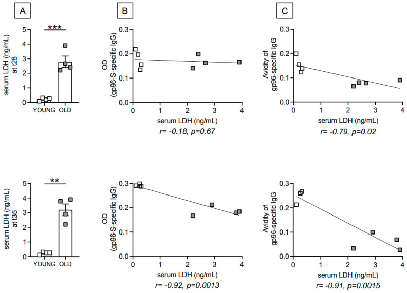
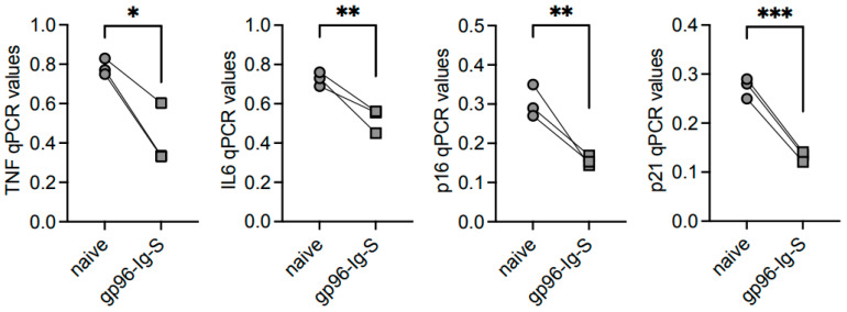
Immune cells undergo metabolic reprogramming to meet the demands associated with immune responses. The effects of aging on these pathways and on the metabolic phenotype of the immune cells participating in antibody responses to vaccines are still largely unknown. Here we used a vaccine for SARS-CoV-2 that utilizes the cellular heat shock chaperone glycoprotein 96 (gp96), engineered to co-express SARS-CoV-2 Spike (spike) protein (gp96-Ig-S). Results show that this vaccine induces comparable B cell primary responses in young and old mice at later time points, but a significantly lesser secondary response in old as compared to young mice, with the antibodies generated in the secondary response being also of lower avidity. This occurs because aging changes the B cell metabolic phenotype and induces hyper-metabolic B cells that are associated with higher intrinsic inflammation and decreased protective antibody responses. However, the gp96-Ig-S vaccine was found to be effective in significantly reducing the metabolic/inflammatory status of B cells from old mice, suggesting the possibility that targeting metabolic pathways may improve immune function in old mice that do not respond adequately to the vaccine.

Keywords: B cells; immunometabolism; inflammation; vaccine.


Conflict of interest statement

The authors declare no conflict of interest.

Figures

Figure 1
Figure 1
Aging induces defects in the antibody response to the gp96-Ig-S vaccine. Young and old mice were vaccinated with the gp96-Ig-S vaccine at t0, boosted with the same dose at t28 and sacrificed 7 days later. (A) Amount of serum antibodies. (B) Avidity of serum antibodies at t28 and t35. Urea 7M (final concentration) was added to the samples in the last 10 min before the detection antibody. Samples without 7 M Urea are the same as those shown in (A). Statistical analyses were performed with one-way ANOVA. * p < 0.05, ** p < 0.001, *** p < 0.001, **** p < 0.0001, ns not significant. Each symbol represents an individual mouse.
Figure 2
Figure 2
Age-dependent increase in serum levels of the metabolic marker LDH are negatively associated with the avidity of gp96-Ig-S vaccine-specific antibodies. (A). Serum levels of LDH were measured by ELISA in the same young and old mice in Figure 1, at t28 (top) and t35 (bottom) after vaccination. Mean comparisons between groups were performed by unpaired Student’s t-test (two-tailed), ** p < 0.01, *** p < 0.001. Correlation of serum levels of LDH and antibody amounts (B) and antibody avidity (C) at t28 (top) and t35 (bottom). Pearson’s correlations and p values are shown at the bottom of each graph. Each symbol represents an individual mouse.
Figure 3
Figure 3
The gp96-Ig-S vaccine induces a metabolic reprogramming of B cells from both young and old mice. B cells were sorted from the spleens of gp96-Ig-S-vaccinated (square symbols) or naïve (round symbols) young mice (white symbols) and old mice (grey symbols). Results are representative of three pairs of young and old mice and are from the t35 time point. After sorting, B cells were left unstimulated and were seeded into the wells of an extracellular flux analyzer at a concentration of 3 × 10⁵/well. (A) ECAR results in B cells from young (left) and old (right) mice. (B) Maximal ECAR. (C) Maximal OCR. (D,E) After sorting, B cells were left unstimulated. qPCR evaluated LDHA and PDHX mRNA expression, respectively. Results show qPCR values (2−ΔCt). Statistical analyses were performed with one-way ANOVA. * p < 0.05, ** p < 0.01, *** p < 0.001, **** p < 0.0001, ns not significant. Each symbol represents an individual mouse.
Figure 4
Figure 4
The gp96-Ig-S vaccine decreases RNA expression of inflammatory and senescent markers in B cells from old mice. B cells from the same mice as shown in Figure 3 were left unstimulated, mRNA was extracted and qPCR was run to evaluate mRNA expression of the indicated markers. Results show qPCR values (2−ΔCt). Statistical analyses were performed with one-way ANOVA. * p < 0.05, ** p < 0.01, *** p < 0.001, **** p < 0.0001, ns not sinificant. Each symbol represents an individual mouse.
Figure 5
Figure 5
The gp96-Ig-S vaccine decreases the frequencies of ABCs and increases those of FO B Cells in old mice, but not in young mice. Splenic B cells from the same mice shown in Figure 3 were membrane stained to evaluate the frequencies of FO, ABCs, and MZ B cells. Statistical analyses were performed with one-way ANOVA. * p < 0.05, ** p < 0.01, **** p < 0.0001, ns not significant. Each symbol represents an individual mouse.
Figure 6
Figure 6
The gp96-Ig-S vaccine decreases RNA expression of inflammatory and senescent markers in ABCs from old mice. ABCs were sorted from the spleens of old mice at t35, the mRNA was extracted and qPCR run to evaluate mRNA expression of the indicated markers. Results show qPCR values (2−ΔCt). Statistical analyses were performed with unpaired Student’s t-test (two-tailed) to evaluate mean comparisons between groups. * p < 0.05, ** p < 0.01, *** p < 0.001. Each symbol represents an individual mouse.

Similar articles

Cited by

References

    1. Frasca D., Diaz A., Romero M., Garcia D., Blomberg B.B. B Cell Immunosenescence. Annu. Rev. Cell Dev. Biol. 2020;36:551–574. doi: 10.1146/annurev-cellbio-011620-034148. - DOI - PMC - PubMed
    1. Franceschi C., Bonafe M., Valensin S., Olivieri F., De Luca M., Ottaviani E., De Benedictis G. Inflamm-aging. An evolutionary perspective on immunosenescence. Ann. N. Y. Acad. Sci. 2000;908:244–254. doi: 10.1111/j.1749-6632.2000.tb06651.x. - DOI - PubMed
    1. Woudstra T., Thomson A.B. Nutrient absorption and intestinal adaptation with ageing. Best Pract. Res. Clin. Gastroenterol. 2002;16:1–15. doi: 10.1053/bega.2001.0262. - DOI - PubMed
    1. Fink R.I., Kolterman O.G., Griffin J., Olefsky J.M. Mechanisms of Insulin Resistance in Aging. J. Clin. Investig. 1983;71:1523–1535. doi: 10.1172/JCI110908. - DOI - PMC - PubMed
    1. Ryan A.S. Insulin resistance with aging: Effects of diet and exercise. Sports Med. 2000;30:327–346. doi: 10.2165/00007256-200030050-00002. - DOI - PubMed

LinkOut - more resources