. 2024 Sep 17;12(9):1058.
doi: 10.3390/vaccines12091058.

Anti-Tumor Immunity to Patient-Derived Breast Cancer Cells by Vaccination with Interferon-Alpha-Conditioned Dendritic Cells (IFN-DC)

Affiliations

Anti-Tumor Immunity to Patient-Derived Breast Cancer Cells by Vaccination with Interferon-Alpha-Conditioned Dendritic Cells (IFN-DC)

Caterina Lapenta et al. Vaccines (Basel). .

Abstract

Background: Breast cancer represents one of the leading causes of death among women. Surgery can be effective, but once breast cancer has metastasized, it becomes extremely difficult to treat. Conventional therapies are associated with substantial toxicity and poor efficacy due to tumor heterogeneity, treatment resistance and disease relapse. Moreover, immune checkpoint blockade appears to offer limited benefit in breast cancer. The poor tumor immunogenicity and the immunosuppressive tumor microenvironment result in scarce T-cell infiltration, leading to a low response rate. Thus, there is considerable interest in the development of improved active immunotherapies capable of sensitizing a patient's immune system against tumor cells.

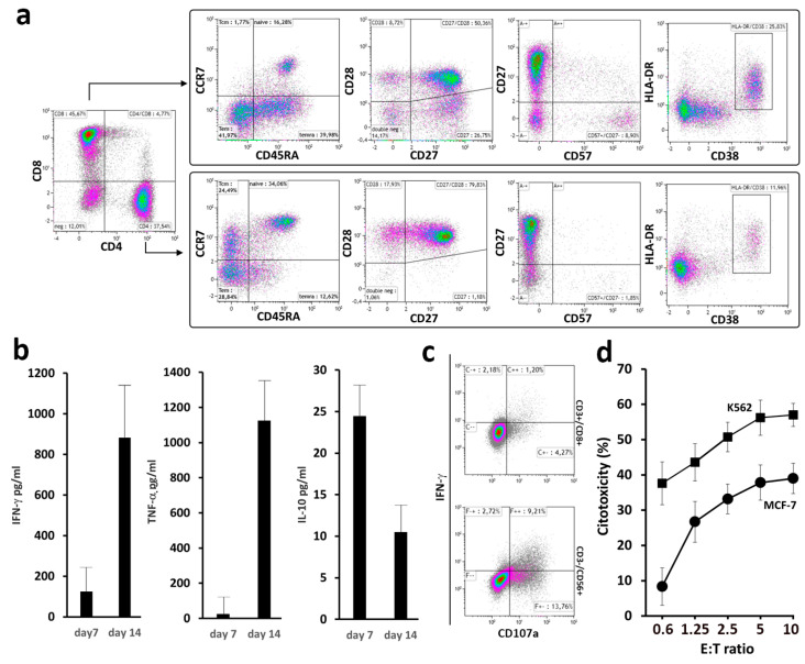
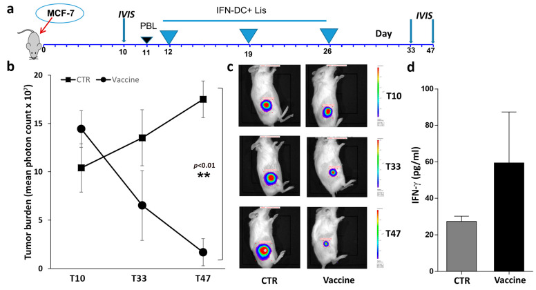
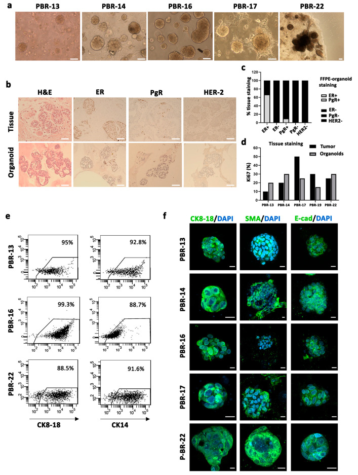
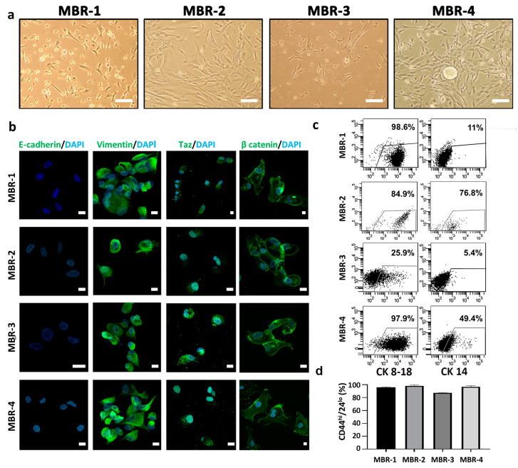
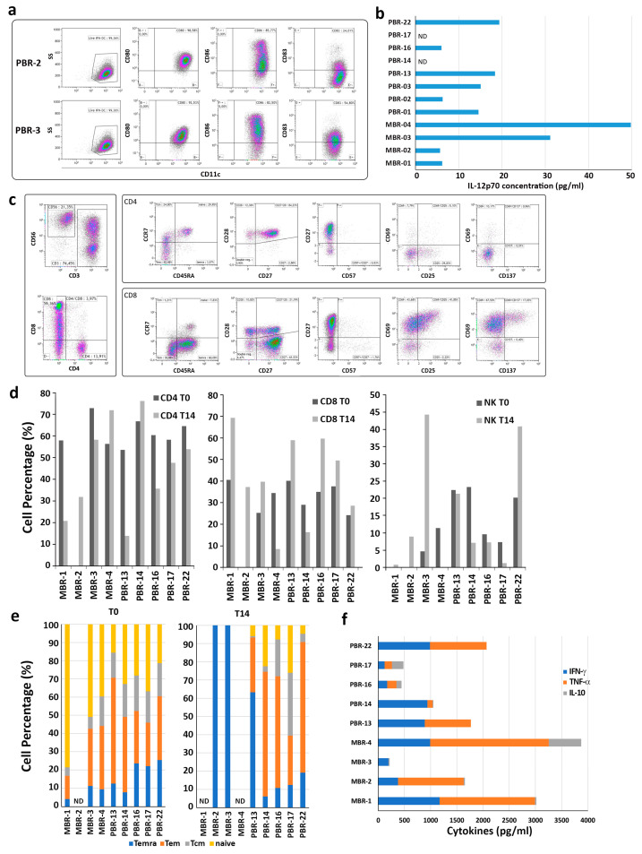
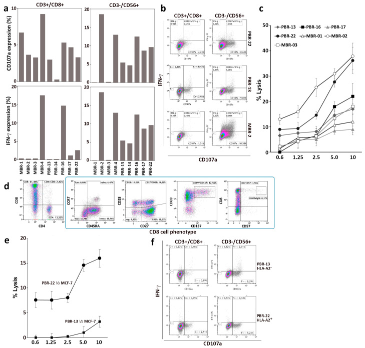
Methods: We evaluated the in vitro anti-tumor activity of a personalized vaccine based on dendritic cells generated in the presence of interferon (IFN)-α and granulocyte-macrophage colony-stimulating factor (IFN-DC) and loaded with an oxidized lysate from autologous tumor cells expanded as 3D organoid culture maintaining faithful tumor antigenic profiles.

Results: Our findings demonstrate that stimulation of breast cancer patients' lymphocytes with autologous IFN-DC led to efficient Th1-biased response and the generation in vitro of potent cytotoxic activity toward the patients' own tumor cells.

Conclusions: This approach can be potentially applied in association with checkpoint blockade and chemotherapy in the design of new combinatorial therapies for breast cancer.

Keywords: breast cancer; cancer vaccines; dendritic cells; immunotherapy.


Conflict of interest statement

The authors declare no conflicts of interest.

Figures

Figure 1
Figure 1
Phenotypic and functional analysis of peripheral blood lymphocytes (PBL) stimulated with HOCl-oxidized MCF-7 breast tumor cell lysate. (a) Dot-plot analysis of CD4+ and CD8+ cells as determined by flow cytometry in PBL stimulated with IFN-DC. PBL isolated from HLA-A2+ healthy blood donors were cultured with autologous IFN-DC (IFN-DC/PBL ratio of 1:4) pulsed with MCF-7 tumor cell lysate, as described in Section 2. Representative results of three independent experiments are shown. (b) Cytokine production (IFN-γ, TNF-α and IL-10) in culture supernatants as evaluated by ELISA on days 7 and 14 of co-culture. (c) Degranulation assay as a surrogate evaluation of cytotoxic activity by detection of CD107a membrane expression and intracellular IFN-γ production in CD8+ and natural killer (NK) cells. Representative dot-plot analysis in electronically gated CD8+CD3+ and CD56+CD3- cells in PBL co-cultured with tumor cell-loaded IFN-DC. On day 21 of culture, PBL were restimulated with MCF-7 or K562 target cell lines for 4 h at 37 °C (E:T ratio of 2:1) (see Section 2). Dot-plots show CD107a membrane exposure and IFN-γ expression in electronically gated CD8+CD3+ and CD56+CD3- lymphocytes in response to the indicated target cells. Results from one representative experiment out of four are shown. (d) Representative cytotoxicity assay against NK-sensitive K562 and MCF-7 target cell lines as evaluated by a Calcein-AM assay (see Section 2) at different E:T ratios. Data are mean ± SD of a triplicate assay of PBL derived from an HLA-A2+ donor.
Figure 2
Figure 2
Evaluation of tumor-growth inhibition by in vivo immunization of hu-PBL-NSG mice with IFN-DC loaded with HOCl-oxidized tumor cell lysate. (a) Vaccination schedule. Mice were reconstituted with HLA-A2+ PBL as soon as the implanted tumors became detectable by in vivo bioluminescence imaging (10–11 days). Humanized mice were then randomized into treatment and control groups. (b) Quantitative evaluation of tumor cell growth by bioluminescence analysis. Tumor burden was detected by in vivo non-invasive imaging of the firefly luciferase expressing MCF-7 cells after intraperitoneal luciferin injection. Tumor bioluminescence intensity was plotted in pseudocolor over black/white photographs and quantified as total flux in photons/seconds. Graph represents mean total flux of MCF-7 cell growth rate in hu-PBL-NSG mice immunized as described. The data are presented as mean ± SEM. The difference in tumor growth was highly statistically significant only at the last time point, day 47 (** p < 0.01 by Mann–Whitney test). (c) Representative tumor burden images of the two groups (CTR vs. vaccine) at different time points by IVIS imaging system. (d) Evaluation of IFN-γ levels in mouse sera collected at the time of sacrifice.
Figure 3
Figure 3
Isolation and characterization of patient-derived breast cancer organoids (PDBCOs). (a) Representative bright-field images of 5 PDBCOs used for the study, showing different structures: cohesive and discohesive organoids, dense and solid (PBR-13, PBR-14, PBR-16, PBR-17), cohesive organoids, cystic and grape-like (P-BR-22). Scale bar, 100 µm. (b) Comparative histological and immunohistochemical images of BC tissues and derived organoid lines. Shown are representative examples of H&E staining and IHC of either HR or HER-2 status for PBR-17. Scale bar, 200 µm. (c) Stacked bar chart indicating the percentage of PDBCO lines found positive (grey) and negative (black) by IHC for the receptor expression grouped per original tumor receptor status. (d) Bar graph displaying proliferation rate percentage of PDBCOs and corresponding parental tumors as quantified by Ki67 immunohistochemical staining. (e) Representative dot-plot graphs showing CK14 and CK8-18 expression in PBR-13, PBR-16 and PBR-22 (f) CLSM analyses of PFA-fixed PDBCOs stained for CK8-18, SMA and E-cadherin (green); 4′-6-Diamidino-2-phenylindole (DAPI) was used to counterstain nuclei (light blue). Several (>50 organoids) were observed for each condition and representative images are shown. Scale bars, 10 µm.
Figure 4
Figure 4
Isolation and characterization of breast cancer patient-derived metastatic cells (PDMCs). (a) Representative phase-contrast images of PDMCs from ascitic fluid (MBR-1, MBR-2) or pleural effusion (MBR-3, MBR-4) cultured in serum-free conditions. Scale bar, 100 µm. (b) CLSM analyses of PFA-fixed PDMCs stained for E-cadherin, vimentin Taz and beta-catenin (green); DAPI was used to counterstain nuclei (light blue). Several fields were observed for each condition and representative images are shown. Scale bars, 10 µm. (c) Dot-plots showing luminal CK8-18 and myoepithelial CK14 expression in PDMC lines. (d) Bar chart reporting percentages of CD44highCD24−/low phenotype in MBR-1, MBR-2, MBR-3 and MBR-4 lines obtained from ascitic fluid or pleural effusion of breast cancer metastatic patients. Percentages, referring to CD44highCD24−/low positive cells, were determined by setting the gate on the isotype control from at least two independent FACS stainings.
Figure 5
Figure 5
Characterization of PBL cultures from breast tumor patients stimulated with IFN-DC loaded with HOCl-oxidized autologous tumor cell lysate. (a). Representative phenotypical analysis of IFN-DC obtained from breast tumor patients. IFN-DC were differentiated from peripheral blood monocytes as described in Section 2. Partially mature CD11c+ IFN-DC ex-pressed high levels of the costimulatory molecules CD80 and CD86, as well as variable levels of the maturation marker CD83. (b) IL-12 release in supernatants collected from IFN-DC cultures on day 3 of differentiation. (c) Representative phenotypic analysis of PBL isolated from breast tumor patients and cultured with autologous IFN-DC loaded with HOCl-oxidized tumor cell lysate for 14 days. (d) Evaluation of CD4, CD8 and NK cell percentages in PBL from breast cancer patients before (T0) and after 14 days of culture (T14) with autologous IFN-DC. (e) Flow cytometric analysis of CD4 and CD8 cell memory subsets of freshly purified PBL from breast cancer patients and after 14 days of culture with autologous IFN-DC loaded with HOCl-oxidized tumor cell lysate. (f) Cytokine release (IFN-γ, TNF-α and IL-10) in culture supernatants as evaluated by ELISA on day 14 of co-cultures.
Figure 6
Figure 6
Antitumor activity of PBL from breast cancer patients co-cultured with IFN-DC loaded with HOCl-oxidized autologous tumor cell lysate. PBL isolated from nine patient blood donors were cultured with IFN-DC pulsed with HOCl-oxidized tumor cell lysate for 14 days. (a) Degranulation activity of expanded effectors cells tested against autologous breast cancer target cells as detected by flow cytometry; analysis of CD107a membrane expression and intracellular IFN-γ. (b) Representative degranulation assay as assessed by dot-plot analysis of ectopic CD107a and IFN-γ expression in CD8+ and CD3-CD56+ NK cells derived from three representative breast cancer patients in response to autologous tumor cells. (c) Cytotoxic assay of PBL culture from breast cancer patients after in vitro culture for 14 days, tested against autologous breast cancer cells. (d) Phenotypic analysis of CD8 cells from patient PBR-22 (HLA-A2+) stimulated with IFN-DC loaded with HOCl-oxidized autologous tumor cell lysate for 14 days. (e) Cytotoxic activity of PBL from PBR-22 (HLA-A2+) as compared to PBR-13 (HLA-A2–) as determined by cytotoxic assay towards HLA-A2+ MCF-7 target cells. (f) Degranulation activity as determined by dot plot analysis of CD107a and IFN-γ expression in CD8+ and CD3-CD56+ NK cells from patients PBR-22 and P-BR13 toward MCF-7 target cells.

Similar articles

Cited by

References

    1. Sung H., Ferlay J., Siegel R.L., Laversanne M., Soerjomataram I., Jemal A., Bray F. Global Cancer Statistics 2020: GLOBOCAN Estimates of Incidence and Mortality Worldwide for 36 Cancers in 185 Countries. CA. Cancer J. Clin. 2021;71:209–249. doi: 10.3322/caac.21660. - DOI - PubMed
    1. Moo T.A., Sanford R., Dang C., Morrow M. Overview of Breast Cancer Therapy. PET Clin. 2018;13:339–354. - PMC - PubMed
    1. Yang J., Ju J., Guo L., Ji B., Shi S., Yang Z., Gao S., Yuan X., Tian G., Liang Y., et al. Prediction of HER2-positive breast cancer recurrence and metastasis risk from histopathological images and clinical information via multimodal deep learning. Comput. Struct. Biotechnol. J. 2022;20:333–342. doi: 10.1016/j.csbj.2021.12.028. - DOI - PMC - PubMed
    1. Locy H., Verhulst S., Cools W., Waelput W., Brock S., Cras L., Schiettecatte A., Jonckheere J., van Grunsven L.A., Vanhoeij M., et al. Assessing Tumor-Infiltrating Lymphocytes in Breast Cancer: A Proposal for Combining Immunohistochemistry and Gene Expression Analysis to Refine Scoring. Front. Immunol. 2022;13:794175. doi: 10.3389/fimmu.2022.794175. - DOI - PMC - PubMed
    1. Garaud S., Buisseret L., Solinas C., Gu-Trantien C., De Wind A., Van Den Eynden G., Naveaux C., Lodewyckx J.N., Boisson A., Duvillier H., et al. Tumor-infiltrating B cells signal functional humoral immune responses in breast cancer. JCI Insight. 2019;4:e129641. doi: 10.1172/jci.insight.129641. - DOI - PMC - PubMed

LinkOut - more resources