. 2024 Oct 28;52(19):12074-12092.
doi: 10.1093/nar/gkae796.

Combining Nanopore direct RNA sequencing with genetics and mass spectrometry for analysis of T-loop base modifications across 42 yeast tRNA isoacceptors

Affiliations

Combining Nanopore direct RNA sequencing with genetics and mass spectrometry for analysis of T-loop base modifications across 42 yeast tRNA isoacceptors

Ethan A Shaw et al. Nucleic Acids Res. .

Abstract

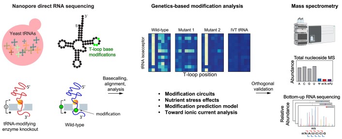
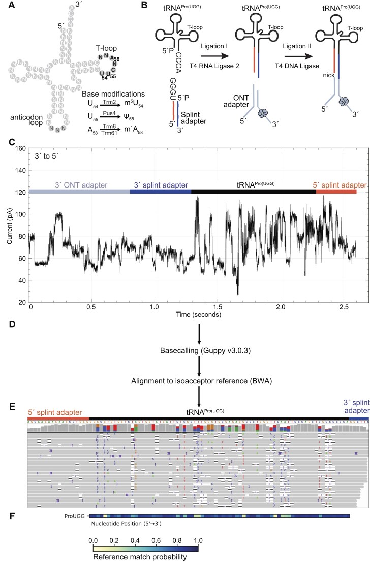
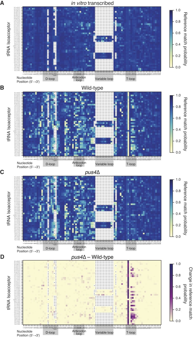
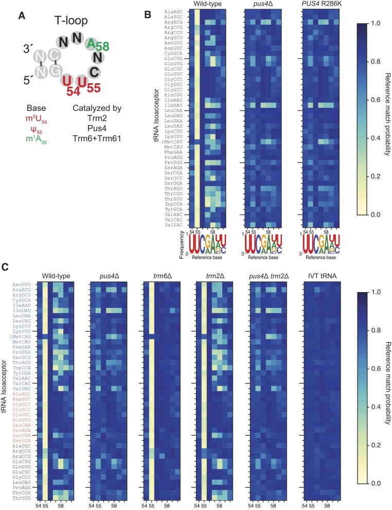
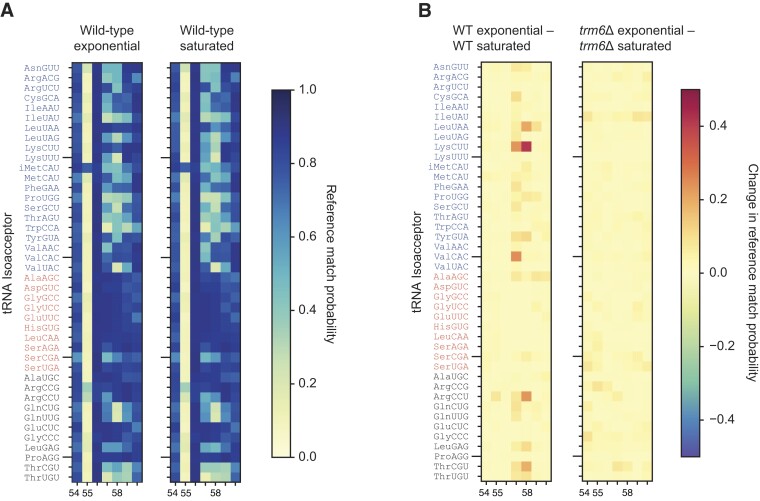
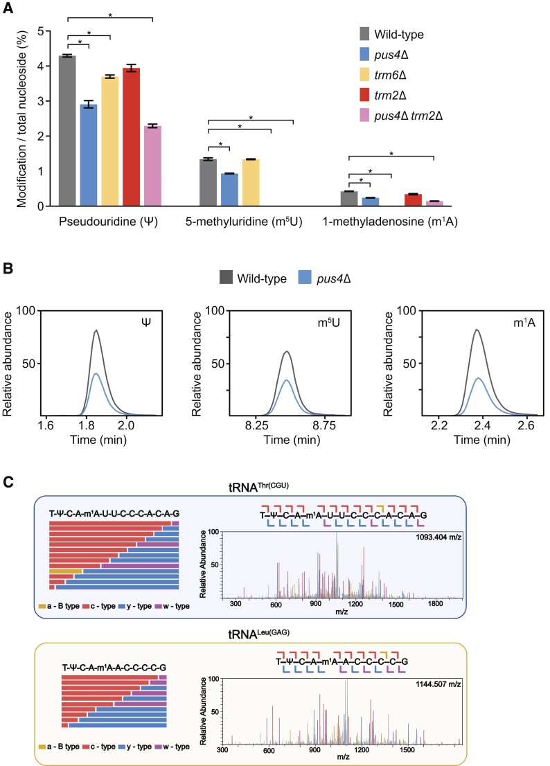
Transfer RNAs (tRNAs) contain dozens of chemical modifications. These modifications are critical for maintaining tRNA tertiary structure and optimizing protein synthesis. Here we advance the use of Nanopore direct RNA-sequencing (DRS) to investigate the synergy between modifications that are known to stabilize tRNA structure. We sequenced the 42 cytosolic tRNA isoacceptors from wild-type yeast and five tRNA-modifying enzyme knockout mutants. These data permitted comprehensive analysis of three neighboring and conserved modifications in T-loops: 5-methyluridine (m5U54), pseudouridine (Ψ55), and 1-methyladenosine (m1A58). Our results were validated using direct measurements of chemical modifications by mass spectrometry. We observed concerted T-loop modification circuits-the potent influence of Ψ55 for subsequent m1A58 modification on more tRNA isoacceptors than previously observed. Growing cells under nutrient depleted conditions also revealed a novel condition-specific increase in m1A58 modification on some tRNAs. A global and isoacceptor-specific classification strategy was developed to predict the status of T-loop modifications from a user-input tRNA DRS dataset, applicable to other conditions and tRNAs in other organisms. These advancements demonstrate how orthogonal technologies combined with genetics enable precise detection of modification landscapes of individual, full-length tRNAs, at transcriptome-scale.


Figures

Graphical Abstract
Graphical Abstract
Figure 1.
Figure 1.
Overview of tRNA library preparation, sequencing, and alignment strategy. (A) Illustration of a generic yeast tRNA, highlighting overall structural segments, three chemical modifications that can occur in the T-loop, and the names of the enzymes that catalyze them. (B) tRNAs are ligated to a double-stranded splint adapter with RNA Ligase 2 by the tRNA’s 3′ NCCA overhang. A second ligation is performed using T4 DNA Ligase with the tRNA and ONT sequencing adapters. (C) Ionic current trace measured in picoamperes for an adapted tRNAPro(UGG) molecule as it translocates from the 3′ to 5′ direction through a Nanopore. (D) Ionic current is basecalled using Guppy v3.0.3. Fastq files are aligned to our S. cerevisiae reference sequences using BWA-MEM. (E) Alignments are visualized in Integrative Genomics Viewer (32). The reference sequence for tRNAPro(UGG) is on the top. Read coverage is designated by the height of the gray bar at that position. In the panel containing gray or colored vertical bars, gray represents a match to the reference base for at least 80% of the reads, and colored represents the relative proportion of alternative nucleotide calls at that position. Colored bars (U/T = red; A = green; C = blue, G = gold) indicate positions where base call differs from the listed reference base. Rows below show alignments of individual reads with the reference, interrupted by: non-reference basecalls (colored letters); deletions (white spaces bisected with a black bar); and insertions (purple spaces bisected with a black bar). (F) Reference match probabilities are calculated with marginCaller (27) and used to generate a heatmap, where the nucleotide position is shown from 5′–3′ along the tRNA. Dark blue indicates that the base-called nucleotide matches the reference nucleotide more frequently given the alignment, and yellow indicates that the base-called nucleotide matches the reference nucleotide less frequently.
Figure 2.
Figure 2.
Heatmaps representing comprehensive alignments of 42 S. cerevisiae cytosolic isoacceptors exhibit miscalls coincident with modified positions in the T-loop and elsewhere. (A) Heatmap representing in vitro transcribed (IVT) tRNA sequences of 42 S. cerevisiae cytosolic isoacceptors. A higher reference match probability (dark blue) corresponds to positions where the base-called nucleotide more frequently concurs with the reference nucleotide given the alignment, and a lower reference match probability (light yellow) corresponds to positions where the base-called nucleotide more frequently disagrees with the reference nucleotide. Sequences were aligned by the D-loop, anticodon loop, variable loop, and T-loop—highlighted with grey boxes, with anticodon positions in bold type; white boxes with dots indicate gaps—following a conventional tRNA base numbering scheme and based on the sequence alignment in Supplementary Figure S3. Note that tRNAHis(GUG) reads were aligned beginning with position 1 for simplicity, however note that this tRNA contains a non-templated G added at 5′ end in the ‘-1’ position (36). (B) Wild-type aligned isoacceptors. Otherwise as described above. (C) pus4Δ aligned isoacceptors. Otherwise as described above. (D) The change in reference match probabilities between pus4Δ and wild-type aligned isoacceptors. As described above except that dark purple squares correspond to large differences in basecalls between tRNAs in the two strains and yellow squares show basecalls that are not different between the two strains.
Figure 3.
Figure 3.
Reference match probabilities identify changes corresponding to modifications in the T-loop across 42 cytosolic isoacceptor tRNAs. (A) Illustration of a generic yeast tRNA T-loop, including its three possible chemical modifications and the names of the enzymes that catalyze them. (B) Reference match probability heat maps of aligned T-loop sequences across 42 cytosolic isoacceptor tRNAs, in wild-type, pus4Δ and PUS4 R286K strains. Conventional tRNA position numbers of modifications (m5U54; Ψ55; m1A58) are shown below each plot. In the sequence logos below, the height of each unmodified nucleotide letter matches its frequency in shown tRNA sequences. (Note that for tRNAiMet, in contrast to other 41 isoacceptors, positions 54 and 60 are adenosines, barely visible in the logo.) (C) Reference match probability heat maps for tRNAs purified from wild-type, pus4Δ, trm6Δ, trm2Δ, pus4Δtrm2Δ strains and in vitro transcribed (IVT) control sequences. tRNAs are ordered differently than in previous panels. tRNAs in blue letters are those annotated in Modomics (14) to contain m1A in the T-loop; tRNAs in red letters are those annotated in Modomics to not contain m1A in the T-loop; tRNAs in black letters are those not included in the current version of Modomics.
Figure 4.
Figure 4.
Nanopore reveals increased m1A58 frequency on some tRNAs upon nutrient depletion. (A) Reference match probability heat maps of the T-loop in wild-type cells growing exponentially (reprinted from Figure 3C) or to saturation. Otherwise as displayed in Figure 3C. (B) Bi-directional, differential reference match probability heat maps of the T-loop in wild-type or trm6Δ cells, comparing changes observed between exponentially growing and saturated cells. A positive change in reference match probability (red squares) indicates an increase in base miscalls during saturated phase. Comparison between heat maps indicates that several isoacceptors, most prominently tRNALys(CUU), had increases in m1A58 during saturation that was catalyzed by Trm6.
Figure 5.
Figure 5.
LC-MS/MS confirms DRS-based modification predictions. (A) Modification abundance in wild-type (grey), pus4Δ (blue), trm6Δ (gold), trm2Δ (red) and pus4Δtrm2Δ (pink) total tRNA quantified using LC-MS/MS ribonucleoside modification profiling. Significant changes (P < 0.01) are noted with an asterisk. (B) Extracted ion chromatograms of m1A and m5U signals in wild-type (grey) and pus4Δ (blue) displaying a decrease in abundance in pus4Δ total tRNA. (C) We used Collision-induced dissociation (CID) fragmentation to confirm the location of m1A found by DRS. CID fragments oligonucleotides at each phosphodiester backbone, resulting in ladder oligonucleotide fragments that can be detected within the MS/MS spectrum. This enables the sequencing of oligonucleotides by MS/MS, in addition to locating post-transcriptional modifications within these specific fragments. Here, we display two MS/MS fragmentation spectra and sequence coverage maps confirming that m1A58 is found within tRNAThr(CGU) and tRNALeu(GAG). (left) Sequence coverage maps displaying sequence informative MS/MS fragmentation ions for m1A containing oligonucleotide digestion products resulting from RNase T1 digestion of tRNAThr(CGU) and tRNALeu(GAG). (right) The respective MS/MS spectra for tRNAThr(CGU) and tRNALeu(GAG) are displayed, where each detected sequence information fragmentation ion is displayed in gold (a-B type), red (c-type), blue (y-type), and purple (w-type). Full MS/MS sequence coverage was achieved with confidence scores greater than 99% by BioPharma Finder, confirming the presence of m1A at the expected nucleotide position in tRNAThr(CGU) and tRNALeu(GAG). Each detected MS/MS fragmentation ion was manually inspected to confirm correct isotopic distributions. The approach, analysis and data presentation are drawn from standards for MS analysis of oligonucleotides (71–75).

References

    1. Yoluç Y., Ammann G., Barraud P., Jora M., Limbach P.A., Motorin Y., Marchand V., Tisné C., Borland K., Kellner S.. Instrumental analysis of RNA modifications. Crit. Rev. Biochem. Mol. Biol. 2021; 56:178–204. - PubMed
    1. RajBhandary U.L., Köhrer C.. Early days of tRNA research: discovery, function, purification and sequence analysis. J. Biosci. 2006; 31:439–451. - PubMed
    1. Hernandez-Alias X., Katanski C.D., Zhang W., Assari M., Watkins C.P., Schaefer M.H., Serrano L., Pan T.. Single-read tRNA-seq analysis reveals coordination of tRNA modification and aminoacylation and fragmentation. Nucleic Acids Res. 2023; 51:e17. - PMC - PubMed
    1. Behrens A., Rodschinka G., Nedialkova D.D.. High-resolution quantitative profiling of tRNA abundance and modification status in eukaryotes by mim-tRNAseq. Mol. Cell. 2021; 81:1802–1815. - PMC - PubMed
    1. Thomas N.K., Poodari V.C., Jain M., Olsen H.E., Akeson M., Abu-Shumays R.L.. Direct nanopore sequencing of individual full length tRNA strands. ACS Nano. 2021; 15:16642–16653. - PMC - PubMed

MeSH terms

LinkOut - more resources