Review
. 2024 Sep 28;52(1):133.
doi: 10.1007/s00240-024-01631-x.

Two distinct phenotypes of calcium oxalate stone formers could imply different long-term risks for renal function

Affiliations
Review

Two distinct phenotypes of calcium oxalate stone formers could imply different long-term risks for renal function

James C Williams Jr et al. Urolithiasis. .

Abstract

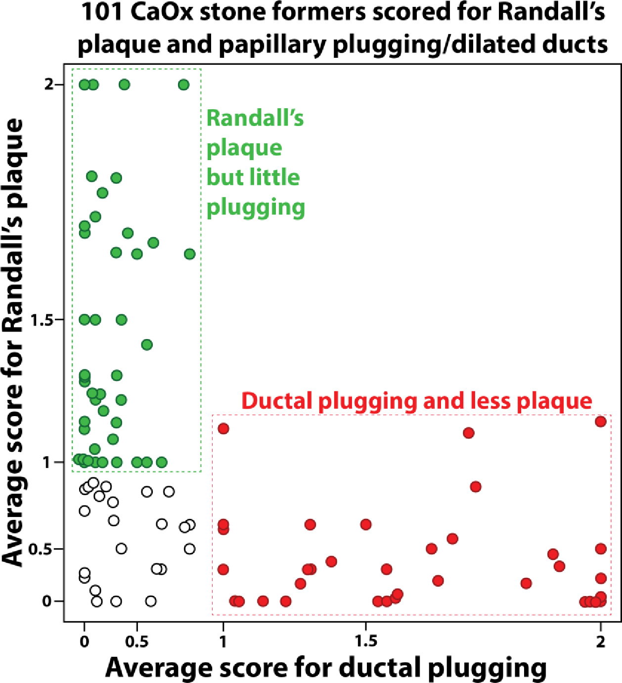
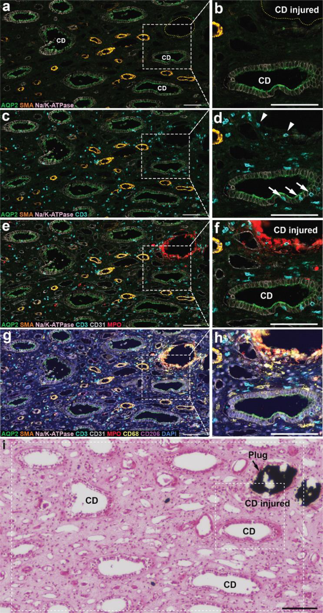
Endoscopic and biopsy findings have identified two distinct phenotypes among individuals with calcium oxalate (CaOx) kidney stones. The first type has normal renal papillae but shows interstitial mineral deposition, known as Randall's plaque. The other phenotype presents with collecting duct plugging and a higher incidence of loss of papilla tissue mass. With Randall's plaque, renal papilla injury involves the loss of small patches of calcified tissue (Randall's plaque detaching with the stone), which likely results in damage to only a few nephrons. In contrast, collecting duct mineral plugs are very large, causing obstruction to tubular flow. Since each terminal collecting duct drains thousands of nephrons, ductal plugs could lead to the degeneration of many nephrons and a significant loss of renal glomeruli. New visualization techniques for immune cells in papillary biopsies have revealed that the Randall's plaque phenotype is marked by the accumulation of macrophages around the plaque regions. In contrast, preliminary data on the plugging phenotype shows collecting duct damage with mineral plugs and increased T-lymphocytes throughout the papilla. These regions also show tubulitis, i.e., T-cell infiltration into nearby collecting duct epithelium. This suggests that while some CaOx stone formers may have some papillary inflammation but with minimal damage to nephrons, others suffer from obstruction to flow for many nephrons that may also include destructive inflammation in the renal tissue. We propose that CaOx stone formers with the plugging phenotype will have a higher long-term risk for loss of renal function.

Keywords: Calcium oxalate; Inflammation; Kidney stones; Nephrolithiasis.


Figures

Figure 1.
Figure 1.
Endoscopic views of representative papillae from two patients. Both patients had majority CaOx stones and no systemic cause for their disease. The patient on the left was a 50 year-old female, and the patient on the right was a 40 year-old female. Left: Two papillae show normal shape, but both have some deposition of Randall’s (interstitial) plaque. Right: The two papillae show many plugs of mineral (yellowish in color). The upper right panel shows a papilla with a large amount of tissue loss at its tip. Lower right panel shows a compound papilla with a dilated duct. (A typical human papilla is about 1 cm wide at its base.) [add scores to the images]
Figure 2.
Figure 2.
Idiopathic calcium oxalate (CaOx) stone formers tend to group into two papillary mineral patterns. Graph shows semi-quantitative grading of papillary mineral using endoscopic video in 101 CaOx stone formers. Axis scales are exponential. Note that a patient plugging score of 1.0 indicates that on average every papilla showed 1–5 plugs or dilated ducts; thus, a score <1.0 means that at least one papilla in that patient had no plugs or dilated ducts. In contrast, a Randall’s plaque score of 1.0 means that the average papilla was moderately covered with plaque, and a score of 0 means ‘mild’ coverage by plaque. Empty circles show patients with average scores <1 for both Randall’s plaque and plugging/dilated ducts (the Neither group in Table 1).
Figure 3.
Figure 3.
Multiplexed imaging on a biopsy from a CaOx stone former with ductal plugging. a. Antibody localization is shown for aquaporin 2 (green), alpha-SMA (orange), and NaK-ATPase (light pink). b. Blow-up showing injured collecting duct (which contained mineral, dashed yellow line), along with a normal appearing collecting duct. c, d. The same regions but with staining for T-cells revealed (cyan). Note T-cells invading the epithelium of the healthy collecting duct (arrows). e, f. Same regions but with visualization of CD31 (endothelium, light gray) and myeloperoxidase (neutrophils, red). Note coating of neutrophils around mineral plug. g, h. Visualization of macrophages (CD68, light yellow) and CD206 (marker of M2 macrophages, pink). Note that many macrophages also surround the mineral plug. i. Yasue stain of consecutive section, verifying the mineral plug in the collecting duct marked ‘injured’ in b and f. Dashed rectangles indicate approximate regions of panels a-h in this consecutive section. Scale bars are 100 μm.
Figure 4.
Figure 4.
Papillary biopsy from a patient who had extensive deposition of Randall’s plaque, as seen by endoscopy. a. H&E staining of the multiplexed-imaged section after all the cycling of reagents and fluorescence imaging was complete; note remarkable preservation of the tissue. b. Multiplex image from that section: DAPI, stain for nuclei; AQP2, aquaporin-2 for collecting duct; aSMA, alpha-smooth muscle actin; CD11c, for antigen-presenting cells; CD206, for resident macrophages; CD31, labelling endothelium; CD3, labelling T-cells; and CD68, a general macrophage marker that is highly expressed in inflammatory states. c, d. Consecutive sections stained with Yasue and H&E, respectively. e. Higher power view of inset shown in b; arrows indicate CD68+ macrophages. f. Same region as e from Yasue stained section (c), showing that macrophages were associated with mineralization (Randall’s plaque, brown-to-black staining; note nearby thin limbs that have only dots of mineral). g. Higher power view of the other inset in b; V is a vessel surrounded by alpha-SMA-positive pericytes (blue), and arrows indicate CD68+ macrophages. h. Same region as g from Yasue stained section (c); again, locations of macrophages (arrows) are at regions of developing Randall’s plaque (brown; again, note variation in mineral deposition among thin limb segments). CD, collecting duct. TL, thin limb. Bars in a-d, 300 um; bars in e and g, 50 um.
Figure 5.
Figure 5.
Extended hypotheses based on the observations that CaOx stone formers tend to have either Randall’s plaque or ductal plugging. The obstructive nature of ductal plugging suggests that it is more likely to lead to significant nephron loss. In addition, preliminary data on cellular inflammation suggests that ductal plugging is associated with an aggressive T-cell infiltrate that could also lead to nephron loss.

Update of

References

    1. Roberson D, Sperling C, Shah A, Ziemba J (2020) Economic Considerations in the Management of Nephrolithiasis. Curr Urol Rep 21: 18. - PubMed
    1. Daudon M, Jungers P, Bazin D, Williams JC Jr. (2018) Recurrence rates of urinary calculi according to stone composition and morphology. Urolithiasis 46: 459–470 - PMC - PubMed
    1. Coe FL, Evan AP, Worcester EM (2005) Kidney stone disease. J Clin Invest 115: 2598–2608 - PMC - PubMed
    1. Williams JC, Al-Awadi H, Muthenini M, Bledsoe SB, El-Achkar T, Evan AP, Coe F, Lingeman JE, Worcester EM (2022) Stone morphology distinguishes two pathways of idiopathic calcium oxalate stone pathogenesis. J Endourol 36: 694–702 - PMC - PubMed
    1. Coe FL, Evan AP, Lingeman JE, Worcester EM (2010) Plaque and deposits in nine human stone diseases. Urol Res 38: 239–247 - PMC - PubMed

Substances

LinkOut - more resources