. 2024 Dec:70:101452.
doi: 10.1016/j.dcn.2024.101452. Epub 2024 Sep 21.

Quantifying brain development in the HEALthy Brain and Child Development (HBCD) Study: The magnetic resonance imaging and spectroscopy protocol

Douglas C Dean 3rd  1 M Dylan Tisdall  2 Jessica L Wisnowski  3 Eric Feczko  4 Borjan Gagoski  5 Andrew L Alexander  6 Richard A E Edden  7 Wei Gao  8 Timothy J Hendrickson  9 Brittany R Howell  10 Hao Huang  11 Kathryn L Humphreys  12 Tracy Riggins  13 Chad M Sylvester  14 Kimberly B Weldon  15 Essa Yacoub  16 Banu Ahtam  17 Natacha Beck  18 Suchandrima Banerjee  19 Sergiy Boroday  18 Arvind Caprihan  20 Bryan Caron  18 Samuel Carpenter  21 Yulin Chang  22 Ai Wern Chung  17 Matthew Cieslak  23 William T Clarke  24 Anders Dale  25 Samir Das  18 Christopher W Davies-Jenkins  7 Alexander J Dufford  26 Alan C Evans  18 Laetitia Fesselier  18 Sandeep K Ganji  27 Guillaume Gilbert  28 Alice M Graham  21 Aaron T Gudmundson  7 Maren Macgregor-Hannah  29 Michael P Harms  30 Tom Hilbert  31 Steve C N Hui  32 M Okan Irfanoglu  33 Steven Kecskemeti  34 Tobias Kober  31 Joshua M Kuperman  35 Bidhan Lamichhane  36 Bennett A Landman  37 Xavier Lecour-Bourcher  18 Erik G Lee  9 Xu Li  7 Leigh MacIntyre  38 Cecile Madjar  18 Mary Kate Manhard  39 Andrew R Mayer  20 Kahini Mehta  23 Lucille A Moore  15 Saipavitra Murali-Manohar  7 Cristian Navarro  40 Mary Beth Nebel  41 Sharlene D Newman  42 Allen T Newton  43 Ralph Noeske  44 Elizabeth S Norton  45 Georg Oeltzschner  7 Regis Ongaro-Carcy  18 Xiawei Ou  46 Minhui Ouyang  11 Todd B Parrish  47 James J Pekar  7 Thomas Pengo  29 Carlo Pierpaoli  33 Russell A Poldrack  48 Vidya Rajagopalan  3 Dan W Rettmann  49 Pierre Rioux  18 Jens T Rosenberg  50 Taylor Salo  23 Theodore D Satterthwaite  23 Lisa S Scott  51 Eunkyung Shin  52 Gizeaddis Simegn  7 W Kyle Simmons  53 Yulu Song  7 Barry J Tikalsky  15 Jean Tkach  39 Peter C M van Zijl  7 Jennifer Vannest  54 Maarten Versluis  27 Yansong Zhao  55 Helge J Zöllner  7 Damien A Fair  56 Christopher D Smyser  57 Jed T Elison  58 HBCD MRI Working Group
Affiliations

Quantifying brain development in the HEALthy Brain and Child Development (HBCD) Study: The magnetic resonance imaging and spectroscopy protocol

Douglas C Dean 3rd et al. Dev Cogn Neurosci. 2024 Dec.

Abstract

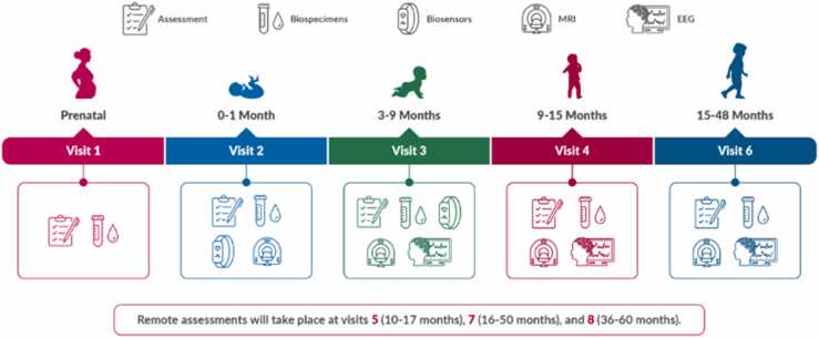
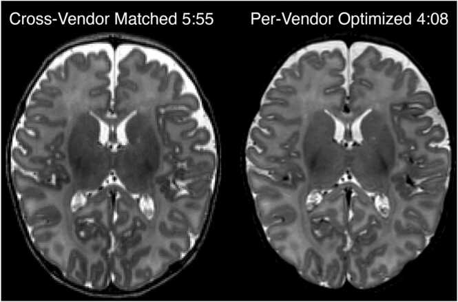
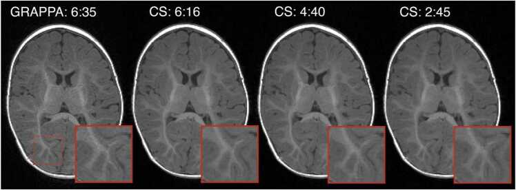
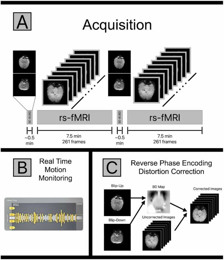
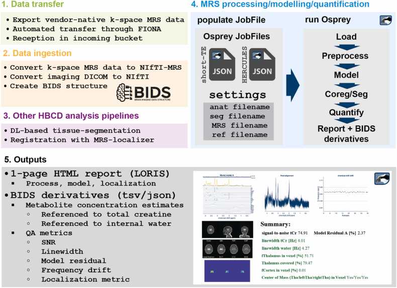
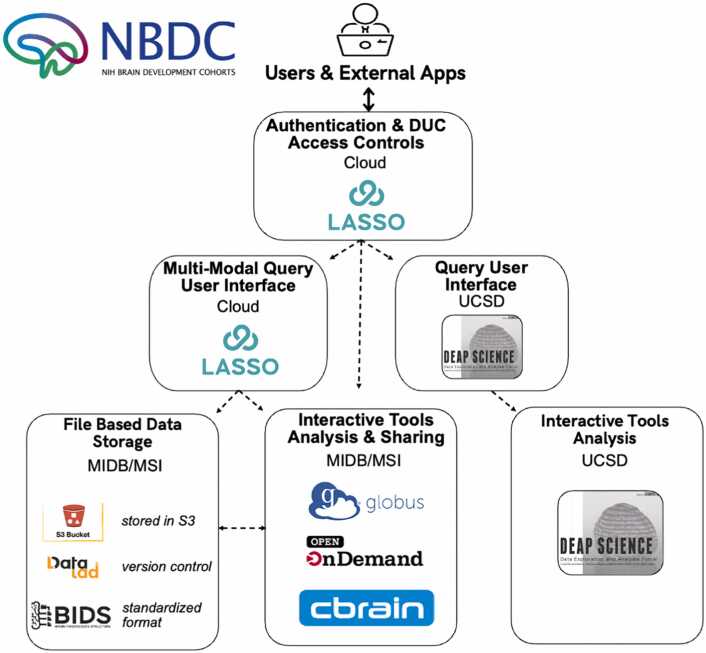
The HEALthy Brain and Child Development (HBCD) Study, a multi-site prospective longitudinal cohort study, will examine human brain, cognitive, behavioral, social, and emotional development beginning prenatally and planned through early childhood. The acquisition of multimodal magnetic resonance-based brain development data is central to the study's core protocol. However, application of Magnetic Resonance Imaging (MRI) methods in this population is complicated by technical challenges and difficulties of imaging in early life. Overcoming these challenges requires an innovative and harmonized approach, combining age-appropriate acquisition protocols together with specialized pediatric neuroimaging strategies. The HBCD MRI Working Group aimed to establish a core acquisition protocol for all 27 HBCD Study recruitment sites to measure brain structure, function, microstructure, and metabolites. Acquisition parameters of individual modalities have been matched across MRI scanner platforms for harmonized acquisitions and state-of-the-art technologies are employed to enable faster and motion-robust imaging. Here, we provide an overview of the HBCD MRI protocol, including decisions of individual modalities and preliminary data. The result will be an unparalleled resource for examining early neurodevelopment which enables the larger scientific community to assess normative trajectories from birth through childhood and to examine the genetic, biological, and environmental factors that help shape the developing brain.

Keywords: Development; HBCD; Infant; MRI; MRS; Protocol.


Conflict of interest statement

Declaration of Competing Interest The authors declare that they have no known competing financial interests or personal relationships that could have appeared to influence the work reported in this paper. Tobias Kober and Tom Hilbert are employees of Siemens Healthineers International AG, Switzerland. Yulin Chang is an employee of Siemens Medical Solutions USA Inc. Dan Rettmann and Ralph Noeske are employed by GE HealthCare. Guillaume Gilbert, Yansong Zhao, Sandeep Ganji, and Maarten Versluis are employed by Philips Healthcare. Carina Lucena, Lucky Heisler-Roman, and Dhruman Goradia are employed by PrimeNeuro Inc. Under a license agreement between Philips and the Johns Hopkins University, Dr. van Zijl and the University are entitled to fees related to an imaging device used in the study discussed for publication. Dr. van Zijl also is a paid lecturer for Philips and receives research support from Philips. This arrangement has been reviewed and approved by the Johns Hopkins University in accordance with its conflict of interest policies. Damien Fair is a patent holder on the Framwise Integrated Real-Time Motion Monitoring (FIRMM) software. He is also a co-founder of Turing Medical Technologies, Inc. The nature of this financial interest and the design of the study have been reviewed by two committees at the University of Minnesota. They have put in place a plan to help ensure that this research is not affected by the financial interest. All other authors report no biomedical financial interests or potential conflicts of interest.

Figures

Fig. 1
Fig. 1
Visit schedule for the HBCD protocol. Of note, MRI scans will be collected at each in person assessment visit (visits 2, 3, 4, and 6). Of note, procedures for Visit 4 and beyond are only current as of the time of this manuscript submission and may change during piloting.
Fig. 2
Fig. 2
: Pilot data, acquired in a 5 week old participant, showing two alternative T2-weighted protocols evaluated during optimization on the Siemens platform. The vendor-matched protocol harmonized TR, TE, and echo train length (ETL) across all vendors. However, due to variations in how each vendor implements variable-flip-angle turbo spin echo, we could achieve equivalent or superior image contrast in shorter time with vendor-specific choices of TR, TE, and ETL.
Fig. 3
Fig. 3
: Pilot data, acquired in a 5 month old participant, showing four acceleration factors that were evaluated for T1-weighted protocols during the optimization on the Siemens platform (either GRAPPA or Compressed SENSE, with total scan duration noted). The region outlined by the red dashed box is zoomed for each volume to highlight the effects of acceleration on fine features. Comparing GRAPPA 6:35 to CS 6:15, we can see that compressed sensing of equivalent scan time preserves fine high-contrast structures with less noise than GRAPPA. However, at CS 2:45 the high degree of acceleration leads to a subtle blurring of fine features. Our final protocol set 4 minutes as the target T1-weighted structural scan duration across all three vendors.
Fig. 4
Fig. 4
(A) A depiction of the HBCD fMRI acquisition protocol. There are two resting state (rs) fMRI acquisitions lasting 7.5 min each. Each rs-fMRI run is preceded by a pair of single shot spin-echo (SE) EPI images with matched bandwidth to the resting state data. One image in the pair of spin-echo images is acquired with reversed polarity phase encoding gradients, allowing for the pair to be used in a blip-up-blip-down (BUBD) distortion correction algorithm. (B) All HBCD fMRI data are monitored in real time at the point of acquisition for motion using FIRMM. If motion is severe enough to prevent further analysis, the pair of SE-BUBD images and a 7.5 min resting state acquisition can be repeated until sufficient data have been acquired. (C) Spin-echo data pairs can be used to estimate the static field as well as the corrections necessary to account for image distortion. Spin echo images are used for this purpose to obtain better estimates in regions of significant inhomogeneity. The calculated corrections are then applied to the gradient echo resting state images, resulting in the distortion corrected rs-fMRI data.
Fig. 5
Fig. 5
: Axial slices of unprocessed diffusion-weighted images from an HBCD acquisition. Slices acquired with AP and PA phase encoding directions are shown in the left and right columns, respectively. Gradient strengths in b (s/mm2) are shown per row with the number of images collected at the b value in parenthesis. The AP and PA images shown at b>0 are not the same gradient direction, as the gradient directions are split across the phase encoding directions.
Fig. 6
Fig. 6
: Representative axial, sagittal, and coronal slices of quantitative T1 and T2 maps acquired in age-matched (3–4 weeks) infants across the three major MRI vendors. Histograms of quantitative T1 and T2 relaxation times of these infants highlight good inter-vendor agreement.
Fig. 7
Fig. 7
: MRS data acquisition is localized to a single voxel (30×23×23 mm3) in the bilateral thalamus as shown in the top panel above. The thalamus is reciprocally interconnected with nearly the entire cerebral cortex and the GABAergic neurons of the thalamus are involved in the generation of normal and abnormal synchronized activity across the various thalamocortical networks. The MRS WG selected the thalamus as the region of interest from which glutamate, GABA and other relevant metabolites would be measured with the goal of determining how differences in the trajectory of thalamic GABA levels related to the trajectory of thalamocortical connectivity and cognitive-behavioral development across the first decade of life. Shown below are averaged spectra (solid lines) and standard deviation (shaded region) across participants acquired during the pilot phase of HBCD on each MRI vendor platform. Note that the pilot GE data were acquired using an older HERMES sequence as the GE ISTHMUS implementation had not yet been finalized at the time.
Fig. 8
Fig. 8
: Schematic representations of hypothetical infant MRI scan sessions. These are example sequences of events that can occur during infant MRI scan sessions. Note the variation in total session duration (2–5+ hrs, with older infants often taking longer to acquire data), times infants are awake and asleep, and amount of usable data acquired. Scan sessions can be short due to time or staff restrictions (Case A), or because the infant falls asleep quickly and stays asleep for the duration of data acquisition (Case B – ideal situation). Infants may wake shortly after data acquisition begins (Case C), or after some data are acquired (Case E), may stay asleep for all data collection after taking a long time to fall asleep (Case F), or may never go to sleep leading to the family or imaging center policy ending the scan without any data being acquired (Case D). Some children will wiggle throughout a scan session (Case G), or a team may rush getting a sleeping infant into the scanner (Case H), when waiting for them to fall into a deeper sleep in the scanner would have been better (Case I). Others may need to be removed from the scanner and comforted multiple times in order to collect adequate data (Cases J and L). Some extended scan sessions will result in adequate data collection (Case K), while others will seem like anything and everything that can go wrong does (Case L), sometimes with data acquired and other times without. The key is to remain positive and to adapt to each family.
Fig. 9
Fig. 9
: Summary of the fully automated MRS data processing workflow. The workflow includes automated data transfer and ingestion, integrates derivatives from the HBCD MRI analysis, performs the MRS analysis, and generates quantitative results and summary reports.
Fig. 10
Fig. 10
: High-level schematic of the CBRAIN user interface, data management and processing components for HBCD study.

References

    1. Alex A.M., Aguate F., Botteron K., Buss C., Chong Y.S., Dager S.R., Donald K.A., Entringer S., Fair D.A., Fortier M.V., Gaab N., Gilmore J.H., Girault J.B., Graham A.M., Groenewold N.A., Hazlett H., Lin W., Meaney M.J., Piven J., Qiu A., Rasmussen J.M., Roos A., Schultz R.T., Skeide M.A., Stein D.J., Styner M., Thompson P.M., Turesky T.K., Wadhwa P.D., Zar H.J., Zollei L., de Los Campos G., Knickmeyer R.C., group EO A global multicohort study to map subcortical brain development and cognition in infancy and early childhood. Nat. Neurosci. 2024;27(1):176–186. doi: 10.1038/s41593-023-01501-6. - DOI - PMC - PubMed
    1. Alexander A.L., Hurley S.A., Samsonov A.A., Adluru N., Hosseinbor A.P., Mossahebi P., Tromp do P.M., Zakszewski E., Field A.S. Characterization of cerebral white matter properties using quantitative magnetic resonance imaging stains. Brain Connect. 2011;1(6):423–446. doi: 10.1089/brain.2011.0071. - DOI - PMC - PubMed
    1. Alexander D.C., Dyrby T.B., Nilsson M., Zhang H. Imaging brain microstructure with diffusion MRI: practicality and applications. NMR Biomed. 2019;32(4) doi: 10.1002/nbm.3841. - DOI - PubMed
    1. Almli C.R., Rivkin M.J., McKinstry R.C., Brain Development Cooperative G The NIH MRI study of normal brain development (Objective-2): newborns, infants, toddlers, and preschoolers. Neuroimage. 2007;35(1):308–325. doi: 10.1016/j.neuroimage.2006.08.058. - DOI - PubMed
    1. Andersen M., Bjorkman-Burtscher I.M., Marsman A., Petersen E.T., Boer V.O. Improvement in diagnostic quality of structural and angiographic MRI of the brain using motion correction with interleaved, volumetric navigators. PLoS One. 2019;14(5) doi: 10.1371/journal.pone.0217145. - DOI - PMC - PubMed

Publication types

Grants and funding