. 2024 Sep 26:14:100991.
doi: 10.1016/j.jshs.2024.100991. Online ahead of print.

Decorin, an exercise-induced secretory protein, is associated with improved prognosis in breast cancer patients but does not mediate anti-tumorigenic tissue crosstalk in mice

Affiliations

Decorin, an exercise-induced secretory protein, is associated with improved prognosis in breast cancer patients but does not mediate anti-tumorigenic tissue crosstalk in mice

Marit Hjorth et al. J Sport Health Sci. .

Abstract

Purpose: Regular exercise can reduce incidence and progression of breast cancer, but the mechanisms for such effects are not fully understood. The purpose of this study was to examine the mechanisms behind the protective effects of exercise.

Methods: We used a variety of rodent and human experimental model systems to determine whether exercise training can reduce tumor burden in breast cancer and to identify mechanism associated with any exercise training effects on tumor burden.

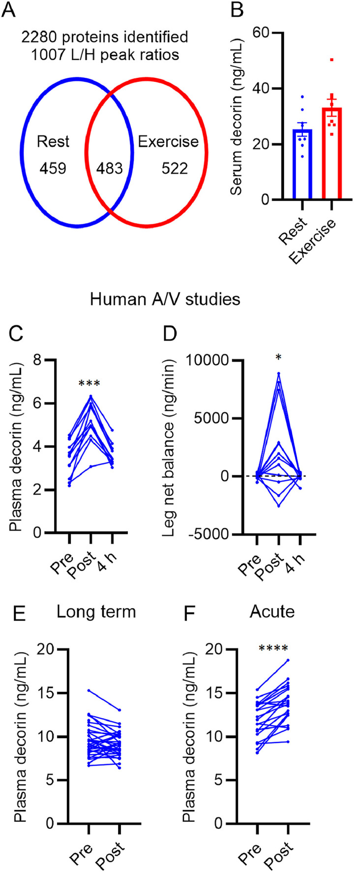
Results: We show that voluntary wheel running slows tumor development in the mammary specific polyomavirus middle T antigen overexpression (MMTV-PyMT) mouse model of breast cancer but only when mice are not housed alone. We identify the proteoglycan decorin as a contraction-induced secretory factor that systemically increases in patients with breast cancer immediately following exercise. Moreover, high expression of decorin in tumors is associated with improved prognosis in patients, while treatment of breast cancer cells in vitro with decorin reduces cell proliferation. Notwithstanding, when we overexpressed decorin in murine muscle or injected recombinant decorin systemically into mouse models of breast cancer, elevated plasma decorin concentrations did not result in higher tumor decorin levels and tumor burden was not improved.

Conclusion: Exercise training is anti-tumorigenic in a mouse model of luminal breast cancer, but the effect is abrogated by social isolation. The proteoglycan decorin is an exercise-induced secretory protein, and tumor decorin levels are positively associated with improved prognosis in patients. The hypothesis that elevated plasma decorin is a mechanism by which exercise training improves breast cancer progression in humans is not, however, supported by our pre-clinical data since elevated circulating decorin did not increase tumor decorin levels in these models.

Keywords: Breast cancer; Exercise training; Muscle secretory factors; Proteoglycans.


Conflict of interest statement

Competing interests MAF is a shareholder and scientific advisor for N-Gene Pharmaceuticals; MAF is the founder and shareholder of Celesta Therapeutics. All the support had no involvement in the study design and writing of the manuscript or the decision to submit it for publication. The authors declare that they have no other competing interests.

Figures

Image, graphical abstract
Graphical abstract
Fig 1
Fig. 1
Voluntary wheel running suppresses tumor growth in dually housed MMTV-PyMT mice. Mice were housed in pairs with 2 running wheels per cage from 6 weeks of age. Wheels were either locked or functional. (A) Body mass, (B and C) gonadal fat mass and spleen mass at termination, (D) lean mass, (E) fat mass, and (F) quadriceps muscle mass at termination, in sedentary (Sed) and running (Run) mice. Tumor burden was estimated by measuring tumor surface area of tumors in (G) all mammary glands and (H) the largest tumor at 11 weeks of age. (I) Tumor burden was measured as total tumor mass relative to body weight (%), and (J) number of lung metastases was quantified as number of metastatic lesions per microtomy section at termination. Statistical testing was done with two-way analyses of variance or mixed effects model for repeated measures or Student's t test. * p < 0.05, ** p < 0.01, between groups; #p < 0.05 group × time effect. Data expressed as means ± SEM (n = 4–11 mice per group). MMTV-PyMT = mammary specific polyomavirus middle T antigen overexpression mouse model; SEM = standard error of the mean; wks = weeks.
Fig 2
Fig. 2
Decorin is an exercise-induced muscle secretory factor. (A) Male C57Bl/6J mice were acutely exercised on a motorized treadmill for 90 min (n = 3) or kept resting in the home cage (n = 3). Quantitative proteomic profiling of m. gastrocnemius using a 13C6 SILAC labeled reference sample. Number of detected proteins and quantified L/H ratios. (B) Female (n = 8 per group), tumor-free MMTV-PyMT-WT mice were exercised on a treadmill for 90 min; Serum decorin immediately after exercise and in resting littermates. (C and D) 12 healthy male participants were catheterized in the femoral artery and vein and subjected to bicycling at increasing intensities for approximately 1 h. Blood samples were taken before (pre), immediately after (post), and after 4 h recovery; (C) Plasma concentration and (D) leg net balance, calculated as arteriovenous difference multiplied by leg blood flow. (E) Plasma decorin in patients (n = 37) from the PACT trail. (F) Patients had undergone treatment for operable Stages I–III breast cancer, and plasma decorin was measured before and after 6 months of counseling and exercise training. Plasma decorin in breast cancer patients (n = 20) before and after 2 h of acute exercise (high-intensity aerobic training and resistance training). Data are expressed as means ± SEM and include individual data points (B) or are expressed as individual data points (C–E). * p < 0.05, *** p < 0.001, **** p < 0.0001. A/V = arteriovenous difference; L/H = light/heavy label ratio; MMTV-PyMT-WT = the mammary specific polyomavirus middle T antigen overexpression mouse model wild type (tumor free); SEM = standard error of the mean.
Fig 3
Fig. 3
Expression of decorin in human tumors. Decorin is a prognostic marker in human mammary tumors. (A–D) Decorin mRNA expression in mammary tumors Grades 1–3, from 4 different breast cancer cohorts. Data were extracted using the ShinyGEO application. (E–H) Survival analyses comparing breast cancer patients with high or low tumor expression of decorin. Data were from Kaplan–Meier plotter containing data from several publicly available transcriptomic datasets. The numbers in parenthesis indicate the number of samples in each group. HR, logrankanalysis from Kaplan-Meier plotter. Black and red numbers on the bottom rows indicate the number at risk in the 2 groups (red: high decorin expression and black: low decorin expression). * p < 0.05, ** p < 0.01, and *** p < 0.001 for difference between the indicated group and the reference group (Grade 1). ###p < 0.001 for overall difference (Kruskal–Wallis test). Dcn = Decorin; GEO = gene expression omnibus; HR = hazard ratio; KMplot = Kaplan-Meier plot; MMTV-PyMT = the mammary specific polyomavirus middle T antigen overexpression mouse model; MMTV-PyMT/FVB = the mammary specific polyomavirus middle T antigen overexpression mouse model on a Friend Virus B NIH Jackson genetic background.
Fig 4
Fig. 4
Expression of decorin in murine tumors. (A) mRNA expression of decorin in MMTV-PyMT tumor and mammary fat pad. (B) Immunostaining of collagen Type I α 1 (Col1a1) and decorin in tumors from MMTV-PyMT/FVB mice. Scale bar represents 100 µm. (C–E) Decorin was quantified with MS/MS in decellularized tissues from BALB/c mice implanted with 4T1 cells (n = 3). (C) Primary mammary tumor and healthy mammary fat pad. (D) Healthy and metastatic lung. (E) Healthy and metastatic lymph node. Represented as LFQ intensities relative to healthy tissue. Data are expressed as means ± SEM. * p < 0.05, *** p < 0.001, Student's t test. Dcn = Decorin; LFQ = label-free quantitation; LN = lymph node; MMTV-PyMT = mammary specific polyomavirus middle T antigen overexpression mouse model.
Fig 5
Fig. 5
Single-cell RNA sequencing of PyMT tumors. (A) Cell composition of MMTV-PyMT tumors. tSNE plot showing cell clusters defined by a k-means-based clustering algorithm. (B) Violin plot of the decorin mRNA expression in cell clusters defined in panel A. (C–H) mRNA expression of Dcn, Bgn, Lum, Lox, Col8a1, and Col12a1 plotted in the tSNE defined in panel A. tSNE = t-distributed stochastic neighbor embedding. MMTV-PyMT = mammary specific polyomavirus middle T antigen overexpression mouse model. Bgn = biglycan; Col = collagen; Dcn = decorin; ECM = extracellular matrix; Lox = lysyl oxidase; LP = luminal progenitor; Lum = lumican; ML = mature luminal; NK = natural killer; UMAP  = uniform manifold approximation and projection.
Fig 6
Fig. 6
Overexpression of decorin in skeletal muscle of MMTV-PyMT mice. Skeletal muscles were injected with recombinant AAVs carrying either FLAG-tagged decorin (AAV:Dcn) or an empty vector control (AVV:Con) at 6 weeks of age. (A) Immunoflourescence staining of decorin and FLAG in skeletal muscles of AAV:Dcn and AAV:Con mice. Muscles from decorin-deficient mice (Dcn-/-) were used as negative controls for decorin staining. Scale bar represents 25 µm. (B) Immunoprecipitation of FLAG from lysates of gastrocnemius muscle, tumor tissue, and mammary fat pad in AAV:Con and AAV:Dcn mice; C- was a protei-free negative control. Immunoprecipitates were subjected to SDS-PAGE and immunoblotted for decorin or FLAG. (C) Decorin concentration in serum from Dcn-/-, mice overexpressing decorin in skeletal muscle (AAV:Dcn), or control mice (AAV:Con). (D) Immunofluorescence staining of decorin and FLAG in tumors from AAV:Dcn and AAV:Con mice. Scale bar represents 75 µm. (E–F) Decorin mRNA expression in tumors and mammary fat pad. (G–H) At 12 weeks of age, tumor burden was measured as total tumor mass relative to body weight (%), and number of lung metastases was quantified as number of metastatic lesions per microtomy section. (I–J) Survival; animals were euthanized when tumor burden was 10% ± 3% or when the largest tumor was >1 cm2. Days of age at termination (I) and tumor burden (J). Data expressed as means ± SEM (C, E, F, G, H, J). (n = 5–13 mice per group). *** p < 0.001. AAV = adeno-associated virus; Con = control; Dcn = decorin; MMTV-PyMT = mammary specific polyomavirus middle T antigen overexpression mouse model; SDS-PAGE = sodium dodecyl-sulfate polyacrylamide gel electrophoresis; SEM = standard error of the mean.
Fig 7
Fig. 7
Systemic delivery of recombinant decorin into SCID mice injected with MDA-MB-231-HM cells. SCID mice injected with the MDA-MB-231-HM cells into mammary pads received 2 different doses of recombinant decorin systemically. (A) Tumor volume, (B) metastatic burden, (C) plasma decorin levels, and (D) tumor decorin levels after the low-dose experiment. Mice were dosed every second day with 100 µg of recombinant decorin or an equal volume of saline (Control) ∼14 days post tumor-cell inoculation. (E) Tumor volume, (F) metastatic burden, (G) plasma decorin levels, and (H) tumor decorin levels after the high-dose experiment. Mice were dosed daily with 10 mg/kg body weight (∼2.5 times the dose of low-dose experiment) of recombinant decorin or an equal volume of saline (Control) ∼7 days post tumor-cell inoculation. Tumor volume was assessed by calliper measurement of primary tumor growth and calculated as (length × width2)/2. Metastatic burden was assessed by bioluminescence imaging detection. Plasma decorin levels were measured by ELISA. Tumor decorin levels were measured by western blot using total protein normalization. Two AAV:Dcn gastrocnemius lysates from the overexpression experiments were used as positive controls. Statistical testing was done with two-way analyses of variance or mixed effects model for repeated measures or Student's t test. Data expressed as means ± SEM. (n = 5–7 mice per group). A.U. = arbitrary unit; con = control; Dcn  = decorin; ELISA = enzyme linked immunosorbent assay.

References

    1. Global Burden of Disease Cancer, Fitzmaurice C, Abate D, et al. Global, regional, and national cancer incidence, mortality, years of life lost, years lived with disability, and disability-adjusted life-years for 29 cancer groups, 1990 to 2017: A systematic analysis for the global burden of disease study. JAMA Oncol. 2019;5:1749–1768. - PMC - PubMed
    1. Jassem J, Buchanan M, Jänicke F, et al. The Hamburg statement: The partnership driving the European agenda on breast cancer. Eur J Cancer. 2004;40:1810–1811. - PubMed
    1. Kyu HH, Bachman VF, Alexander LT, et al. Physical activity and risk of breast cancer, colon cancer, diabetes, ischemic heart disease, and ischemic stroke events: Systematic review and dose–response meta-analysis for the global burden of disease study 2013. BMJ. 2016;354:i3857. doi: 10.1136/bmj.i3857. - DOI - PMC - PubMed
    1. Pizot C, Boniol M, Mullie P, et al. Physical activity, hormone replacement therapy and breast cancer risk: A meta-analysis of prospective studies. Eur J Cancer. 2016;52:138–154. - PubMed
    1. Cormie P, Zopf EM, Zhang X, Schmitz KH. The impact of exercise on cancer mortality, recurrence, and treatment-related adverse effects. Epidemiol Rev. 2017;39:71–92. - PubMed

LinkOut - more resources