. 2024 Sep 28;15(1):8420.
doi: 10.1038/s41467-024-52587-w.

QSOX2 Deficiency-induced short stature, gastrointestinal dysmotility and immune dysfunction

Affiliations

QSOX2 Deficiency-induced short stature, gastrointestinal dysmotility and immune dysfunction

Avinaash V Maharaj et al. Nat Commun. .

Abstract

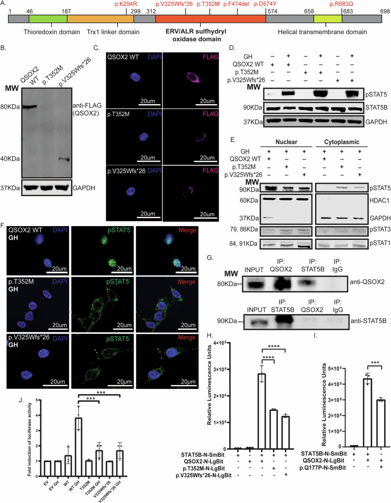
Postnatal growth failure is often attributed to dysregulated somatotropin action, however marked genetic and phenotypic heterogeneity exist. We report five patients from three families who present with short stature, immune dysfunction, atopic eczema and gastrointestinal pathology associated with recessive variants in QSOX2. QSOX2 encodes a nuclear membrane protein linked to disulphide isomerase and oxidoreductase activity. Loss of QSOX2 disrupts Growth hormone-mediated STAT5B nuclear translocation despite enhanced Growth hormone-induced STAT5B phosphorylation. Moreover, patient-derived dermal fibroblasts demonstrate Growth hormone-induced mitochondriopathy and reduced mitochondrial membrane potential. Located at the nuclear membrane, QSOX2 acts as a gatekeeper for regulating stabilisation and import of phosphorylated-STAT5B. Altogether, QSOX2 deficiency modulates human growth by impairing Growth hormone-STAT5B downstream activities and mitochondrial dynamics, which contribute to multi-system dysfunction. Furthermore, our work suggests that therapeutic recombinant insulin-like growth factor-1 may circumvent the Growth hormone-STAT5B dysregulation induced by pathological QSOX2 variants and potentially alleviate organ specific disease.


Conflict of interest statement

The authors declare no competing interests.

Figures

Fig. 1
Fig. 1. Pedigree charts of all families and anthropometric analyses of Kindred 1.
A Inheritance of QSOX2 variants delineated across two generations for each respective kindred. B Height, weight, and BMI centile growth charts (2–9 yrs) of probands 1 and 2, generated by Growth XP (PC PAL version 2.8). GH indicates when recombinant growth hormone therapy (0.025 mg/kg/day) was commenced. Most recent measurements suggest a modest improvement in height trajectories. C Colonic marker transit studies for probands 1 and 2 were performed after bowel dis-impaction. Patients ingested 10 differently shaped markers for three consecutive days. Plain abdominal X-rays were performed on days 4 and 6 post-first marker ingestion. Colonic marker transit study of proband 1 was indicative of rectal outlet dysfunction. D Abdominal X-rays for colonic marker transit study in proband 2 indicate a mixed type of rectal outlet dysfunction and slow colonic transit (retention of innumerable markers).
Fig. 2
Fig. 2. Loss of function variants in QSOX2 negatively impact STAT5B functions.
A Schematic of QSOX2 protein with key domains, including the relative location of QSOX2 variants identified in probands P1-P4. B Immunoblotting of FLAG-tagged QSOX2 cDNA constructs showed that expression of both variants was reduced compared to wild-type (WT)-QSOX2, with expected truncated protein due to early protein termination observed for p.V325Wfs*26. C Immunofluorescent microscopy demonstrated a reduction in QSOX2 peri-nuclear expression for both variants when compared to WT-QSOX2. D Immunoblot analyses of transfected HEK 293-hGHR cell lysates demonstrated that p-STAT5 was markedly enhanced in the presence of both variants following GH stimulation. E Nuclear and cytoplasmic fractions of transfected HEK 293-hGHR cells demonstrated a reduction of p-STAT5 in nuclear fractions of both variants with concomitant cytoplasmic abundance of p-STAT5 when compared to wild type. Nuclear levels of p-STAT3 and p-STAT1 were indistinguishable between both variants and the wild type. F Immunofluorescent microscopic analysis of GH-stimulated transfected HEK 293-hGHR cells showed nuclear translocation impairment of p-STAT5 for both QSOX2 variants but not with WT-QSOX2. G Co-immunoprecipitation and immunoblot analysis of WT-QSOX2-STAT5B interactions showed a direct protein-protein interaction between unstimulated WT-QSOX2 and STAT5B. H NanoBit complementation assays showed that the robust interaction seen between QSOX2-WT and STAT5B-WT was attenuated for both p.T352M (p < 0.0001) and p.V325Wfs*26 (p < 0.0001). Ordinary one-way-ANOVA was used for statistical analysis with multiple testing corrections performed using Sidak’s test. I NanoBit complementation assays showed a significant reduction in interaction affinity for the pathogenic variant p.Q177P known to abrogate nuclear STAT5B import (p = 0.0004), supporting the importance of QSOX2 for STAT5B nuclear localisation. Ordinary one-way-ANOVA was used for statistical analysis with multiple testing corrections performed using Dunnett’s test. J In vitro STAT5B transcriptional activities were evaluated by dual luciferase growth hormone response element (GHRE) reporter assay. The 4-fold increase in GH-induced luciferase activities in the presence of WT-QSOX2 (WT), was significantly blunted in the presence of QSOX2 variants (“T352M”, p = 0.0001; “V325Wfs*26”, p = 0.0001). Ordinary one-way-ANOVA was used for statistical analysis with multiple testing corrections performed using Sidak’s test. Source data are provided as a Source Data file. Data are presented as the mean ± SD of three repeated measurements (3 independent replicates).
Fig. 3
Fig. 3. QSOX2 p.F474del variant function analogous to p.T352M and p.V325Wfs*26.
A Expression of p.F474del was reduced compared to wild-type (WT)-QSOX2. Molecular weights (MW), in kiloDaltons, indicated left of the immunoblot. B Immunofluorescent microscopy demonstrated a reduction in QSOX2 peri-nuclear expression when compared to WT-QSOX2. C Immunoblot analyses of transfected HEK 293-hGHR cell lysates showed that in the presence of p.F474del, STAT5 was robustly phosphorylated following GH stimulation. D Immunofluorescent microscopic analysis of GH-stimulated transfected HEK 293-hGHR cells revealed nuclear translocation impairment of p-STAT5 and perinuclear accumulation for p.F474del when compared to WT-QSOX2. E Nuclear and cytoplasmic fractionation of transfected HEK 293-hGHR cells were probed by immunoblotting for p-STAT5, nuclear marker HDAC1, and cytoplasmic GAPDH. A reduction of p-STAT5 in p.F474del nuclear fractions was noted when compared to wild type. F NanoBit complementation assays demonstrated blunted interaction between unstimulated STAT5B and QSOX2 p.F474del (p = 0.0019). Ordinary one-way-ANOVA was used for statistical analysis with multiple testing corrections performed using Sidak’s test. Source data are provided as a Source Data file. Data are presented as the mean ± SD of three repeated measurements (3 independent replicates).
Fig. 4
Fig. 4. QSOX2 deficient patient (P2) dermal fibroblasts demonstrate STAT5B nuclear localisation defects and distinct mitochondrial dysfunction.
A Immunoblot analysis of control (C), proband 2 (P2) and parental dermal fibroblasts (M, F) revealed a global reduction in QSOX2 protein in patient-derived fibroblasts. B Robust tyrosine phosphorylation of STAT5 was elicited in patient fibroblasts when compared to control and heterozygote parents. C Immunofluorescent microscopy indicated GH-stimulated p-STAT5 translocated to the nucleus in (C), M and F fibroblasts but not in P2 fibroblasts. P2 fibroblasts demonstrated diffused cytoplasmic staining for p-STAT5 with nuclear sparing. D Immunoblot analysis of IGF-1 stimulated (100 ng/ml, 30 min) signalling pathways. IGF-1 activated pAKT, and pERK1/2 was comparable between P2, C and parental fibroblasts. E MitoTracker immunostaining of patient P2 fibroblasts, compared to control fibroblasts, indicate disrupted mitochondrial morphology upon GH, but not IGF-1, stimulation. Alterations in patient mitochondrial morphology seen with GH stimulation were consistent with mitochondrial fragmentation. F Immunofluorescent microscopy of P2 fibroblasts demonstrated an increase in GH-induced phospho-S616-DRP1 when compared to control (C). G Cytoplasmic accumulated p-STAT5B appeared to localise to the mitochondria in P2 fibroblasts co-immunostained for outer mitochondrial membrane marker, Tom20 and p-STAT5B. H Unstimulated and GH stimulated control (C), patient (P2), and parental (M, F) fibroblasts were immunoblot analysed for expression of mitochondrial oxidative phosphorylation complexes I–V. In P2 fibroblasts, a stark reduction in complex profiles was observed upon GH stimulation. IGF-1 stimulation, in contrast, did not alter complex profiles. I Mitochondrial membrane potential measurements of untreated and GH-treated primary fibroblasts with carbonyl cyanide p-triflouromethoxyphenylhydrazone, FCCP (20 μM) added to control fibroblasts as a depolarisation control. Reproducible reduction in mitochondrial membrane potential was detected in GH-treated patient fibroblasts compared to control fibroblasts (p = 0.0013). FCCP depolarisation control effectively showed reduced mitochondrial membrane potential when compared to GH-treated control fibroblasts (p = 0.0105). Ordinary one-way-ANOVA was used for statistical analysis with multiple testing corrections performed using Sidak’s test. Source data are provided as a Source Data file. Data are presented as the mean ± SD of three repeated measurements (3 independent replicates).

References

    1. Murray, P. G., Clayton, P. E. & Chernausek, S. D. A genetic approach to evaluation of short stature of undetermined cause. Lancet Diabetes Endocrinol.6, 564–574 (2018). - PubMed
    1. Sovio, U. et al. Genetic determinants of height growth assessed longitudinally from infancy to adulthood in the Northern Finland Birth Cohort 1966. PLoS Genet.5, e1000409 (2009). - PMC - PubMed
    1. Storr, H. L. et al. Nonclassical GH insensitivity: Characterization of mild abnormalities of GH action. Endocr. Rev.40, 476–505 (2019). - PMC - PubMed
    1. Klammt, J. et al. Dominant-negative STAT5B mutations cause growth hormone insensitivity with short stature and mild immune dysregulation. Nat. Commun.9, 2105 (2018). - PMC - PubMed
    1. Vidarsdottir, S. et al. Clinical and biochemical characteristics of a male patient with a novel homozygous STAT5b mutation. J. Clin. Endocrinol. Metab.91, 3482–3485 (2006). - PubMed

Publication types

MeSH terms

LinkOut - more resources