. 2024 Sep 28;9(1):258.
doi: 10.1038/s41392-024-01961-7.

Epigenetic reader ZMYND11 noncanonical function restricts HNRNPA1-mediated stress granule formation and oncogenic activity

Affiliations

Epigenetic reader ZMYND11 noncanonical function restricts HNRNPA1-mediated stress granule formation and oncogenic activity

Cheng Lian et al. Signal Transduct Target Ther. .

Abstract

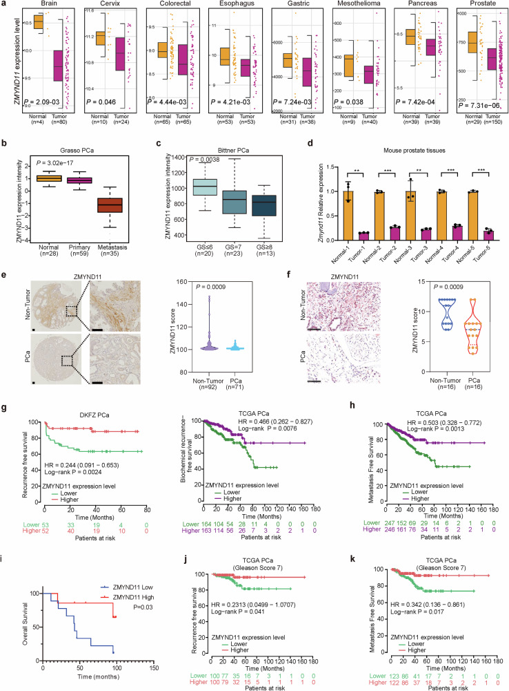
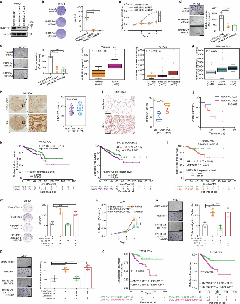
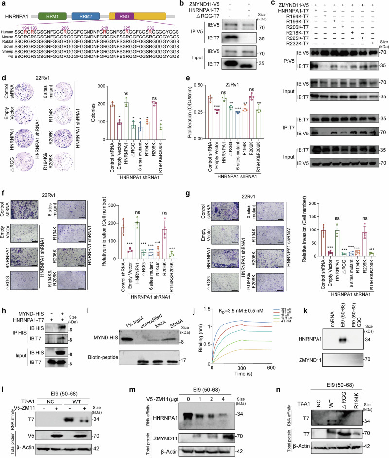
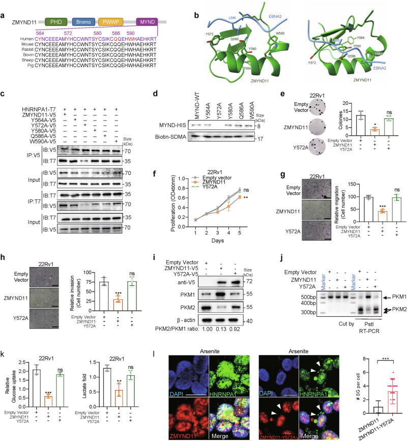
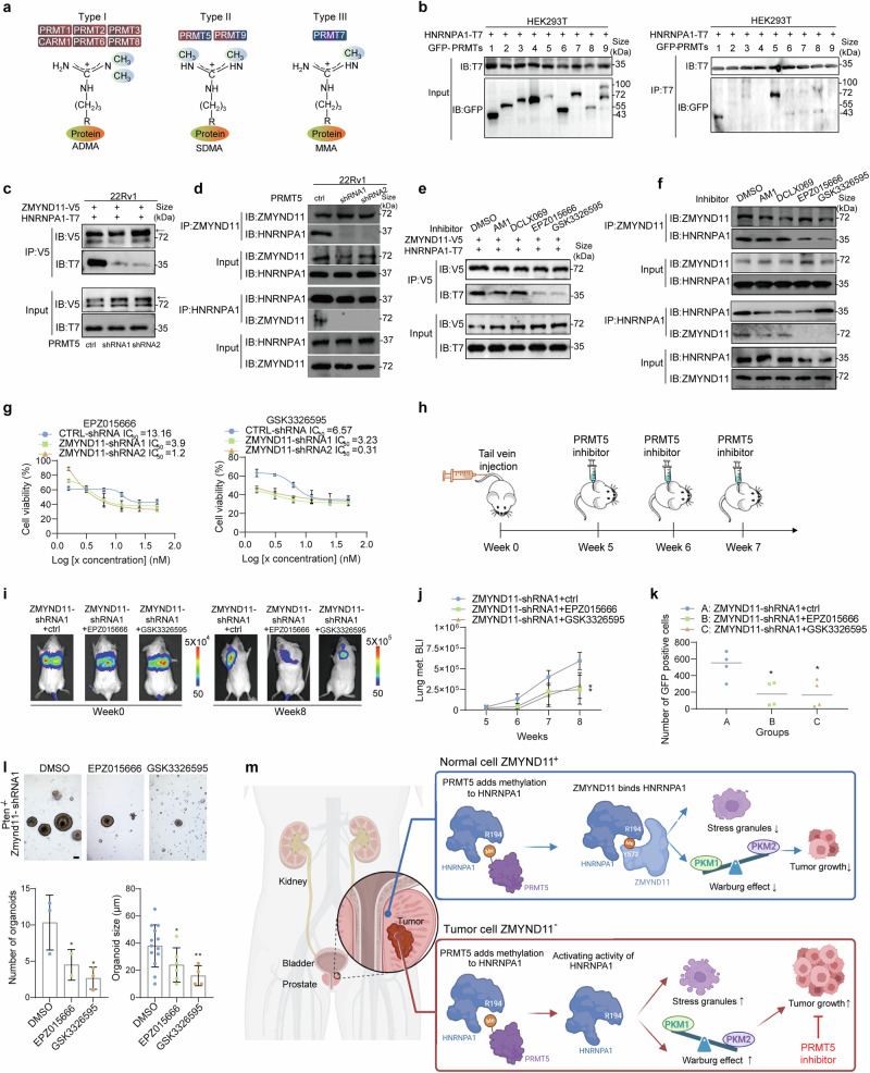
Epigenetic readers frequently affect gene regulation, correlate with disease prognosis, and hold significant potential as therapeutic targets for cancer. Zinc finger MYND-type containing 11 (ZMYND11) is notably recognized for reading the epigenetic marker H3.3K36me3; however, its broader functions and mechanisms of action in cancer remain underexplored. Here, we report that ZMYND11 downregulation is prevalent across various cancers and profoundly correlates with poorer outcomes in prostate cancer patients. Depletion of ZMYND11 promotes tumor cell growth, migration, and invasion in vitro, as well as tumor formation and metastasis in vivo. Mechanistically, we discover that ZMYND11 exhibits tumor suppressive roles by recognizing arginine-194-methylated HNRNPA1 dependent on its MYND domain, thereby retaining HNRNPA1 in the nucleus and preventing the formation of stress granules in the cytoplasm. Furthermore, ZMYND11 counteracts the HNRNPA1-driven increase in the PKM2/PKM1 ratio, thus mitigating the aggressive tumor phenotype promoted by PKM2. Remarkably, ZMYND11 recognition of HNRNPA1 can be disrupted by pharmaceutical inhibition of the arginine methyltransferase PRMT5. Tumors with low ZMYND11 expression show sensitivity to PRMT5 inhibitors. Taken together, our findings uncover a previously unexplored noncanonical role of ZMYND11 as a nonhistone methylation reader and underscore the critical importance of arginine methylation in the ZMYND11-HNRNPA1 interaction for restraining tumor progression, thereby proposing novel therapeutic targets and potential biomarkers for cancer treatment.


Conflict of interest statement

The authors declare no competing interests.

Figures

Fig. 1
Fig. 1
ZMYND11 is profoundly downregulated in cancers and its downregulation correlates with adverse events and poor prostate cancer patient outcomes. a Downregulation of ZMYND11 gene expression in several types of cancers compared to healthy tissues (yellow). P values were assessed by the Wilcoxon test while effect sizes were estimated using Cohen’s distance: dbrain = 1.96, dcervix = 0.86, dcolon = 0.56, desophagus = 0.64, dgastric = 0.7, dmesothelioma = 0.64, dpancreas = 0.77, dprostate = 0.99. b ZMYND11 mRNA expression was significantly downregulated in human metastatic prostate tumors. c Association of ZMYND11 downregulation with high Gleason grade cancer. P values examined by the Kruskal-Wallis test in (b, c). d Expression of Zmynd11 in five paired prostate cancer specimens and adjacent normal tissues of murine prostate. e Protein expression levels of ZMYND11 in paraffinized prostate tumor tissue microarrays (TMA) of Tongji prostate cancer cohort determined by immunostaining. Original magnification, 100×; insets, 400×; Scale bar, 100 μm. f ZMYND11 protein expression were determined in Fudan cohort of prostate cancer using immunohistochemistry. The IHC scores calculated with staining areas (see Methods). Original magnification, 400×; Scale bar, 100 μm. g Kaplan-Meier analysis of biochemical recurrence-free survival in prostate tumors with high or low levels of ZMYND11 in two independent cohorts of prostate cancer. h Metastasis free survival analysis of 493 prostate cancer patient with tumors expressing high or low mRNA levels of ZMYND11. i Kaplan-Meier overall-survival analysis in a TMA cohort of patients with prostate cancer tumors having higher protein expression levels of ZMYND11 (top 50%; n = 35) or lower (bottom 50%; n = 35). Lower expression levels of ZMYND11 indicates predictive values for recurrence (j) and metastasis-free (k) survival in patient group with Gleason score 7 (intermediate-risk prostate cancer). In (df), statistical significance assessed using the two-tailed Student’s t test. **p < 0.01, ***p < 0.001. In (gk), p values examined by the log-rank test
Fig. 2
Fig. 2
Dampened ZMYND11 expression promotes prostate cancer cell proliferation and metastasis in vitro and in vivo. a Efficiency of shRNA-mediated ZMYND11 knockdown was determined by immunoblotting. b Colony-forming units of 22Rv1 cells stably expressing control or shRNAs against ZMYND11. c Proliferation rates of 22Rv1 cells measured at indicated times (absorbance at 450 nm; mean ± SD of three independent experiments). Representative images and quantification of relative migration (d) and invasion (e) for the cells stably expressing the indicated shRNAs. f Representative images of the tumor xenografts harvested 6 weeks after subcutaneous injection of 1 × 107 22Rv1 cells with the indicated control or shRNA-mediated stable silencing of ZMYND11 into the nude mouse (left). Growth curves of 22Rv1 xenografts at the indicated time points. g NOD-SCID mice treated with tail vein injection of 22Rv1 cells having stable knockdown of ZMYND11 and stable transfection of the firefly luciferase. Representative images at the experimental endpoint and weekly quantitation of bioluminescent imaging (BLI) analyses of mouse lung metastasis. h Fluorescence activated cell sorting (FACS) analysis of the circulating GFP-positive 22Rv1 cell variants (CTCs) in the blood of SCID mice. The scatter plot shows the number of CTCs recovered from each mouse injected with control or shRNA-mediated ZMYND11 knockdown cells. i Ectopic expression efficiency of ZMYND11 was measured by Western blot analysis. j Colony-forming units of 22Rv1 cells ectopically expressing empty vector control or ZMYND11. k Proliferation rates of 22Rv1 cells measured at indicated times (absorbance at 450 nm; mean ± SD of three independent experiments). Representative images and quantification of relative migration (l) and invasion (m) for cells with ectopic expression of either the empty vector control or ZMYND11. n Representative images of tumor xenografts harvested four weeks after subcutaneous injection of 22Rv1 cells with either the control vector or ectopically expressing ZMYND11 into nude mouse (left). Showing on the right are the growth curves of the xenografts at the specified time points. o NOD-SCID mice treated with a tail vein injection of 22Rv1 cells ectopically expressing control vector or ZMYND11, along with stable transfection of firefly luciferase. Representative images at the experimental endpoint and weekly quantitation of bioluminescent imaging (BLI) analyses of mouse lung metastasis. p The efficiency of shRNA-mediated mouse Zmynd11 knockdown was determined by immunoblotting. q Representative images of tumor xenografts harvested 6 weeks after orthotopic implantation of 5 × 105 RM1 cells with either control or shRNA-mediated stable silencing of ZMYND11 into the prostate of C57BL/6J mice (left). Tumor volume of xenografts at the 6-week endpoint is shown on the right. r Representative images of organoids derived from prostatic Pten−/− mice with or without shZMYND11 treatment. Quantitative results from three experiments are shown on the right. Scale bar: 40 μm. In (br), data shown are mean ± SD Error bars, *p < 0.05, **p < 0.01, ***p < 0.001, two-tailed Student’s t test
Fig. 3
Fig. 3
ZMYND11 physically interacts with HNRNPA1 in vitro and in vivo and inhibits HNRNPA1-mediated formation of stress granules. a Venn diagram showing the overlap between three replicates of the identified ZMYND11-interaction proteins in LNCaP cells using co-immunoprecipitation coupled to mass spectrometry (Co-IP/MS). b Table showing top-scoring ZMYND11-interaction proteins reproducibly identified by Co-IP/MS as indicated in (a). c Immunoprecipitation (IP)-Western blot for analyzing the interaction between ZMYND11 and HNRNPA1 in HEK293T cells cotransfected with the indicated V5- or T7-tagged plasmids. IB immunoblot. d Immunoprecipitation-Western blot analysis of an endogenous interaction of ZMYND11 with HNRNPA1 in 22Rv1 cells. IgG immunoglobulin, IB immunoblot. e Immunofluorescence staining and colocalization analysis of ZMYND11 and HNRNPA1 in 22Rv1 cells. Scale bar, 25 μm. f Schematic illustration of the full-length ZMYND11 and the indicated domain deletion mutants. g Immunoprecipitation-Western blot analysis of the interactions between wild type ZMYND11 or its mutants and HNRNPA1. Crude total cell lysates were extracted from HEK293T cells co-expressing T7-tagged HNRNPA1 and the indicated V5-tagged ZMYND11 variants. h 22Rv1 cells stably expressing control or shRNAs against ZMYND11, treated with sodium arsenite (SA, 0.5 mM) for 1 h followed by immunostaining analysis of stress granule (SG) formation. Scale bar, 25 μm. i The number of SGs per cell and ZMYND11 or HNRNPA1 colocalized SGs each cell were quantified. **p < 0.01, ***p < 0.001 evaluated by two-tailed Student’s t test. j Immunoblotting analysis of ZMYND11 or HNRNPA1 in the nuclear and cytoplasmic fractions of the tested prostate cancer cell lines 22Rv1 and LNCaP, respectively. k Apoptosis of 22Rv1 cells stably expressing control or shRNAs targeting ZMYND11. ***P < 0.001 assessed by two-tailed Student’s t test
Fig. 4
Fig. 4
ZMYND11 antagonizes oncogenic function of HNRNPA1 in prostate cancer dependent on its MYND domain. a Knockdown efficiency of shRNAs against HNRNPA1 determined by western blot analysis. b Colony-forming units of 22Rv1 cells stably expressing control or shRNAs against HNRNPA1. c Proliferation capacity of 22Rv1 cells measured at the indicated times. Absorbance at 450 nm; mean ± SD of three independent experiments. Representative images and quantification of relative migration (d) and invasion (e) for the cells stably expressing the indicated shRNAs. Error bars, ±SD of triplicate experiments. f HNRNPA1 expression levels are significantly upregulated in human metastatic prostate tumors. P values determined by the Wilcoxon and Kruskal-Wallis tests, respectively. g HNRNPA1 upregulation correlates with higher Gleason grade cancer. P values assessed by the Kruskal-Wallis test. h Immunohistochemistry on the expression of HNRNPA1 and evaluation of HNRNPA1 staining intensity in a paraffinized prostate tissue microarray consisting of 163 samples. Original magnification, 100×; insets, 400×; Scale bar, 100 μm. i Immunostaining for HNRNPA1 was performed on another cohort of prostate specimens. Representative images and quantification of HNRNPA1 protein staining are shown. Original magnification, 400×; Scale bar, 100 μm. j Kaplan-Meier analysis of overall survival in the Tongji cohort of patients with prostate cancer. Patients were stratified into two groups according to the state of HNRNPA1 expression, higher (top 50%; n = 35) or lower (bottom 50%; n = 35). k Kaplan-Meier analysis showed that HNRNPA1 upregulation is significantly associated with elevated risk of biochemical recurrence or metastasis in prostate cancer subsets within the TCGA database. l High expression levels of HNRNPA1 indicates predictive values for recurrence-free survival in patient group with Gleason sum score 7 prostate cancer (intermediate-risk). Representative images and quantification of colony formation (m), cell proliferation (n), migration (o), and invasion (p) for 22Rv1 cells transfected with empty vector, HNRNPA1 alone, or together with the indicated full-length ZMYND11 or MYND-domain deletion mutant. Scale bar, 400 μm. q Kaplan-Meier analysis of biochemical recurrence or metastasis-free survival in the TCGA cohort of prostate cancer patients stratified into two groups with ZMYND11low/HNRNPA1high and ZMYND11high/HNRNPA1low expression, respectively. Error bars, ±SD (be, mp). ns not significant, *p < 0.05, **p < 0.01, ***p < 0.001, two-tailed Student’s t tests
Fig. 5
Fig. 5
ZMYND11 regulates HNRNPA1-mediated alternative splicing of PKM and counteracts PKM2-induced aggressive cellular phenotype in prostate cancer. RNA (a, c) or protein (b, d) was extracted from 22Rv1 cells either transfected with full-length ZMYND11 or the indicated mutant (a, b) or stably expressing the ZMYND11-targeting shRNAs (c, d) followed by the examination of PKM isoforms both at the mRNA (a, c) and protein levels (b, d). 22Rv1 cells transfected with HNRNPA1 alone or together with either full-length ZMYND11 or the MYND-domain-deletion mutant were assayed for PKM splicing at the mRNA (e) and protein levels (f). Representative images and quantification of colony forming (g), cell proliferation (h), migration (i), and invasion (j) in 22Rv1 cells co-transfected with PKM2 alone or together with either the full-length ZMYND11 or the MYND-domain-deletion mutant. Mean ± SD of triplicate experiments. *p < 0.05, **p < 0.01, ***p < 0.001, Student’s t test. Scale bar, 400 μm. Relative glucose uptake and lactate production in 22Rv1 cells overexpressing ZMYND11 or its MYND-domain-deletion mutant (k) and stably expressing ZMYND11 shRNA (l) compared to control cells. Error bars, ±SD (kl), n = 3 biological replicates. *p < 0.05, **p < 0.01, ***p < 0.001, Student’s t tests. m Visualization of PKM splicing using LeafViz examined by RNA sequencing in 22Rv1 cells stably expressing either ZMYND11-targeting or control shRNAs. Statistic assessment via two-tailed Student’s t test. n Visualization of PKM isoform expression in 134 patients of prostate tumors and matched normal tissues in CPGEA cohort by RNA-seq profiling [PMID: 32238934]. Differential expression analysis of PKM1 (o) or PKM2 (p) in this patient cohort of 134 tumor-normal paired prostate specimens. The data were examined by the Wilcoxcon test compares two paired groups
Fig. 6
Fig. 6
HNRNPA1 arginine 194 methylation is required for its oncogenic function and recognition by ZMYND11. a Schematic diagram showing the protein domain organization of HNRNPA1. Shown are amino acid sequence conservation of the glycine-arginine-rich (RGG) motif with highlighted arginine (R) residues at the indicated position. RRM: RNA recognition motif. b Whole-cell lysates from HEK293T cells expressing V5-tagged ZMYND11 and T7-tagged HNRNPA1 or its mutant lacking the RGG-box were subjected to co-immunoprecipitation (co-IP) with anti-V5 antibody followed by immunoblotting. c The lysates from HEK293T cells expressing V5-tagged ZMYND11 and T7-tagged HNRNPA1 or the indicated mutant constructs, were immunoprecipitated with anti-V5 antibody followed by immunoblotting, and the reciprocal co-IP shown in the bottom. Representative images and quantification of colony-formation (d), cell proliferation (e), migration (f), and invasion (g) of 22Rv1 cells with stable knockdown of HNRNPA1 and rescue by co-transfection with the indicated constructs. Scale bar, 400 μm. Statistical significance was assessed using two-tailed Student’s t test. ns: not significant, *: P < 0.05, **: P < 0.01, ***: P < 0.001. h His-tag pull-down assay investigating direct interactions between the MYND domain of ZMYND11 and HNRNPA1. i Pull-down assays between the MYND domain of ZMYND11 and the biotin-labeled peptides carrying R194 monomethylation (MMA) or symmetric demethylation (SDMA). j Bio-layer interferometry (BLI) binding assay of ZMYND11-MYND and HNRNPA1-SDMA-R194. k The 22Rv1 nuclear extract (NE) were affinity-purified using the indicated biotin-labeled RNAs, and the eluted proteins were detected using anti-HNRNPA1 and ZMYND11 antibodies. l 22Rv1 cells were co-transfected with ZMYND11-V5 and HNRNPA1-T7 plasmid and RNA affinity purification was performed as in a using biotin-labeled RNA E19 (50–68). m 22Rv1 cells were transfected with ZMYND11-V5 plasmid at the indicated doses and RNA affinity purification was performed as in (k) using biotin-labeled RNA E19. n 22Rv1 cells were transfected with the indicated HNRNPA1-truncated constructs or HNRNPA1 R194K mutant and RNA affinity purification was performed as in (k) using biotin-labeled RNA E19 (50–68)
Fig. 7
Fig. 7
Tyrosine residue 572 of ZMYND11 is functionally important and essential for specifically reading SDMA on HNRNPA1. a Schematic representation of the MYND domain of ZMYND11. Shown is MYND domain organization along with potential candidate amino acid residues likely to be involved in binding with HNRNPA1. MYND domain conserved across species is shown. b Two views of the crystal structure (PDB:5HDA) for the MYND domain of ZMYND11 complexes with EBNA2 (blue). Potential candidate amino acid residues responsible for recognition with HNRNPA1 are show in sticks. c Co-immunoprecipitation assay in HEK293T cells expressing T7-tagged HNRNPA1 and V5-tagged ZMYND11 or the indicated mutants. d Pull-down assays demonstrated protein-protein interactions between HIS-tagged MYND domain of ZMYND11 or the mutants and the biotinylated peptide containing R194 SDMA. Representation and quantification of colony-forming (e), cell proliferation (f), migration (g), and invasion (h) of 22Rv1 cells overexpressing ZMYND11 or the Y572A mutant. Scale bar, 400 μm. *P < 0.05, **P < 0.01, ***P < 0.001, were examined by two-tailed Student’s t test. PKM splicing assay (i) and immunoblots for the indicated PKM isoforms (j) were performed in 22Rv1 cells ectopically expressing ZMYND11 or the Y572A mutant. k Relative glucose uptake and lactate production were measured in 22Rv1 cells with an ectopical expression of ZMYND11 or Y572A mutant. ns, not significant, *P < 0.05, **P < 0.01, ***P < 0.001, two-tailed Student’s t test. l Fluorescence colocalization microscopy analysis of ZMYND11 or the Y572A mutant with HNRNPA1 in 22Rv1 cells treated with sodium arsenite or control. Note that stress-induced cytoplasmic accumulation of HNRNPA1 was substantially attenuated by wild-type ZMYND11, but not the Y572A mutant. Arrows, stress granules in cells. Mean ± SD, was assessed using two-tailed Student’s t test. *** represents p < 0.005. Scale bars, 25 μm
Fig. 8
Fig. 8
Pharmacological inhibition of PRMT5 impairs ZMYND11-HNRNPA1 interaction and suppresses in vivo metastatic capacity of prostate cancer cells with ZMYND11-low expression. a Schematics of arginine methylation states by PRMT family of enzymes. b Co-immunoprecipitation assay of HEK293T cells expressing T7-tagged HNRNPA1 and the indicated GFP-tagged PRMT family member. c Co-immunoprecipitation assays using 22Rv1 cell protein extracts co-expressing V5-tagged ZMYND11 and T7-tagged HNRNPA1 while having stable expression of control or shRNAs against PRMT5. d Immunoprecipitation-Western blot analysis of an endogenous interaction between ZMYND11 and HNRNPA1 in 22Rv1 cells stably expressing control or ZMYND11-targeting shRNAs. e Immunoprecipitation using V5-tag antibodies from 22Rv1 cells ectopically co-expressing ZMYND11 and HNRNPA1 while treated with PRMT1 inhibitors (AM1 or DCLX069, 50 μM, 48 h) or PRMT5 inhibitors (EPZ015666 or GSK3326595, 10 μM, 48 h). f Co-immunoprecipitation assay of an endogenous interaction between ZMYND11 and HNRNPA1 in 22Rv1 cells treated with the indicated PRMT1/5 inhibitor. g Half-inhibitory concentration (IC50) test of two PRMT5 inhibitors (EPZ015666 or GSK3326595, 10 μM, 48 h) for ZMYND11 knockdown or control cells in 22Rv1. h Schematics of in vivo mouse experiments with PRMT5-Selective Inhibitors. 22Rv1 cells with stable knockdown of ZMYND11 while expressing luciferase were injected into the tail-vein of male NOD-SCID mice. At 5, 6, and 7 weeks after injection, mice were subject to intraperitoneal treatment with the indicated PRMT5 inhibitors. Shown are the representative images at indicated time points (i) and weekly quantification of BLI photon flux of lung metastasis in mice (j). Errors bar, ±SD, n = 5. *p < 0.05, **p < 0.01, ***p < 0.001, Student’s t tests. k FACS-analysis of GFP-positive 22Rv1 cells (CTCs) in the peripheral blood of SCID mice (n = 4). The scatter plot indicated the number of CTCs recovered from each mouse treated with vesicles or PRMT5 inhibitors. Statistical significance was assessed using two-tailed Student’s t test. * p < 0.05. l Organoid images derived from prostatic ZMYND11 knockdown Pten−/− mice treated with PRMT5 inhibitors (EPZ015666 or GSK3326595, 10 μM); quantitative results are representative of 3 experiments shown at the right. Scale bar: 40 μm. Errors bar, ±SD, n = 5. *p < 0.05, **p < 0.01, Student’s t tests. m A model illustrating how ZMYND11 recognition of arginine methylation constrains HNRNPA1-mediated tumor progression is proposed. Top: In normal cells with higher expression of ZMYND11, the protein can specifically recognize the R194 methylation of HNRNPA1, which is catalyzed by PRMT5. This interaction inhibits HNRNPA1’s involvement in stress granule formation and the alternative splicing of PKM, thereby mitigating tumor aggressiveness. Bottom: In tumor contexts with reduced ZMYND11 expression, HNRNPA1 promotes the formation of stress granules and shifts the PKM isoform balance towards a higher PKM2/PKM1 ratio, contributing to tumor progression. In this scenario, PRMT5 inhibitors emerge as potential therapeutic agents capable of curtailing cancer progression by disrupting the methylation-dependent interaction between ZMYND11 and HNRNPA1, ultimately impairing HNRNPA1’s tumor-promoting activities

References

    1. Hanahan, D. & Weinberg, R. A. Hallmarks of cancer: the next generation. Cell144, 646–674 (2011). - PubMed
    1. Ge, R. et al. Epigenetic modulations and lineage plasticity in advanced prostate cancer. Ann. Oncol.31, 470–479 (2020). - PubMed
    1. Coe, B. P. et al. Refining analyses of copy number variation identifies specific genes associated with developmental delay. Nat. Genet.46, 1063–1071 (2014). - PMC - PubMed
    1. Wen, H. et al. ZMYND11 links histone H3.3K36me3 to transcription elongation and tumour suppression. Nature508, 263–268 (2014). - PMC - PubMed
    1. Guo, R. et al. BS69/ZMYND11 reads and connects histone H3.3 lysine 36 trimethylation-decorated chromatin to regulated pre-mRNA processing. Mol. Cell56, 298–310 (2014). - PMC - PubMed

Publication types

MeSH terms