. 2024 Sep 28;14(1):22487.
doi: 10.1038/s41598-024-72989-6.

Dual activity of Minnelide chemosensitize basal/triple negative breast cancer stem cells and reprograms immunosuppressive tumor microenvironment

Affiliations

Dual activity of Minnelide chemosensitize basal/triple negative breast cancer stem cells and reprograms immunosuppressive tumor microenvironment

Fulya Koksalar Alkan et al. Sci Rep. .

Abstract

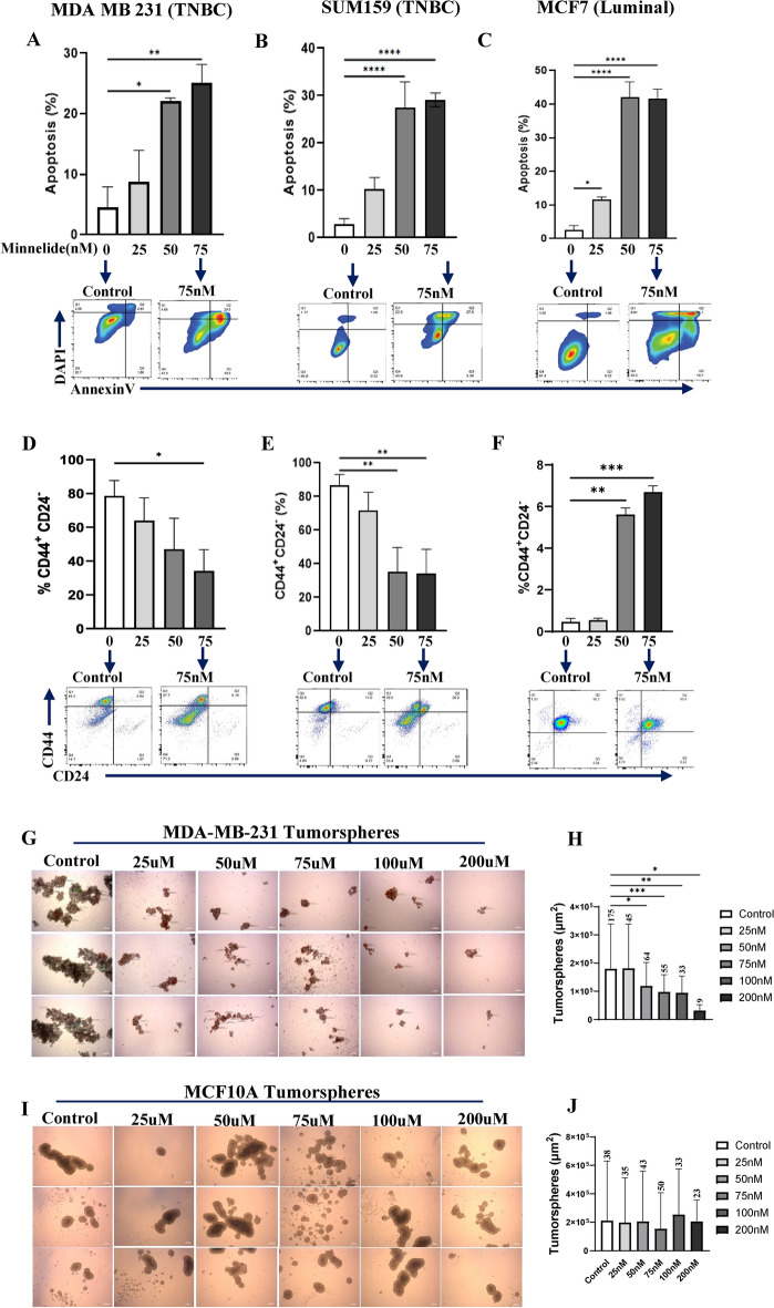
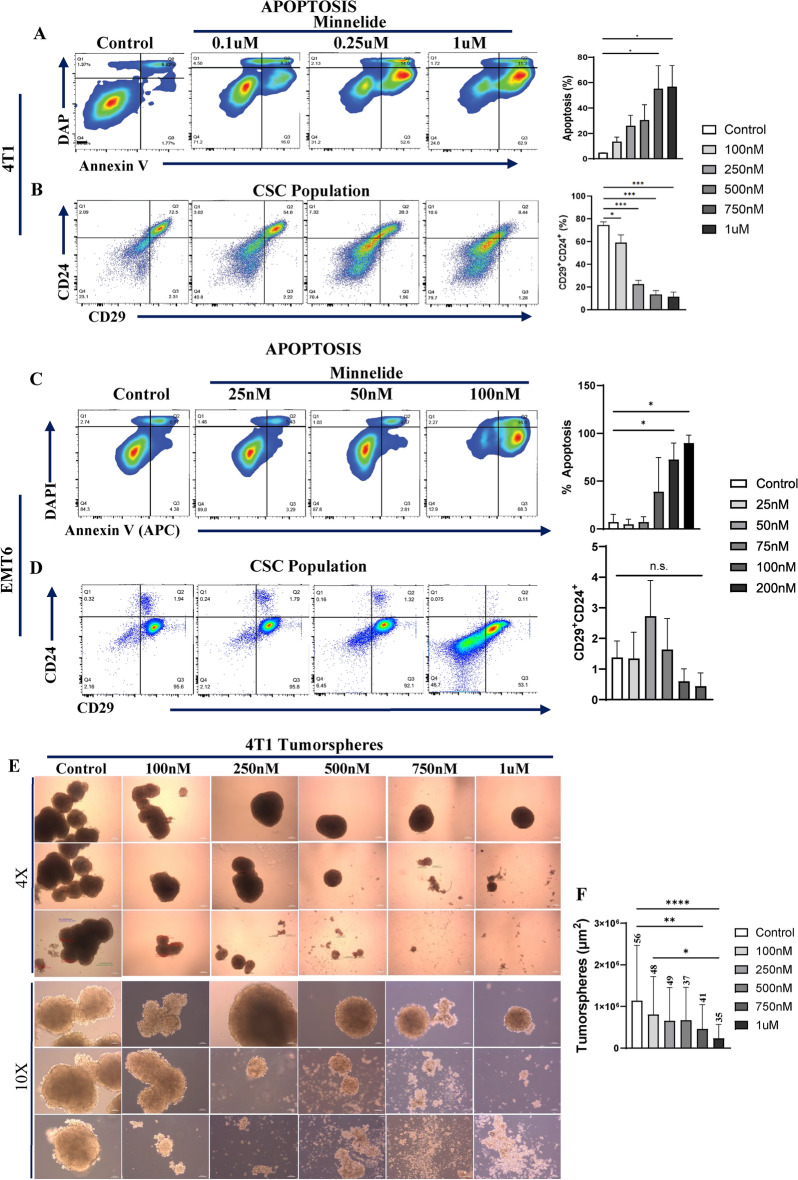
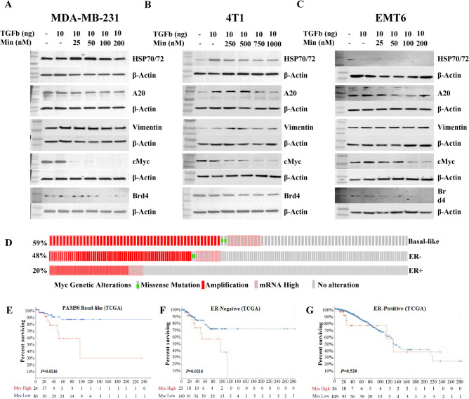
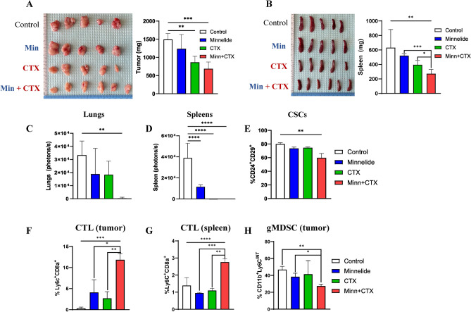
Triple negative breast cancer (TNBC) subtype is characterized with higher EMT/stemness properties and immune suppressive tumor microenvironment (TME). Women with advanced TNBC exhibit aggressive disease and have limited treatment options. Although immune suppressive TME is implicated in driving aggressive properties of basal/TNBC subtype and therapy resistance, effectively targeting it remains a challenge. Minnelide, a prodrug of triptolide currently being tested in clinical trials, has shown anti-tumorigenic activity in multiple malignancies via targeting super enhancers, Myc and anti-apoptotic pathways such as HSP70. Distinct super-enhancer landscape drives cancer stem cells (CSC) in TNBC subtype while inducing immune suppressive TME. We show that Minnelide selectively targets CSCs in human and murine TNBC cell lines compared to cell lines of luminal subtype by targeting Myc and HSP70. Minnelide in combination with cyclophosphamide significantly reduces the tumor growth and eliminates metastasis by reprogramming the tumor microenvironment and enhancing cytotoxic T cell infiltration in 4T1 tumor-bearing mice. Resection of residual tumors following the combination treatment leads to complete eradication of disseminated tumor cells as all mice are free of local and distant recurrences. All control mice showed recurrences within 3 weeks of post-resection while single Minnelide treatment delayed recurrence and one mouse was free of tumor. We provide evidence that Minnelide targets tumor intrinsic pathways and reprograms the immune suppressive microenvironment. Our studies also suggest that Minnelide in combination with cyclophosphamide may lead to durable responses in patients with basal/TNBC subtype warranting its clinical investigation.


Conflict of interest statement

The authors declare no competing interests.

Figures

Fig. 1
Fig. 1
Minnelide activity on tumor cell proliferation of human and murine breast cancer cell lines: Human breast cancer cell lines, MDA-MB-231, Sum159, ZR75-1, MCF7 AND murine breast cancer cell lines, EMT6, E0771, AT-3 and 4T1 were incubated with indicated doses of Minnelide for 48 h and cell viability was measured by MTT assay. (A-D) Minnelide reduces proliferation in a dose dependent manner in TNBC lines and luminal subtype. Experiments were done 2 independent times in triplicate and cell viability was shown in percentages (± SD). One-Way ANOVA is used to compare test groups to control. *P < 0.05, **P < 0.01, ***P < 0.001, ****P < 0.0001.
Fig. 2
Fig. 2
Minnelide significantly reduces CSC population in MDA-MB-231 via inducing apoptotic cell death. Figure 2. Minnelide significantly reduces CSC population in MDA-MB-231 via inducing apoptotic cell death. Breast cancer cell lines, MDA-MB-231 and MCF7 were incubated with indicated doses of Minnelide for 48 h and apoptotic cell death and CSC population was evaluated by flow cytometer. (A, B) Minnelide induces a dose dependent cytotoxic cell death particularly in CSC (D, E) population of MDA-MB-231 and Sum159 TNBC cell lines in vitro. (C, F) Minnelide had no activity on the CSC population of luminal MCF7 cell lines while it induced a significant apoptotic cell death in these cells with increasing doses of Minnelide. Experiments were done in triplicates and values were shown in percentages (± SD). (G,H) Treatment of MDA-MB-231 tumorspheres with different doses of Minnelide and quantification of their number and size. (I,J) MCF10A spheroids were treated with the same doses of Minnelide as control. Scale bar 250μm. One-Way ANOVA is used to compare test groups to control. *P < 0.05, **P < 0.01, ***P < 0.001, ****P < 0.0001.
Fig. 3
Fig. 3
Minnelide targets CSC population in murine 4T1 tumor cells. Murine 4T1 and EMT6 tumor cells were incubated with indicated doses of Minnelide for 48 h and apoptotic cell death and CSC population was evaluated by flow cytometer. (A, B) Minnelide induces a dose dependent apoptotic cell death in 4T1 cells in vitro in addition to the reduced capacity of cancer stemness. (C, D) Apoptotic cell death was increased by the increasing doses of minnelide while CSC population is not affected in vitro in EMT6 cells for 48 h. Experiments were done in triplicates and values were shown in percentages (± SD). (E, F) 4T1 tumorspheres were treated with increasing doses of Minnelide and the size and number of spheroids were quantified. Scale bar for 4X and 10X is 250μm and 100μm, respectively. One-Way ANOVA is used to compare test groups to control. *P < 0.05, **P < 0.01, ***P < 0.001.​
Fig. 4
Fig. 4
Minnelide suppresses TGFβ-induced upregulation of signaling pathways. (A-C) Protein expression of HSP70/72, A20, Vimentin, MYC and BRD4 was shown in human MDA-MB-231 and murine 4T1 and EMT6 cell lines treated with increased dose of Minnelide in the presence and absence of recombinant human TGFβ. Experiments were done in duplicates. (D) TCGA data showing the genetic alterations of myc in Basal-like, ER- and ER + breast cancer subtypes. (E–G) Kaplan–Meier TCGA data showing the association between the Myc expression levels and survival rates among the patients with high gene expression (red) in Basal-like, ER- and ER + breast cancer compared to patients with low expression (blue), respectively.
Fig. 5
Fig. 5
Minnelide in combination with CTX significantly reduces tumor growth and metastasis by reducing gMDSCs and enhancing T cell infiltration in 4T1 tumor bearing mice. Mice bearing 4T1 tumors were treated with single Minnelide (n = 9, 0.5mg/kg, daily), single CTX (n = 9, 150mg/kg, weekly) and in combination (n = 9) for 5 weeks compared to controls (n = 9). (A) Size of primary tumors and their weights at the end point of the experiment. (B) Size of matching spleens their weights at the end point of the experiment. (C) Bioluminescent imagings of lungs from each treatment group compared to controls. (D) Bioluminescent imagings of spleens from each treatment group compared to controls. (E) Reduced cancer stemness in the tumors of combination group measured by flow cytometry (n = 8 per group). (F) Increased tumor infiltrating CTLs (CD8a + Ly6C +) in the tumors of combination group determined by flow cytometry (n = 8 per group). (G) increased systemic CTLs from the spleens of combination group (n = 8 per group). (H) Low gMDSC (CD11b + Ly6Cint) levels in the tumors of combination group(n = 8). Values were shown in percentages (± SD). One-Way ANOVA is used to compare test groups to control and t-test was applied between the conditions. *P < 0.05, **P < 0.01, ***P < 0.001.
Fig. 6
Fig. 6
Minnelide in combination with CTX eliminates residual 4T1 tumors following surgery in syngeneic mouse model. Mice bearing 4T1 tumors were treated with Minnelide (0.5mg/kg) daily for 3 weeks and primary tumors were resected, and treatment continued another 2 weeks. (A-C) Minnelide alone significantly reduced the relapse after surgery, and it eliminated all residual tumors when combined with CTX. (D)Survival rates of 4T1 tumor bearing mice before and after the resections. (E) Staining for Ly6CHI and Ly6CINT among CD11b + myeloid cells from circulating MDSCs from control (n = 2), single Minnelide (n = 3) and combination (n = 3) groups were measure by flow cytometry one week after the resection surgery. (F) Bar graphs for immunosuppressive MDSCs showing lowering effects in combination group compared to control and Minnelide. Values were shown in percentages (± SD). One-Way ANOVA is used to compare test groups to control. *P < 0.05.​
Fig. 7
Fig. 7
Minnelide induces monocyte-driven CD11c expansion in monocyte/macrophage-like cells in vitro. Raw264.7 cells were treated with IL-4 (10ng/ml), LPS (500ng/ml) in the presence or absence of Minnelide (100 uM) for 96 h in vitro. (A) Staining for CD11c and CD11b among live cells from 4 days of culture with indicated differentiating factors were measured by flow cytometer. Experiments were done in triplicates. (B) Bar graphs for monocyte-driven differentiation to CD11c + cells among CD11b- cells were increased by Minnelide. One-Way ANOVA applied for values compared to control and t-test was applied between the conditions. *P < 0.05, **P < 0.01, ***P < 0.001, ****P < 0.0001. (C) Staining for CD11c and CD86 among live cells from 4 days of culture with indicated differentiating factors were measured by flow cytometer. Experiments were done in triplicates. (D) Bar graphs for CD11c + CD86 + cells were increased by Minnelide. (E). Expressions of immune activating genes on differentiated monocytes. (F, G) Expressions of Cebpd transcription factor and type I IFN alpha gene. Experiments were done in 2 independent times in triplicate and values were shown in log scale (± SD). One-Way ANOVA is used to compare test groups to control and t-test was applied between the conditions. *P < 0.05, **P < 0.01, ***P < 0.001, ****P < 0.0001.​

Update of

Similar articles

Cited by

  • Harnessing the tumor microenvironment: targeted cancer therapies through modulation of epithelial-mesenchymal transition.
    Glaviano A, Lau HS, Carter LM, Lee EHC, Lam HY, Okina E, Tan DJJ, Tan W, Ang HL, Carbone D, Yee MY, Shanmugam MK, Huang XZ, Sethi G, Tan TZ, Lim LHK, Huang RY, Ungefroren H, Giovannetti E, Tang DG, Bruno TC, Luo P, Andersen MH, Qian BZ, Ishihara J, Radisky DC, Elias S, Yadav S, Kim M, Robert C, Diana P, Schalper KA, Shi T, Merghoub T, Krebs S, Kusumbe AP, Davids MS, Brown JR, Kumar AP. Glaviano A, et al. J Hematol Oncol. 2025 Jan 13;18(1):6. doi: 10.1186/s13045-024-01634-6. J Hematol Oncol. 2025. PMID: 39806516 Free PMC article. Review.
  • Natural anti-cancer products: insights from herbal medicine.
    Cui D, Zhang C, Zhang L, Zheng J, Wang J, He L, Jin H, Kang Q, Zhang Y, Li N, Sun Z, Zheng W, Wei J, Zhang S, Feng Y, Tan W, Zhong Z. Cui D, et al. Chin Med. 2025 Jun 9;20(1):82. doi: 10.1186/s13020-025-01124-y. Chin Med. 2025. PMID: 40490812 Free PMC article. Review.

References

    1. Dent, R. et al. Triple-negative breast cancer: clinical features and patterns of recurrence. Clin. Cancer Res.13, 4429–4434. 10.1158/1078-0432.CCR-06-3045 (2007). - PubMed
    1. Prat, A. et al. Phenotypic and molecular characterization of the claudin-low intrinsic subtype of breast cancer. Breast Cancer Res.12, R68. 10.1186/bcr2635 (2010). - PMC - PubMed
    1. Lee, E. et al. The pleiotropic effects of TNFalpha in breast cancer subtypes is regulated by TNFAIP3/A20. Oncogene10.1038/s41388-018-0472-0 (2018). - PMC - PubMed
    1. Korkaya, H. et al. Activation of an IL6 inflammatory loop mediates trastuzumab resistance in HER2+ breast cancer by expanding the cancer stem cell population. Mol. Cell47, 570–584. 10.1016/j.molcel.2012.06.014S1097-2765(12)00509-6[pii] (2012). - PMC - PubMed
    1. Dalerba, P., Cho, R. W. & Clarke, M. F. Cancer stem cells: models and concepts. Ann. Rev. Med.58, 267–284 (2007). - PubMed

MeSH terms

LinkOut - more resources