Comparative Study
. 2024 Sep 28;14(1):22453.
doi: 10.1038/s41598-024-73184-3.

Comparison of Franseen and novel tricore needles for endoscopic ultrasound-guided fine-needle biopsy in a porcine liver model

Affiliations
Comparative Study

Comparison of Franseen and novel tricore needles for endoscopic ultrasound-guided fine-needle biopsy in a porcine liver model

Yubeen Park et al. Sci Rep. .

Abstract

Endoscopic ultrasound-guided fine needle biopsy is an effective method for obtaining tissue samples from various organs; however, challenges such as inadequate specimens persist. This study compared a newly designed Tricore needle with a Franseen needle for endoscopic ultrasound-guided fine needle biopsy of porcine liver. Both needles were tested on four male Yorkshire pigs. Specimens were obtained with an 100% (36/36) success rate with no procedure-related adverse effects. The Tricore needle experienced significantly less resistance during puncture than Franseen needle (3.83 vs. 5.97 N, P < 0.001) and better ultrasound visibility (168.97 vs. 125.04, P = 0.004). The Tricore needle also achieved faster specimen acquisition time (48.94 vs. 59.90 s, P = 0.038), larger total specimen area (6.67 vs. 4.68 mm2, P = 0.049), fewer fragments (23.94 vs. 31.94, P = 0.190), lager fragment area (0.28 vs. 0.15 mm2, P < 0.001), and more the number of complete portal tracts (15.44 vs. 9.33, P = 0.017) compared to the Franseen needle. The newly designed Tricore needle showed enhanced procedural performance and specimen quantity and quality compared to commercially available Franseen needle. Although further clinical studies are required, the Tricore needle may represent a favorable option for endoscopic ultrasound-guided fine-needle biopsy procedures.

Keywords: Animal model; Endoscopic ultrasonography; Fine-needle biopsy; Image-guided biopsy; Liver.


Conflict of interest statement

The authors declare no competing interests.

Figures

Fig. 1
Fig. 1
Characterization of fine-needle biopsy needles. (a) Puncture ability was measured using the test machine. (b) The angles at the end-tip of all three needle cutting edges were measured before and after endoscopic ultrasound-guided fine-needle biopsy (θ1, θ2, and θ3).
Fig. 2
Fig. 2
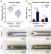
Characterization of the two FNB needles. (a) The resistance to the Franseen needle was significantly higher than that to the Tricore needle. (b) Needle angle changed significantly after the procedure compared with before for both needle types; however, the Franseen needle became more significantly bent than the Tricore needle. (c) Representative images of needles before and after procedures. The white arrowheads indicate the bent needle tips. ** P < 0.01; *** P < 0.001. EUS-FNB, endoscopic ultrasound-guided fine needle biopsy; CI, confidence interval.
Fig. 3
Fig. 3
Ultrasound findings during EUS-FNB in the porcine liver. (a) Representative ultrasound images of the two FNB needles. The Tricore group needle (arrows) produced relatively fewer metal artifacts than the Franseen group needle (arrow heads). (b) The pixel intensity of the Tricore group was significantly higher than that of the Franseen group. ** P < 0.01. CI, confidence interval; EUS-FNB, endoscopic ultrasound-guided fine needle biopsy.
Fig. 4
Fig. 4
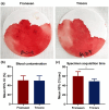
Intraoperative findings. (a) Blood contamination was quantified as the percentage of the total area of the filter paper contaminated with blood following specimen release. (b) Blood contamination did not differ between the Franseen and Tricore groups. (c) The mean specimen acquisition time was significantly shorter in the Tricore group than in the Franseen group. * P < 0.05. CI, confidence interval.
Fig. 5
Fig. 5
Representative histological findings. (a) Hematoxylin and eosin staining was performed to evaluate the sufficiency of the tissue for diagnosis. The number and area of each fragment in the (b) Franseen group and (c) Tricore group were measured using ImageJ. (d) Masson’s trichrome staining was performed to identify complete portal tracts, composed of a PV, HA, and BD. BD, bile duct; HA, hepatic artery; PV, portal vein.

Similar articles

References

    1. Mohan, B. P. et al. Comparison of Franseen and fork-tip needles for EUS-guided fine-needle biopsy of solid mass lesions: A systematic review and meta-analysis. Endosc. Ultrasound. 8, 382–391. 10.4103/eus.eus_27_19 (2019). - PMC - PubMed
    1. Tanisaka, Y. et al. Comparison of endoscopic ultrasound-guided fine-needle aspiration and Biopsy device for Lymphadenopathy. Gastroenterol. Res. Pract.2021(6640862). 10.1155/2021/6640862 (2021). - PMC - PubMed
    1. Gheorghiu, M. et al. Endoscopic ultrasound-guided fine-needle biopsy versus fine-needle aspiration in the diagnosis of focal liver lesions: prospective head-to-head comparison. Diagnostics (Basel). 12. 10.3390/diagnostics12092214 (2022). - PMC - PubMed
    1. Levine, I. & Trindade, A. J. Endoscopic ultrasound fine needle aspiration vs fine needle biopsy for pancreatic masses, subepithelial lesions, and lymph nodes. World J. Gastroenterol.27, 4194–4207. 10.3748/wjg.v27.i26.4194 (2021). - PMC - PubMed
    1. Song, T. J. et al. The prospective randomized, controlled trial of endoscopic ultrasound-guided fine-needle aspiration using 22G and 19G aspiration needles for solid pancreatic or peripancreatic masses. Am. J. Gastroenterol.105, 1739–1745. 10.1038/ajg.2010.108 (2010). - PubMed

Publication types

LinkOut - more resources