. 2024 Sep 29;14(1):22539.
doi: 10.1038/s41598-024-73579-2.

MiRNA expression profiling reveals a potential role of microRNA-148b-3p in cerebral vasospasm in subarachnoid hemorrhage

Affiliations

MiRNA expression profiling reveals a potential role of microRNA-148b-3p in cerebral vasospasm in subarachnoid hemorrhage

Jee-Yeon Ryu et al. Sci Rep. .

Abstract

Cerebral vasospasm (CVS) is an important contributor to delayed cerebral ischemia following aneurysmal subarachnoid hemorrhage (aSAH), leading to high morbidity and long-term disability. While several microRNAs (miRNAs) have been implicated in vasospasm, the underlying mechanisms for CVS remain poorly understood. Our study aims to identify miRNAs that may contribute to the development of CVS. Whole-blood samples were obtained during or outside of vasospasm from aSAH patients whose maximal vasospasm was moderate or severe. MiRNAs were isolated from serial whole-blood samples, and miRNA sequencing was performed. Differentially expressed miRNAs were identified and the expression levels in patients' samples were verified using real-time qPCR. The biological functions of identified miRNA were evaluated in human brain endothelial cells (HBECs). MiRNA profiling revealed significant upregulation of miR-148b-3p in patients during CVS. We demonstrated that miR-148b-3p directly targeted and decreased the expression of ROCK1, affecting cell proliferation, migration, and invasion of HBECs through the ROCK-LIMK-Cofilin pathway. We propose that the upregulation of miRNA-148b-3p plays a role in the development of CVS by regulating actin cytoskeletal dynamics in HBECs, which is crucial for vascular function. Our study highlights miR-148b-3p as a potential diagnostic marker as well as therapeutic target for CVS following aSAH.

Keywords: Aneurysmal subarachnoid hemorrhage; Cerebral vasospasm; MiRNA profiling; MiRNA-148-3p; ROCK-LIMK-Cofilin pathway.


Conflict of interest statement

MAA: Proctor for Covidien and Codman. The other authors declare no competing interests.

Figures

Fig. 1
Fig. 1
The expression profiles of miRNAs (a) The volcano plot displays significant differentially expressed miRNAs (DEMis) between CVS+ and CVS samples. Red dots represent upregulated miRNAs, blue dots indicate downregulated miRNAs, and grey dots show no change. (b) Biological function network of DEMis with gene ontology (GO) terms as nodes linked. The node color depicts the significant enrichment. DEMis differentially expressed miRNAs, CVS+ blood samples with CVS,  CVS blood samples without CVS, GO gene ontology.
Fig. 2
Fig. 2
Enriched biological processes of miR-148b-3p. (a) Gene network summarizes the miRNA targets belonging to the gene ontology (GO) term “regulation of microtubule cytoskeleton organization” as linked nodes. (b) Cell type enrichment analysis predicts the cell types regulated by miR-148b-3p. The bars indicate the transformed P-values of highly enriched cell types for each cell cluster (P-value < 0.05). (c) Chord diagram illustrates enriched biological processes of miR-148b-3p, linking each gene cluster to its corresponding processes. (d) miRNA expression levels of miR-148b-3p in patients’ samples using RT-qPCR. Error bars indicate mean ± S.D. of three independent experiments. miRNAs expressions were normalized to U6 expression. ***P < 0.001 when compared with CVS. GO gene ontology, CVS+ blood samples with CVS, CVS blood samples without CVS.
Fig. 3
Fig. 3
MiR-148b-3p inhibits cell proliferation, migration, and invasion. (a) Effects of miR-148b-3p on cell proliferation in human brain endothelial cells (HBECs). The cell growth rates were determined for 5 days using a WST-1 assay in untreated HBECs (HBECs group), HBECs treated with miRNA-negative control mimic (miR-NC mi group), and HBECs treated with miRNA-148b-3p mimic (miR-148b-3p mi group). (b) Representative microscopic images of cell migration and invasion by miR-148b-3p in HBECs using a wound healing assay and a trans-well assay, respectively. (c) Percentage of the wound closure area at 24 h. (d) Quantification of the number of invasive cells per field. Error bars indicate mean ± S.D. of three independent experiments. **P < 0.01, ***P < 0.001 when compared with untreated HBECs or HBECs treated with miR-NC. miR-NC mi miRNA-negative control mimic, miR-148b-3p mi miRNA-148b-3p mimic.
Fig. 4
Fig. 4
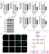
MiR-148b-3p affects ROCK-LIMK-Cofilin pathway in human brain endothelial cells (HBECs). (a) mRNA expression levels of RhoA and ROCK1 in untreated HBECs, and HBECs treated with miR-NC or miR-148b-3p mimic using RT-qPCR. (b) Representative western blot images of protein expression of markers associated with ROCK-LIMK-Cofilin pathway. (c) Quantification of protein expression levels using ImageJ. (d) Representative confocal images of untreated HBECs, and HBECs treated with miR-NC or miR-148b-3p mimic after immunostaining for G-actin (red), F-actin (green), and nuclei (blue). (e) Schematic diagram of this study. Overall, we discovered that miRNA-148b-3p is highly expressed in aSAH patients during vasospasm, and that it regulates cell proliferation, migration, and invasion in human brain endothelial cells. Error bars indicate mean ± S.D. of three independent experiments. mRNAs expressions were normalized to GAPDH expression. **P < 0.01, ***P < 0.001 when compared with untreated HBECs or HBECs treated with miR-NC. miR-NC mi miRNA-negative control mimic, miR-148b-3p mi miRNA-148b-3p mimic, ROCK Rho-associated protein kinase, LIMK LIM kinase, G-actin globular actin, F-actin fibrous actin, DAPI 4’,6-diamidino-2-phenylindole.

References

    1. de Rooij, N. K., Linn, F. H., van der Plas, J. A., Algra, A. & Rinkel, G. J. Incidence of subarachnoid haemorrhage: a systematic review with emphasis on region, age, gender and time trends. J. Neurol. Neurosurg. Psychiatry. 78, 1365–1372. 10.1136/jnnp.2007.117655 (2007). - PMC - PubMed
    1. Foreman, B. The pathophysiology of delayed cerebral ischemia. J. Clin. Neurophysiol. 33, 174–182. 10.1097/WNP.0000000000000273 (2016). - PubMed
    1. Geraghty, J. R. & Testai, F. D. Delayed cerebral ischemia after subarachnoid hemorrhage: beyond vasospasm and towards a multifactorial pathophysiology. Curr. Atheroscler Rep. 19, 50. 10.1007/s11883-017-0690-x (2017). - PubMed
    1. Kassell, N. F. et al. The International Cooperative Study on the timing of aneurysm surgery. Part 1: overall management results. J. Neurosurg. 73, 18–36. 10.3171/jns.1990.73.1.0018 (1990). - PubMed
    1. Dorsch, N. W. & King, M. T. A review of cerebral vasospasm in aneurysmal subarachnoid haemorrhage part I: incidence and effects. J. Clin. Neurosci. 1, 19–26. 10.1016/0967-5868(94)90005-1 (1994). - PubMed

MeSH terms

LinkOut - more resources