. 2025 Apr;97(5):1696-1705.
doi: 10.1038/s41390-024-03582-w. Epub 2024 Sep 28.

Amyloid precursor protein as a fibrosis marker in infants with biliary atresia

Affiliations

Amyloid precursor protein as a fibrosis marker in infants with biliary atresia

Jan C Kamp et al. Pediatr Res. 2025 Apr.

Abstract

Background: Biliary atresia (BA) is a rare condition of unknown origin in newborns with jaundice. In BA bile ducts are non-functional, causing neonatal cholestasis and following liver fibrosis and failure.

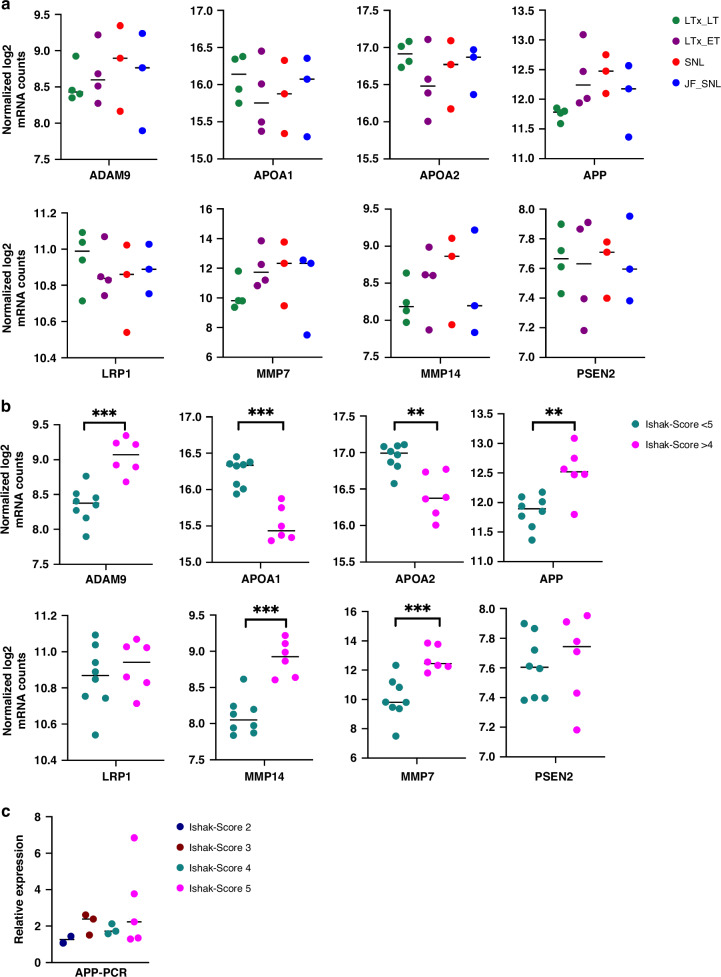
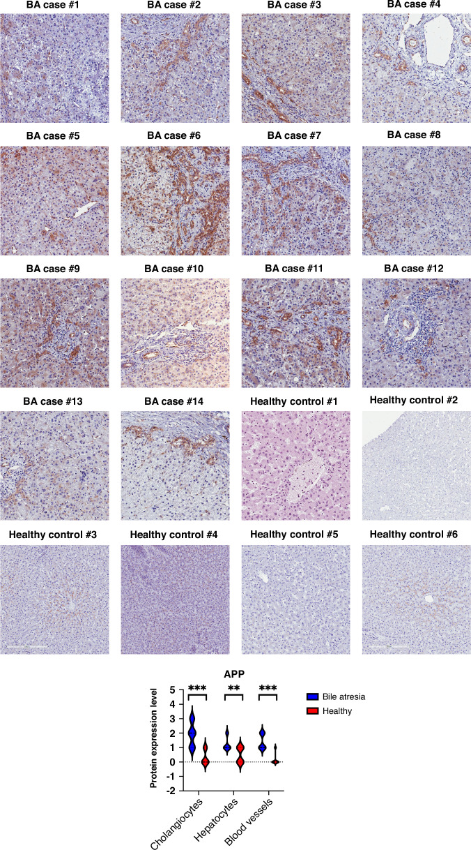
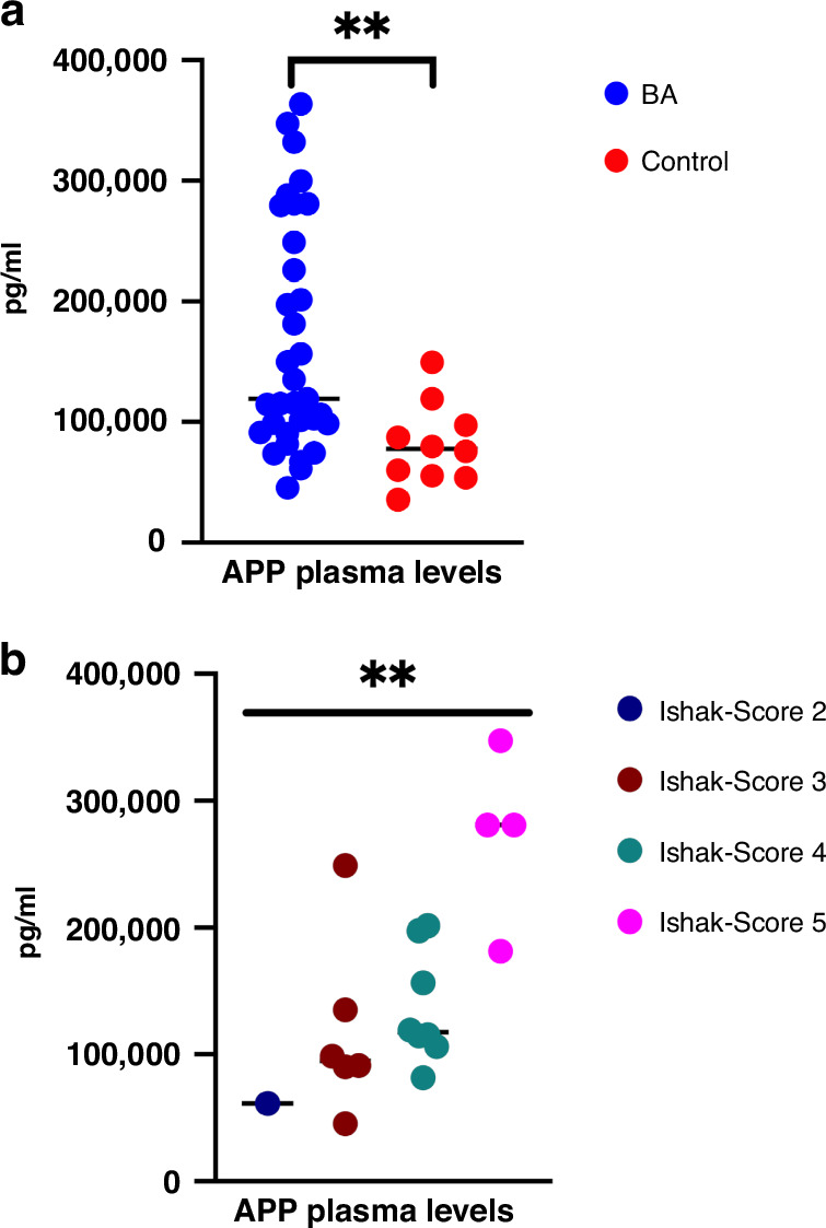
Methods: This retrospective study included liver biopsies of 14 infants with BA aged [mean ± SD] 63 ± 23 days. Patients were grouped according to the clinical course (jaundice-free vs recurrent jaundice vs required liver transplantation or liver fibrosis (Ishak fibrosis score)) and followed for 1.61-5.64 years (mean 4.03). Transcriptome profiles were assessed using a panel of 768 fibrosis-specific genes, reanalyzed via qRT-PCR, and confirmed via immunostaining. Plasma from an additional 30 BA infants and 10 age-matched controls were used for amyloid precursor protein (APP) quantification by ELISA.

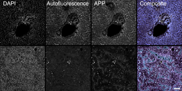
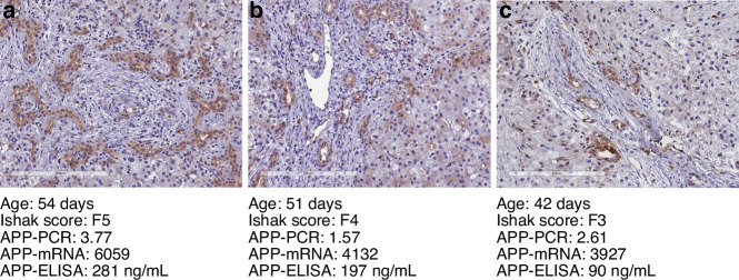
Results: Different clinical outcome groups showed a homogeneous mRNA expression. Altered amyloid-metabolism-related gene expression was found between cases with Ishak fibrosis score greater than 4. Immunostaining confirmed a distinct presence of APP in the livers of all BA subjects. APP plasma levels were higher in BA than in age-matched controls and correlated with the histological fibrosis grade.

Conclusions: These results suggest that amyloidosis may contribute to BA and liver fibrosis, indicating that APP could serve as a potential liquid biomarker for these conditions.

Impact: Biliary atresia patients with higher fibrosis scores according to Ishak have higher hepatic expression of amyloid-related genes while amyloid precursor protein accumulates in the liver and increases in the circulation. After a recent study revealed beta-amyloid deposition as a mechanism potentially involved in biliary atresia, we were able to correlate amyloid-metabolism-related transcript levels as well as amyloid precursor protein tissue and plasma levels with the degree of hepatic fibrosis. These findings suggest that amyloid precursor protein is a fibrosis marker in infants with biliary atresia, reinforcing the role of amyloid metabolism in the pathogenesis of this serious disease.


Conflict of interest statement

Competing interests: The authors declare no competing interests. Consent for publication: All subjects included in this study or their parents gave written informed consent.

Figures

Fig. 1
Fig. 1. Amyloidosis-related gene expression.
Dot plots showing normalized log2 mRNA counts of amyloid-metabolism-related genes in 4 different outcome groups (a) and in subjects with severe (Ishak fibrosis score >4) vs. less severe (Ishak fibrosis score <5) liver fibrosis (b). Additionally, APP-levels are compared between different Ishak fibrosis scores (c). LTx_LT, late liver transplantation (≥1 year); LTx_ET, early liver transplantation (<1 year); SNL, survival with the native liver; JF_SNL, jaundice-free survival with the native liver. Statistical significance was categorized as *(p < 0.05), **(p < 0.01), ***(p < 0.001), and ****(p < 0.0001).
Fig. 2
Fig. 2. Immunostaining of liver APP performed using rabbit monoclonal antibody to APP.
Bound antibodies were visualized using HRP-DAB (3,3′-diaminobenzidine tetrahydrochloride) staining. APP-positive areas are specifically stained in brown. Images were taken after automated whole-slide imaging using the APERIO CS2 scanner (Leica Biosystems, Wetzlar, Germany) and ImageScope software version 12.3.3.5048 (Leica Biosystems). Healthy liver tissue was used as a control. BA bile atresia. APP amyloid precursor protein. Statistical significance was categorized as *(p < 0.05), **(p < 0.01), ***(p < 0.001), and ****(p < 0.0001).
Fig. 3
Fig. 3. Representative immunofluorescence imaging of APP in control and biliary atresia liver.
Figure showing immunofluorescence imaging of control liver (upper row) and bile atresia liver (lower row) of amyloid precursor protein (APP). Note the expression of APP in small intrahepatic bile ducts in bile atresia (arrowhead lower row), while there was no detectable expression of APP in intrahepatic bile ducts of the control liver (arrowhead upper row). APP was visualized using an Alexa Fluor 555 fluorescent secondary antibody. Autofluorescence imaging was generated using a GFP filter cube. Nuclei were stained with DAPI. Magnification 200×, scale bar equals 100 µm.
Fig. 4
Fig. 4. Plasma quantification of APP.
a Dot plots showing plasma levels of APP in bile atresia (BA) subjects vs. age-matched children with inguinal hernia (control). b Dot plots showing APP plasma levels in subjects with different fibrosis severity assessed by Ishak fibrosis score. Data presented as mean (SD), and P values ≤ 0.05 were considered statistically significant. Statistical significance was categorized as *(p < 0.05), **(p < 0.01), ***(p < 0.001), and ****(p < 0.0001).
Fig. 5
Fig. 5. APP expression in exemplary subjects.
Comparison of three exemplary BA cases (ac) that were available for all analyses. Immunostaining of liver APP performed using rabbit monoclonal antibody to APP. Bound antibodies were visualized using HRP-DAB (3,3′-diaminobenzidine tetrahydrochloride) staining. APP-positive areas within hepatocytes are brown in color. BA bile atresia, APP amyloid precursor protein, PCR polymerase chain reaction, mRNA messenger ribonucleic acid, ELISA enzyme-linked immunosorbent assay.

References

    1. Jiang, J. et al. Epidemiological characteristics and risk factors of biliary atresia: a case-control study. BMJ Open.11, e049354 (2021). - PMC - PubMed
    1. Livesey, E. et al. Epidemiology of biliary atresia in England and Wales (1999–2006). Arch. Dis. Child Fetal Neonatal Ed.94, F451–F455 (2009). - PubMed
    1. The, N. S. et al. National Birth Defects Prevention Study. Risk factors for isolated biliary atresia, National Birth Defects Prevention Study, 1997–2002. Am. J. Med. Genet. A.143A, 2274–2284 (2007). - PubMed
    1. Medappil, N. et al. Kasai portoenterostomy for biliary atresia - surgical precautions for better outcomes. J. Pediatr. Surg.54, 868–869 (2019). - PubMed
    1. Davenport, M. et al. Surgical and medical aspects of the initial treatment of biliary atresia: position paper. J. Clin. Med.11, 6601 (2022). - PMC - PubMed

MeSH terms

LinkOut - more resources