

Antimicrobial Sub-MIC induces Staphylococcus aureus biofilm formation without affecting the bacterial count
- PMID: 39342123
- PMCID: PMC11438285
- DOI: 10.1186/s12879-024-09790-3
Antimicrobial Sub-MIC induces Staphylococcus aureus biofilm formation without affecting the bacterial count
Erratum in
-
Correction: Antimicrobial Sub-MIC induces Staphylococcus aureus biofilm formation without affecting the bacterial count.BMC Infect Dis. 2024 Oct 22;24(1):1194. doi: 10.1186/s12879-024-10038-3. BMC Infect Dis. 2024. PMID: 39438809 Free PMC article. No abstract available.
Abstract
Background: Biofilm formation is an essential virulence factor that creates a highly protected growth mode for Staphylococcus aureus (S. aureus) to survive in any hostile environment. Antibiotic sub-minimal inhibitory concentration (sub-MIC) may modulate the biofilm formation ability of bacterial pathogens, thereby affecting bacterial pathogenesis and infection outcomes. Intense antimicrobial therapy to treat biofilm-associated infections can control the pathogenic infection aggravation but cannot guarantee its complete eradication.
Objective: This study aimed to assess the sub-MICs effect of 5 different antimicrobial classes on biofilm-forming capacity among Staphylococcus aureus clinical isolates using three different biofilm quantitation techniques.
Methods: In this study, the effects of 5 different antimicrobial agents, namely, azithromycin, gentamicin, ciprofloxacin, doxycycline, and imipenem, at sub-MICs of 12.5%, 25%, and 50% were tested on 5 different clinical isolates of S. aureus. The biofilms formed in the absence and presence of different antimicrobial sub-MICs were then assessed using the following three different techniques: the crystal violet (CV) staining method, the quantitative PCR (qPCR) method, and the spread plate method (SPM).
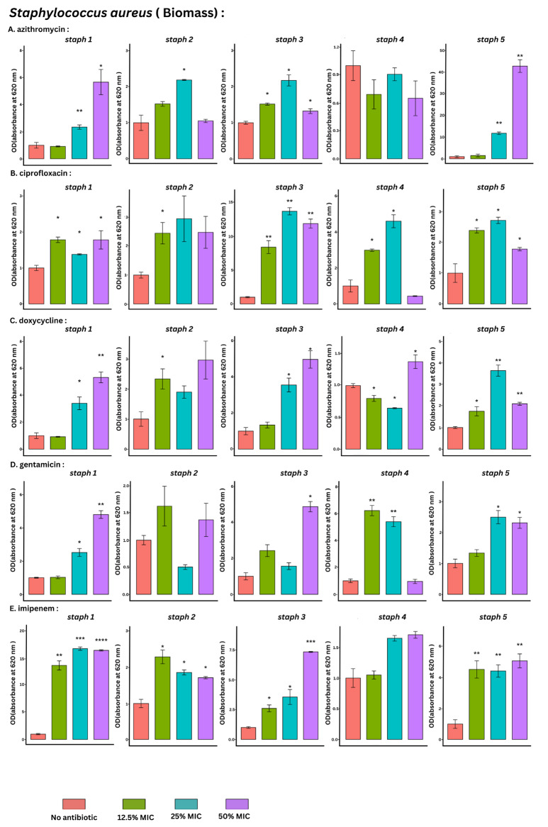
Results: Biofilm formation was significantly induced in 64% of the tested conditions using the CV technique. On the other hand, the qPCR quantifying the total bacterial count and the SPM quantifying the viable bacterial count showed significant induction only in 24% and 17.3%, respectively (Fig. 1). The difference between CV and the other techniques indicates an increase in biofilm biomass without an increase in bacterial growth. As expected, sub-MICs did not reduce the viable cell count, as shown by the SPM. The CV staining method revealed that sub-MICs of imipenem and ciprofloxacin had the highest significance rate (80%) showing an inductive effect on the biofilm development. On the other hand, doxycycline, azithromycin, and gentamicin displayed lower significance rates of 73%, 53%, and 47%, respectively.
Conclusion: Exposure to sub-MIC doses of antimicrobial agents induces the biofilm-forming capacity of S. aureus via increasing the total biomass without significantly affecting the bacterial growth of viable count.
Keywords: S. aureus; Biofilm biomass; Spread plate technique; Subinhibitory concentration; qPCR.
© 2024. The Author(s).
Conflict of interest statement
The authors declare no competing interests.
Figures


References
-
- Lebeaux D, Ghigo J-M. Management of biofilm-associated infections: what can we expect from recent research on biofilm lifestyles? Med Stud. 2012;28(8–9):727–39. - PubMed
-
- Flemming HC, Wingender J. The biofilm matrix. Nat Rev Microbiol. 2010;8(9):623–33. - PubMed
-
- Davies J, Spiegelman GB, Yim G. The world of subinhibitory antibiotic concentrations. Curr Opin Microbiol. 2006;9(5):445–53. - PubMed
-
- Svarcova V, Zdenkova K, Sulakova M, Demnerova K, Pazlarova J. Contribution to determination of extracellular DNA origin in the biofilm matrix. J Basic Microbiol. 2021;61(7):652–61. - PubMed
MeSH terms
Substances
LinkOut - more resources
Full Text Sources
Medical
Molecular Biology Databases
Research Materials
