. 2024 Sep 28;25(1):353.
doi: 10.1186/s12931-024-02962-4.

Single-cell transcriptomics reveals e-cigarette vapor-induced airway epithelial remodeling and injury

Affiliations

Single-cell transcriptomics reveals e-cigarette vapor-induced airway epithelial remodeling and injury

Weitao Cao et al. Respir Res. .

Abstract

Background: In recent years, e-cigarettes have been used as alternatives among adult smokers. However, the impact of e-cigarette use on human bronchial epithelial (HBE) cells remains controversial.

Methods: We collected primary HBE cells of healthy nonsmokers and chronic obstructive pulmonary disease (COPD) smokers, and analyzed the impact of e- cigarette vapor extract (ECE) or cigarette smoke extract (CSE) on HBE cell differentiation and injury by single-cell RNA sequencing, immunostaining, HE staining, qPCR and ELISA. We obtained serum and sputum from healthy non- smokers, smokers and e-cigarette users, and analyzed cell injury markers and mucin proteins.

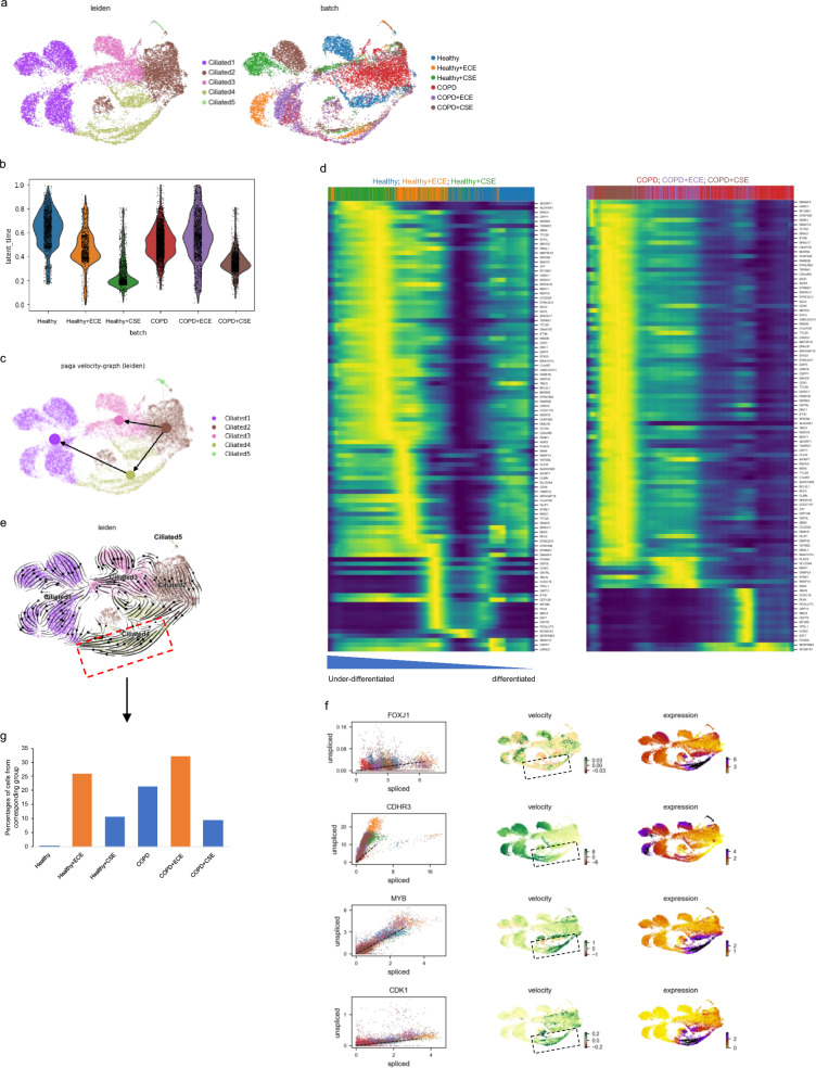
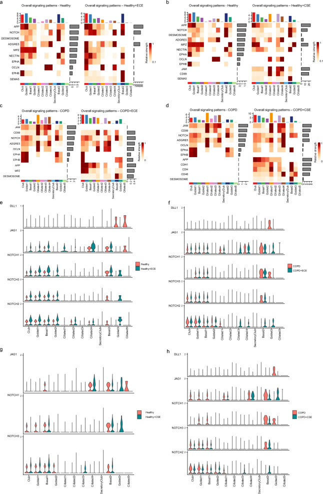
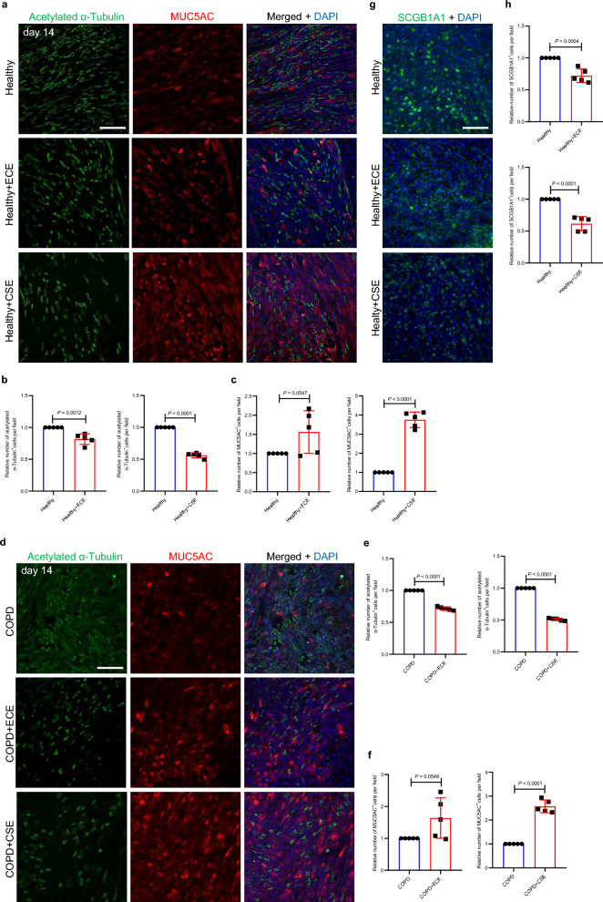
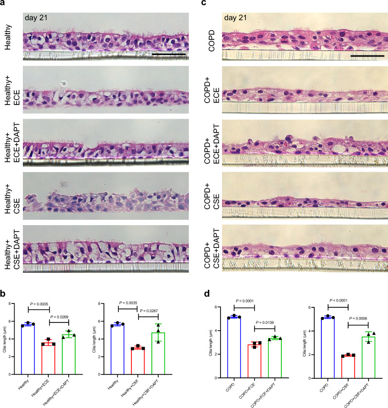
Results: ECE treatment led to a distinct differentiation program of ciliated cells and unique patterns of their cell-cell communications compared with CSE. ECE treatment caused increased Notch signaling strength in a ciliated cell subpopulation, and HBE cell remodeling and injury including hypoplasia of ciliated cells and club cells, and shorter cilia. ECE-induced hypoplasia of ciliated cells and shorter cilia were ameliorated by the Notch signaling inhibition.

Conclusions: This study reveals distinct characteristics in e-cigarette vapor-induced airway epithelial remodeling, pointing to Notch signaling pathway as a potential targeted intervention for e-cigarette vapor-caused ciliated cell differentiation defects and cilia injury. In addition, a decrease in SCGB1A1 proteins is associated with e- cigarette users, indicating a potential lung injury marker for e-cigarette users.

Keywords: COPD; Cilia; Ciliated cells; E-cigarette; Notch signaling.


Conflict of interest statement

The authors declare no competing interests. At the time of the research, Zehong Wu and Xingtao Jiang were employees of RELX Science Center, Shenzhen RELX Tech.

Figures

Fig. 1
Fig. 1
Single-cell RNA-seq data analysis reveals changes in HBE cell populations upon ECE or CSE treatment. a UMAP embedding representation of single-cell transcriptomes of ALI cultured HBE cells from healthy nonsmokers and COPD smokers treated with ECE and CSE, respectively, colored by treatments (the left panel) and cell types (the right panel). b Bubble plot showing the top5 marker genes for each cell type. Colors and size represented the log2fold change and percentage of cells expressing the gene, respectively. c Ratio of each ALI cultured HBE cell type from healthy nonsmokers and COPD smokers treated with ECE and CSE, respectively. d UMAP projections of cells from ECE-treated or CSE-treated ALI cultures from healthy nonsmoker or COPD smokers. Colors represented cell types. UMAP: Uniform Manifold Approximation and Projection; COPD: chronic obstructive pulmonary disease; CSE: cigarette smoke extract; ECE: e-cigarette vapor extract. HBE: human bronchial epithelial
Fig. 2
Fig. 2
Pseudo-time analysis uncovers abnormal ciliated cell differentiation paths upon ECE or CSE treatment. a UMAP plot representing ciliated cell populations, color-coded by ciliated cell subsets (left panel) and treatment groups (right panel). b Violin plot shows inferred latent time indicating differentiation status for ciliated cells from each treatment group. c PAGA velocity graph showing the major ciliated cells differentiation paths. d Heatmaps showing the dynamics of gene expression levels along pseudo-time trajectory for ciliated cells in each treatment group, color-coded by expression level. From left to right: under-differentiated to differentiated ciliated cell stages. Color bars represent the ciliate cell cluster in (a). e RNA velocity vectors projected onto the UMAP embedding, reflecting ciliated cells differentiation paths. f Key genes (FOXJ1, CDHR3, MYB and CDK1) unspliced/spliced mRNA read ratios and expression levels projected onto UMAP embedding. Unspliced/spliced mRNA ratios for each cell, colored by major ciliated subsets (the left panel); key genes with color-coded by upspliced/spliced mRNA ratios projected onto UMAP embedding (the middle panel); key genes expression levels projected onto UMAP embedding (the right panel). g Histograms showing ECE treatment specific ciliated cell percentage from six groups. PAGA: Partition- based graph abstraction
Fig. 3
Fig. 3
Cell–cell communication analysis of HBE cells. a-d CellChat analysis showing comparisons of cell–cell communication pathways for each cell cluster a between healthy and healthy + ECE; b between healthy and healthy + CSE; c between COPD and COPD + ECE; d between COPD and COPD + CSE. e–h Expression levels of Notch signaling components including DLL1, JAG1, NOTCH1, NOTCH2 and NOTCH3in each cell cluster e between healthy and healthy + ECE; f between COPD and COPD + ECE; g healthy and healthy + CSE; h between COPD and COPD + CSE
Fig. 4
Fig. 4
ECE exposure causes reduced numbers of ciliated cells and club cells from healthy nonsmokers or COPD smokers. a Immunostaining for MUC5AC (red), acetylated α-tubulin (green) and DAPI staining (blue) in HBE cells from healthy nonsmokers at the ALI after 9 day DMEM/F-12 medium (n = 5), ECE (n = 5) or CSE (n = 5) treatment. b Quantification of the relative number of acetylated α-tubulin+ cells in HBE cells from healthy nonsmokers at the ALI after 9 day DMEM/F-12 medium (n = 5), ECE (n = 5) or CSE (n = 5) treatment. 4752, 3392 and 1701 acetylated α-tubulin+ cells were analyzed for controls, ECE and CSE treatment, respectively. c Quantification of the relative number of MUC5AC+cellsinHBE cells from healthy nonsmokers at the ALI after 9 day DMEM/F-12 medium (n = 5), ECE (n = 5) or CSE (n = 5) treatment.1604, 2304 and 3592 MUC5AC+ cells were analyzed for controls, ECE and CSE treatment, respectively. d Immunostaining for MUC5AC (red), acetylated α-tubulin (green) and DAPI staining (blue) in HBE cells from COPD smokers at the ALI after 9 day DMEM/F-12 medium (n = 5), ECE (n = 5) or CSE (n = 5) treatment. e Quantification of the relative number of acetylated α-tubulin+ cells in HBE cells from COPD smokers at the ALI after 9 day DMEM/F-12 medium (n = 5), ECE (n = 5) or CSE (n = 5) treatment.3504, 2260 and 1296 acetylated α-tubulin+ cells were analyzed for controls, ECE and CSE treatment, respectively. f Quantification of the relative number of MUC5AC+cells in HBE cells from COPD smokers at the ALI after 9 day DMEM/F-12 medium (n = 5), ECE (n = 5) or CSE (n = 5) treatment. 1952, 2702 and 3552 MUC5AC+ cells were analyzed for controls, ECE and CSE treatment, respectively. g Immunostaining for SCGB1A1 (green) and DAPI staining (blue) in HBE cells from healthy nonsmokers at the ALI after 9 day DMEM/F-12 medium. (n = 5), ECE (n = 5) or CSE (n = 5) treatment. h Quantification of the relative number of SCGB1A1+ cells in HBE cells from healthy nonsmokers at the ALI after 9 day DMEM/F-12 medium (n = 5), ECE (n = 5) or CSE (n = 5) treatment. 8752, 5592 and 4684 SCGB1A1+cells were analyzed for controls, ECE and CSE treatment, respectively. Scale bars: 100 μm. Unpaired Student’s t-test, mean ± s.d
Fig. 5
Fig. 5
ECE exposure leads to cilia shortening in HBE cells from healthy nonsmokers or COPD smokers. a Hematoxylin and eosin staining for ALI culture sections of HBE cells from healthy nonsmokers after 9 day DMEM/F-12 medium (n = 5), ECE (n = 5) or CSE (n = 5) treatment. b Immunostaining for acetylated α- tubulin (green) and DAPI staining (blue)for ALI culture sections of HBE cells from healthy nonsmokers after 9 day DMEM/F-12 medium (n = 5), ECE (n = 5) or CSE (n = 5) treatment. c Quantification of cilia length in HBE cells from healthy. Nonsmokers at the ALI after 9 day DMEM/F-12 medium (n = 5), ECE (n = 5) or CSE (n = 5) treatment. d Hematoxylin and eosin staining for ALI culture sections of HBE cells from COPD smokers after 9 day DMEM/F-12 medium (n = 5), ECE (n = 5) or CSE (n = 5) treatment. e Immunostaining for acetylated α-tubulin (green) and DAPI staining (blue) for ALI culture sections of HBE cells from COPD smokers after 9day DMEM/F-12 medium (n = 5), ECE (n = 5) or CSE (n = 5) treatment. f Quantification of cilia length in HBE cells from COPD smokers at the ALI after 9 day DMEM/F-12 medium (n = 5), ECE (n = 5) or CSE (n = 5) treatment. g, h RT- qPCR analysis of mRNA levels of FOXJ1 in HBE cells from g healthy nonsmokers or h COPD smokers at the ALI after 9 day DMEM/F-12 medium (n = 3), ECE (n = 3) or CSE (n = 3) treatment. i, j ELISA measurements of SCGB1A1 levels in supernatant of HBE cells from i healthy nonsmokers or j COPD smokers at the ALI after 9 day DMEM/F-12 medium (n = 3), ECE (n = 3) or CSE (n = 3) treatment. Scale bars: 50 μm.Unpaired Student’s t-test, mean ± s.d
Fig. 6
Fig. 6
Notch signaling functions as mediators of ECE treatment-induced remodeling of HBE cells from in healthy nonsmokers or COPD smokers. a Timeline for ECE or CSE and DAPT administration. b Immunostaining for acetylatedα-tubulin (green), MUC5AC (red) and DAPI staining (blue) in HBE cells from healthy nonsmokers at the ALI after 16 day DMEM/F-12 medium (n = 3), ECE (n = 3) or CSE (n = 3) treatment, or 16 day ECE and 7 day 10 μM DAPT (n = 3), or 16 day CSE and 7 day 10 μM DAPT (n = 3) treatment. c Quantification of the relative number of acetylated α-tubulin+ cells. d Quantification of the relative number of MUC5AC+ cells. e Immunostaining for acetylated α-tubulin (green), MUC5AC (red) and DAPI staining (blue) in HBE cells from COPD smokers at the ALI after 16 day DMEM/F-12 medium (n = 3), ECE (n = 3) or CSE (n = 3) treatment, or 16 day ECE and 7 day 10 μM DAPT (n = 3), or 16 day CSE and 7 day 10 μM DAPT (n = 3) treatment. f Quantification of the relative number of acetylated α-tubulin+ cells. g Quantification of the relative number of MUC5AC+cells. Scale bars: 100 μm. One- way ANOVA with Tukey’s multiple comparison test for comparisons among three groups and Unpaired Student’s t-test for comparisons between two groups, mean ± s.d
Fig. 7
Fig. 7
Notch signaling inhibition partially rescues cilia shortening and mucus hypersecretion induced by ECE treatment in HBE cells from healthy nonsmokers or COPD smokers. a Hematoxylin and eosin staining for ALI culture sections of HBE cells from healthy nonsmokers at the ALI after 16F day DMEM/F-12 medium (n = 3), CSE (n = 3) or ECE (n = 3) treatment, or 16 day CSE and 7 day 10 μM DAPT (n = 3), or 16 day ECE and 7 day 10 μM DAPT (n = 3) treatment. b Quantification of cilia length in HBE cells. c Hematoxylin and eosin staining for ALI culture sections of HBE cells from COPD smokers at the ALI after 16 day DMEM/F-12 medium (n = 3), CSE (n = 3) or ECE (n = 3) treatment, or 16 day CSE and 7 day 10 μM DAPT (n = 3), or 16 day ECE and 7 day 10 μM DAPT (n = 3) treatment. d Quantification of cilia length in HBE cells. Scale bars: 50 μm. One- way ANOVA with Tukey’s multiple comparison test, mean ± s.d
Fig. 8
Fig. 8
SCGB1A1 levels are decreased in the serum and sputum of E- cigarette users. a ELISA measurements of SCGB1A1 levels in serum of healthy nonsmokers, e-cigarette users and tobacco smokers. b ELISA measurements of SCGB1A1 levels in sputum of healthy nonsmokers, e-cigarette users and tobacco smokers. c ELISA measurements of MUC5AC levels in serum of healthy nonsmokers, e-cigarette users, tobacco smokers and COPD smokers. d ELISA measurements of MUC5AC levels in sputum of healthy nonsmoker, e-cigarette users and tobacco smokers. Unpaired Student’s t-test, mean ± s.d

Similar articles

Cited by

References

    1. Reidel B, Radicioni G, Clapp PW, Ford AA, Abdelwahab S, Rebuli ME, et al. E-cigarette use causes a unique innate immune response in the lung, involving increased neutrophilic activation and altered mucin secretion. Am J Respir Crit Care Med. 2018;197:492–501. - PMC - PubMed
    1. Ghosh A, Coakley RD, Ghio AJ, Muhlebach MS, Esther CR Jr, Alexis NE, et al. Chronic E-cigarette use increases neutrophil elastase and matrix metalloprotease levels in the lung. Am J Respir Crit Care Med. 2019;200:1392–401. - PMC - PubMed
    1. Gellatly S, Pavelka N, Crue T, Schweitzer KS, Day BJ, Min E, et al. Nicotine-Free e-Cigarette Vapor exposure stimulates il6 and mucin production in human primary small airway epithelial cells. J Inflamm Res. 2020;13:175–85. - PMC - PubMed
    1. Garcia-Arcos I, Geraghty P, Baumlin N, Campos M, Dabo AJ, Jundi B, et al. Chronic electronic cigarette exposure in mice induces features of COPD in a nicotine- dependent manner. Thorax. 2016;71:1119–29. - PMC - PubMed
    1. Madison MC, Landers CT, Gu BH, Chang CY, Tung HY, You R, et al. Electronic cigarettes disrupt lung lipid homeostasis and innate immunity independent of nicotine. J Clin Invest. 2019;129:4290–304. - PMC - PubMed

MeSH terms

Substances

LinkOut - more resources