

Morphological CBCT parameters for an accurate differentiation between nasopalatine duct cyst and the normal nasopalatine canal
- PMID: 39342234
- PMCID: PMC11438411
- DOI: 10.1186/s13005-024-00458-6
Morphological CBCT parameters for an accurate differentiation between nasopalatine duct cyst and the normal nasopalatine canal
Abstract
Background: The incisive foramen width was a traditional imaging criterion for diagnosing nasopalatine duct (NPD) cysts. Recent CBCT studies demonstrated significant dimensional variations of the nasopalatine canal, which raised questions about the accuracy of this criterion. This study investigated whether nasopalatine canal diameters assessed on CBCT images can accurately differentiate NPD cysts from normal nasopalatine canals.
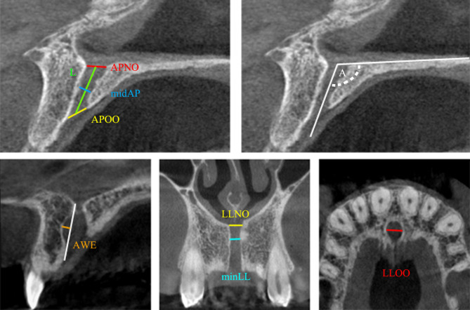
Methods: The study included 19 patients with NPD cysts (12 (63.2%) males, 7 (36.8%) females, mean age 44.7 ± 13.3), and a control group of 164 patients (72 (43.9%) males, 92 (56.1%) females, mean age 47.25 ± 17.74). CBCT images were retrospectively evaluated. The following nasopalatine canal diameters were measured on reference sagittal, coronal, and axial reformation images: nasal opening anteroposterior (AP) and mediolateral (ML) diameter, oral opening AP (APOO) and ML (MLOO) diameter, nasopalatine canal length, minimum ML (minML) diameter, anterior wall expansion (AWE), nasopalatine canal angle, and the mid-level AP diameter (midAP). All parameters were compared between groups. Discriminant functional analysis (DFA) was applied to detect CBCT parameters that best differentiate the NPD cyst from the normal canal.
Results: Patients with NPD cyst had significantly greater values of APOO (7.06 ± 2.09 vs. 5.61 ± 1.70), MLOO (6.89 ± 2.95 vs. 3.48 ± 1.24), minML (2.88 ± 1.53 vs. 2.25 ± 1.09), AWE (2.15 ± 0.65 vs. 0.41 ± 0.67), and midAP (4.58 ± 1.61 vs. 2.48 ± 0.96). DFA showed MLOO, AWE, and midAP as the most accurate in distinguishing NPD cyst from the normal canal. When combined in the discriminant function equation X = 0.390·MLOO + 1.010·AWE + 0.288·midAP (cut score 1.669), the differentiation can be performed with a sensitivity and specificity of 98.8% and 76.9%, respectively.
Conclusion: NPD cysts can be accurately distinguished from the normal nasopalatine canal by measuring MLOO, AWE, and midAP diameter on CBCT images.
Keywords: CBCT; Discriminant functional analysis; Morphology; Nasopalatine canal; Nasopalatine duct cyst.
© 2024. The Author(s).
Conflict of interest statement
The authors declare no competing interests.
Figures

References
-
- Langlais RP, Langland OE, Nortjé CJ, editors. Diagnostic imaging of the jaws. Baltimore: Williams & Wilkins; 1995.
MeSH terms
LinkOut - more resources
Full Text Sources
Miscellaneous
