. 2024 Sep 28;23(1):347.
doi: 10.1186/s12933-024-02444-3.

N6-Methyladenosine-mediated phase separation suppresses NOTCH1 expression and promotes mitochondrial fission in diabetic cardiac fibrosis

Affiliations

N6-Methyladenosine-mediated phase separation suppresses NOTCH1 expression and promotes mitochondrial fission in diabetic cardiac fibrosis

Zhi-Yan Liu et al. Cardiovasc Diabetol. .

Abstract

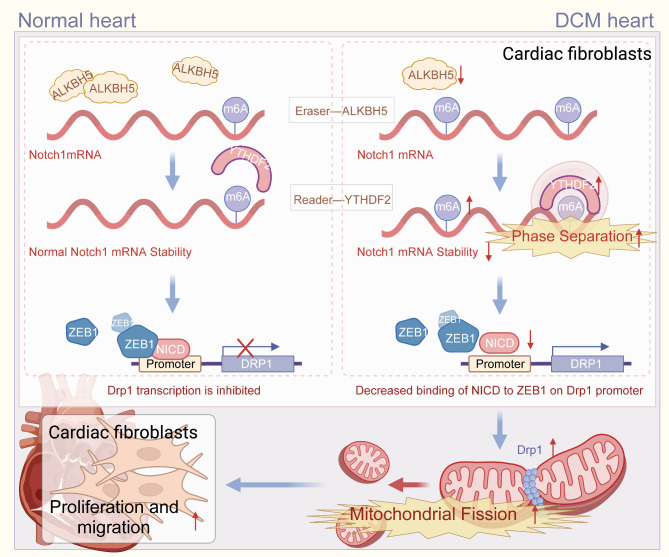
Background: N6-methyladenosine (m6A) modification of messenger RNA (mRNA) is crucial for liquid-liquid phase separation in mammals. Increasing evidence indicates that liquid-liquid phase separation in proteins and RNAs affects diabetic cardiomyopathy. However, the molecular mechanism by which m6A-mediated phase separation regulates diabetic cardiac fibrosis remains elusive.

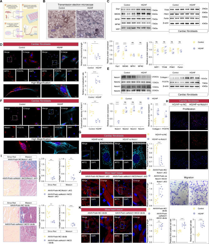
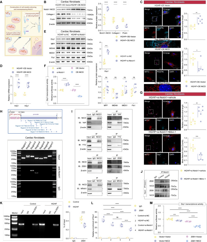
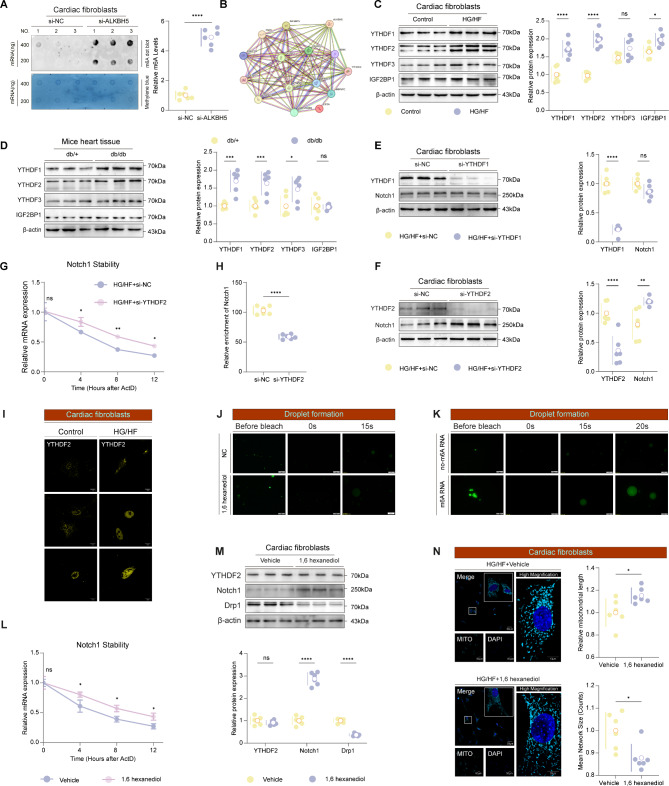
Methods: Leptin receptor-deficient mice (db/db), cardiac fibroblast-specific Notch1 conditional knockout (POSTN-Cre × Notch1flox/flox) mice, and Cre mice were used to induce diabetic cardiac fibrosis. Adeno-associated virus 9 carrying cardiac fibroblast-specific periostin (Postn) promoter-driven small hairpin RNA targeting Alkbh5, Ythdf2, or Notch1, and the phase separation inhibitor 1,6-hexanediol were administered to investigate their roles in diabetic cardiac fibrosis. Histological and biochemical analyses were performed to determine how Alkbh5 and Ythdf2 regulate Notch1 expression in diabetic cardiac fibrosis. NOTCH1 was reconstituted in ALKBH5- and YTHDF2-deficient cardiac fibroblasts and mouse hearts to study its effects on mitochondrial fission and diabetic cardiac fibrosis. Heart tissue samples from patients with diabetic cardiomyopathy were used to validate our findings.

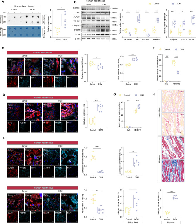
Results: In mice with diabetic cardiac fibrosis, decreased Notch1 expression was accompanied by high m6A mRNA levels and mitochondrial fission. Fibroblast-specific deletion of Notch1 enhanced mitochondrial fission and cardiac fibroblast proliferation and induced diabetic cardiac fibrosis in mice. Notch1 downregulation was associated with Alkbh5-mediated m6A demethylation in the 3'UTR of Notch1 mRNA and elevated m6A mRNA levels. These elevated m6A levels in Notch1 mRNA markedly enhanced YTHDF2 phase separation, increased the recognition of m6A residues in Notch1 mRNA by YTHDF2, and induced Notch1 degradation. Conversely, epitranscriptomic downregulation rescues Notch1 expression, resulting in the opposite effects. Human heart tissues from patients with diabetic cardiomyopathy were used to validate the findings in mice with diabetic cardiac fibrosis.

Conclusions: We identified a novel epitranscriptomic mechanism by which m6A-mediated phase separation suppresses Notch1 expression, thereby promoting mitochondrial fission in diabetic cardiac fibrosis. Our findings provide new insights for the development of novel treatment approaches for patients with diabetic cardiac fibrosis.

Keywords: Diabetic cardiac fibrosis; Diabetic cardiomyopathy; Mitochondrial fission; Phase separation; RNA methylation.


Conflict of interest statement

The authors declare no competing interests.

Figures

Fig. 1
Fig. 1
Decreased NOTCH1 expression accompanies the dysregulation of mitochondrial morphological dynamics in mice with diabetic cardiac fibrosis. A Construction diagram of two animal models of diabetic cardiac fibrosis. B Western blotting analyzed the expression of Notch family in two animal models of diabetic cardiac fibrosis (two-tailed Student t test; n = 6). C Construction diagram of the animal model of diabetic cardiac fibrosis in Notch1 conditional knockout (cKO) mice with HFD/STZ. D Western blotting analyzed the expression of various markers related to mitochondrial fission, fusion, autophagy, and biogenesis in STZ + HFD + cKO-Notch1 model and control group (two-tailed Student t test; n = 6). E Mitochondrial morphodynamics of heart tissue in STZ + HFD + cKO-Notch1 mouse model observed by transmission electron microscopy (two-tailed Student t test; n = 6). F Confocal microscope images showing double staining results of POSTN (pseudo-colored light blue) and Mito Tracker in cardiac fibrotic tissue of STZ + HFD + cKO-Notch1 group and control group mice (two-tailed Student t test; n = 6). Scale bars: 10 μm. G Western blotting analyzed the expression of POSTN, Fibronectin, Collagen I, and Collagen III proteins in STZ + HFD + cKO-Notch1 model and control group (two-tailed Student t test; n = 6). H Confocal microscope images showing double staining results of POSTN (pseudo-colored light blue) and Notch1 in cardiac fibrotic tissue of STZ + HFD + cKO-Notch1 group and control group mice (two-tailed Student t test; n = 6). Scale bars: 10 μm. I Confocal microscope images showing triple staining results of POSTN (pseudo-colored light blue), CTnT (pseudo-colored yellow) and Mito Tracker in cardiac fibrotic tissue of STZ + HFD + cKO-Notch1 group and control group mice (two-tailed Student t test; n = 6). Scale bars: 10 μm and 20 μm
Fig. 2
Fig. 2
Fibroblast-specific NOTCH1-NICD complementation inhibits mitochondrial fission and ameliorates diabetic cardiac fibrosis. A Schematic diagram of a cell model simulating type 2 diabetes in vitro. B Mitochondrial morphodynamics in myocardial fibroblasts treated with HG/HF were observed using transmission electron microscopy (two-tailed Student’s t-test; n = 6). C Western blotting analyzed the expression of various markers related to mitochondrial fission, fusion, autophagy, and biogenesis in cardiac fibroblasts treated with HG/HF (two-tailed Student t test; n = 6). D Confocal microscope images showing the changes of double staining results of POSTN (pseudo-colored light blue) and Mito Tracker in cardiac fibroblasts treated with HG/HF (two-tailed Student t test; n = 6). Scale bars: 10 μm and 50 μm. E Western Blotting analyzed the changes of Notch1 family expression in cardiac fibroblasts treated with HG/HF (two-tailed Student t test; n = 6). F Confocal microscope images showing the changes of double staining results of POSTN (pseudo-colored light blue) and Notch1 in cardiac fibroblasts treated with HG/HF (two-tailed Student t test; n = 6). Scale bars: 10 μm and 50 μm. G Confocal microscope images showed changes in mitochondrial fission (pseudo-colored light blue) in HGHF-treated cardiac fibroblasts after Notch1 knockout (two-tailed Student t test; n = 6). Scale bars: 10 μm and 50 μm. H Using Edu assay to detect the proliferation ability of HGHF-treated cardiac fibroblasts after notch1 knockout (two-tailed Student t test; n = 6; Scale bars: 50 μm); The effect of Notch1 knockdown on HGHF-treated cardiac fibroblasts migration and invasion was determined by transwell assays (two-tailed Student t test; n = 6; Scale bars: 20 μm). I Masson trichromatic staining and Sirius red staining were performed on the cardiac fibrosis tissue sections of Notch1-cKO mice or db/db mice injected with AAV9-Postn-oeNotch1-NICD (two-tailed Student t test; n = 6). JK: Confocal microscope images showed changes in mitochondrial fission in cardiac fibrosis tissue sections of Notch1-cKO mice or db/db mice injected with AAV9-Postn-oeNotch1-NICD (two-tailed Student t test; n = 6). Scale bars: 10 μm
Fig. 3
Fig. 3
Notch1 modulates Drp1 expression and mitochondrial fission through transcriptional modulation. A Schematic diagram of cardiac fibroblasts treated with either Notch1-NICD plasmid overexpression or Notch1 knockdown. B Western blotting verified the overexpression of NICD in HGHF-treated cardiac fibroblasts (two-tailed Student t test; n = 6). C Confocal microscope images showing the changes of double staining results of POSTN (pseudo-colored light blue) and Mito Tracker in cardiac fibroblasts treated with HG/HF after the overexpression of NICD (two-tailed Student t test; n = 6). Scale bars: 50 μm and 10 μm. D The expression of Hes1 and Hey1 in cardiac fibroblasts treated with HG/HF after the overexpression of NICD were analyzed by RT-qPCR (two-tailed Student t test; n = 6). E Western blotting analyzed the expression of various markers related to mitochondrial fission in HGHF-treated cardiac fibroblasts after notch1 knockout (two-tailed Student t test; n = 6). F Dual luciferase assay on 3T3 fibroblasts cotransfected with firefly luciferase constructs containing the Drp1 promoter and si-Notch1 or NICD plasmid (two-tailed Student t test; n = 6). G Confocal microscope images showing the changes of double staining results of Drp1 (pseudo-colored light blue) and Mito Tracker in Notch1 knockdown cardiac fibroblasts after Mdivi-1-treated (two-tailed Student t test; n = 6). Scale bars: 50 μm. H ChIP experiment analyzed the binding of NICD to the Drp1 promoter region, n = 3. I CO-IP experiment analyzed the binding of Notch1-NICD and Drp1 associated transcriptional cofactor, include Zeb1, YAP, YY2, and NR4A1, n = 3. J CO-IP experiment analyzed the binding of NICD and ZEB1 in the HG/HF environment, n = 3. K ChIP experiment analyzed the binding of ZEB1 to the Drp1 promoter region in the HG/HF environment, n = 3. L ChIP-qPCR experiment analyzed the binding of ZEB1 to the Drp1 promoter region in Notch1 knockdown treated and HG/HF-treated cardiac fibroblasts (two-tailed Student t test; n = 6). M A dual luciferase assay was performed on 3T3 fibroblasts co-transfected with firefly luciferase constructs containing the Drp1 promoter and vectors expressing either ZEB1, NICD, or both (two-tailed Student t test; n = 6)
Fig. 4
Fig. 4
Decreases in NOTCH1 expression and mitochondrial fission are caused by ALKBH5-dependent increases in m6A methylation of Notch1 mRNA. A m6A peak enrichment of Notch1 predicted by SRAMP software and RMBase. B Western blotting analyzed the changes of m6A modification enzyme expression in HG/HF treated cardiac fibroblasts (two-tailed Student t test; n = 6). C Western blotting analyzed the expression of m6A modifying enzyme in diabetic cardiac fibrosis tissue of db/db mice (two-tailed Student t test; n = 6). DE The changes of m6A levels in the genome of type 2 diabetes models in vitro and in vivo were detected by dot blotting (two-tailed Student t test; n = 6). F Western blotting analyzed the changes of Notch1 expression after ALKBH5 overexpression in HGHF-treated cardiac fibroblasts (two-tailed Student t test; n = 6). G The changes of m6A levels in the in HGHF-treated cardiac fibroblasts after ALKBH5 overexpression were detected by dot blotting (two-tailed Student t test; n = 6). H The stability of Notch1 transcripts in HG/HF-treated and ALKBH5-overexpressing cardiac fibroblasts, post-ActD treatment, was assessed by RT-qPCR (two-tailed Student t test; n = 3). I MeRIP-qPCR analyzed the changes of m6A levels at three different sites of Notch1 mRNA from HG/HF-treated cardiac fibroblasts after ALKBH5 overexpression (two-tailed Student t test; n = 6). J Schematic illustration of a point mutation at m6A-site 1 of Notch1 mRNA. K RIP analysis revealed the altered interaction between ALKBH5 and Notch1 in HG/HF-treated cardiac fibroblasts following ALKBH5 overexpression (two-tailed Student t test; n = 6). L RNA pull-down assays were performed to assess the binding capability of wild-type and mutant Notch1 to ALKBH5, followed by western blot analysis using an anti-ALKBH5 antibody (two-tailed Student t test; n = 3). M RIP analysis revealed the altered interaction between ALKBH5 and wild-type or mutant Notch1 (two-tailed Student t test; n = 6). N The changes of m6A level of mutant Notch1 mRNA after ALKBH5 overexpression in cardiac fibroblasts were analyzed by MeRIP-qPCR (two-tailed Student t test; n = 6). O Confocal microscope images showed changes in mitochondrial fission (pseudo-colored light blue) in HGHF-treated cardiac fibroblasts following ALKBH5 overexpression (two-tailed Student t test; n = 6). Scale bars: 50 μm and 10 μm
Fig. 5
Fig. 5
Increased m6A of Notch1 mRNA enhances YTHDF2 phase separation and subsequent binding of YTHDF2 to Notch1 mRNA, thereby increasing its degradation. A The changes of m6A levels in cardiac fibroblasts after Notch1 knockout were detected by dot blotting (two-tailed Student t test; n = 6). B The PPI network performed proteomic analysis of the reader (n = 14) most closely associated with m6A -mediated Notch1 modification. C Western blotting analyzed the changes of m6A readers-associated gene expression in HG/HF treated cardiac fibroblasts (two-tailed Student t test; n = 6). D Western blotting analyzed the changes of m6A readers-associated gene expression in diabetic cardiac fibrosis tissue of db/db mice (two-tailed Student t test; n = 6). E F Western blotting analyzed the changes of notch1 expression after YTHDF1/2 knockout in HGHF-treated cardiac fibroblasts (two-tailed Student t test; n = 6). G The stability of Notch1 transcripts in HG/HF-treated and YTHDF2-knockout cardiac fibroblasts, post-ActD treatment, was assessed by RT-qPCR (two-tailed Student t test; n = 3). H RIP analysis revealed the altered interaction between ALKBH5 and Notch1 in HG/HF-treated cardiac fibroblasts following YTHDF2 knockout (two-tailed Student t test; n = 6). I Confocal microscope images showing the distribution of YTHDF2 (pseudo-colored yellow) in the CFs. Scale bars: 10 μm. J Representative fluorescence microscopy images in vitro phase separation assay of YTHDF2 following treatment with 1,6-hexanediol (1,6-HD) (n = 3). Scale bars: 50 μm. K Representative fluorescence microscopy images in vitro phase separation assay on YTHDF2 following treatment with m6A nucleotides or non-m6A nucleotides (n = 3). Scale bars: 50 μm. L The stability of Notch1 transcripts in HG/HF-treated and 1,6-hexanediol-treated cardiac fibroblasts, post-ActD treatment, was assessed by RT-qPCR (two-tailed Student t test; n = 3). M Western blotting analyzed the expression changes of YTHDF2, Notch1, and Drp1 proteins in HGHF-treated cardiac fibroblasts following 1,6-HD treatment (two-tailed Student t test; n = 6). N Confocal microscope images showed changes in mitochondrial fission (pseudo-colored light blue) in HGHF-treated cardiac fibroblasts following 1,6-HD treatment (two-tailed Student t test; n = 6). Scale bars: 50 μm and 10 μm
Fig. 6
Fig. 6
Rescuing NOTCH1 expression by epitranscriptomic downregulation ameliorates diabetic cardiac fibrosis. A Schematic diagram of animal models with adeno-associated virus targeting cardiac fibroblasts, injected via the tail vein in db/db mice. BC Western blotting analyzed the expression changes of ALKBH5/YTHDF2, Collagen I, POSTN, PCNA, Notch1, and Drp1 proteins in cardiac fibrosis tissue of db/db mice injected with AAV9-Postn-oeALKBH5 or AAV9-Postn-shYTHDF2 (two-tailed Student t test; n = 6). DE: Confocal microscope images showed changes in mitochondrial fission in cardiac fibrosis tissue sections of db/db mice injected with AAV9-Postn-oeALKBH5 or AAV9-Postn-shYTHDF2 (two-tailed Student t test; n = 6). Scale bars: 10 μm. FG: Confocal microscope images showing double staining results of Notch1 and Drp1 (pseudo-colored light blue) in cardiac fibrosis tissue sections of db/db mice injected with AAV9-Postn-oeALKBH5 or AAV9-Postn-shYTHDF2 (two-tailed Student t test; n = 6). Scale bars: 20 μm and 10 μm. H: The changes of m6A levels in cardiac fibrosis tissue of db/db mice injected with AAV9-Postn-oeALKBH5 were detected by dot blotting (two-tailed Student t test; n = 6). I: MeRIP-qPCR analyzed the changes of m6A levels in cardiac fibrosis tissue of db/db mice injected with AAV9-Postn-oeALKBH5 (two-tailed Student t test; n = 6). J: RIP analysis revealed the altered interaction between YTHDF2 and Notch1 in cardiac fibrosis tissue of db/db mice injected with AAV9-Postn-shYTHDF2 (two-tailed Student t test; n = 6). KL: Confocal microscope images showed changes in mitochondrial fission in cardiac fibrosis tissue sections of ALKBH5 overexpression or YTHDF2 knockdown in diabetic cardiac fibrosis mice following Notch1-knockdown (two-tailed Student t test; n = 6). Scale bars: 10 μm
Fig. 7
Fig. 7
In patients with DCM, NOTCH1 expression is decreased, mitochondrial fission enhanced, ALKBH5 expression downregulated, and YTHDF2 expression upregulated. A The changes of m6A levels in the fibrotic heart tissues of patients with DCM were detected by dot blotting (two-tailed Student t test; n = 6). B Western blotting analyzed the expression changes of Notch1, Drp1, ALKBH5, YTHDF2, Collagen I, POSTN, and PCNA proteins in the fibrotic heart tissues of patients with DCM (two-tailed Student t test; n = 6). C Confocal microscope images showing double staining results of Mito Tracker and Drp1 (pseudo-colored light blue) in the fibrotic heart tissue sections of patients with DCM (two-tailed Student t test; n = 6). Scale bars: 20 μm. D Confocal microscope images showing double staining results of Mito Tracker and POSTN (pseudo-colored light blue) in the fibrotic heart tissue sections of patients with DCM (two-tailed Student t test; n = 6). Scale bars: 20 μm. E Confocal microscope images showing double staining results of ALKBH5 and YTHDF2 (pseudo-colored light blue) in the fibrotic heart tissue sections of patients with DCM (two-tailed Student t test; n = 6). Scale bars: 20 μm. FG: RIP analysis revealed the altered interaction between ALKBH5/YTHDF2 and Notch1 in the fibrotic heart tissues of patients with DCM (two-tailed Student t test; n = 6). H Masson trichromatic staining and Sirius red staining were performed in the fibrotic heart tissue sections of patients with DCM (two-tailed Student t test; n = 6). I Confocal microscope images showing double staining results of Notch1 and POSTN (pseudo-colored light blue) in the fibrotic heart tissue sections of patients with DCM (two-tailed Student t test; n = 6). Scale bars: 20 μm
Fig. 8
Fig. 8
A novel epitranscriptomic mechanism for Alkbh5 and Ythdf2 as gatekeeper regulators of Notch1 expression, promoting mitochondrial fission and aggravating diabetic cardiac fibrosis

References

    1. Malfitano C, Barboza CA, Mostarda C, da Palma RK, dos Santos CP, Rodrigues B, Freitas SC, Belló-Klein A, Llesuy S, Irigoyen MC, et al. Diabetic hyperglycemia attenuates sympathetic dysfunction and oxidative stress after myocardial infarction in rats. Cardiovasc Diabetol. 2014;13:131. - PMC - PubMed
    1. Zeng H, Pan T, Zhan M, Hailiwu R, Liu B, Yang H, Li P. Suppression of PFKFB3-driven glycolysis restrains endothelial-to-mesenchymal transition and fibrotic response. Signal Transduct Target Therapy. 2022;7(1):303. - PMC - PubMed
    1. Du CX, Huang C, Lu Y, Spatz ES, Lipska KJ, Krumholz HM. Revisiting ACCORD: Should blood pressure targets in people with and without type 2 diabetes be different? Am J Med. 2023;136(1):6–8. - PubMed
    1. Yan M, Li Y, Luo Q, Zeng W, Shao X, Li L, Wang Q, Wang D, Zhang Y, Diao H, et al. Mitochondrial damage and activation of the cytosolic DNA sensor cGAS-STING pathway lead to cardiac pyroptosis and hypertrophy in diabetic cardiomyopathy mice. Cell Death Discov. 2022;8(1):258. - PMC - PubMed
    1. Sangha V, Nargesi AA, Dhingra LS, Khunte A, Mortazavi BJ, Ribeiro AH, Banina E, Adeola O, Garg N, Brandt CA, et al. Detection of left ventricular systolic dysfunction from Electrocardiographic images. Circulation. 2023;148(9):765–77. - PMC - PubMed

Publication types

MeSH terms

LinkOut - more resources