. 2024 Sep 28;22(1):256.
doi: 10.1186/s12957-024-03532-0.

Integrated bioinformatics analysis identifies PCSK9 as a prognosticator correlated with lipid metabolism in pancreatic adenocarcinoma

Affiliations

Integrated bioinformatics analysis identifies PCSK9 as a prognosticator correlated with lipid metabolism in pancreatic adenocarcinoma

Siqi Zhou et al. World J Surg Oncol. .

Abstract

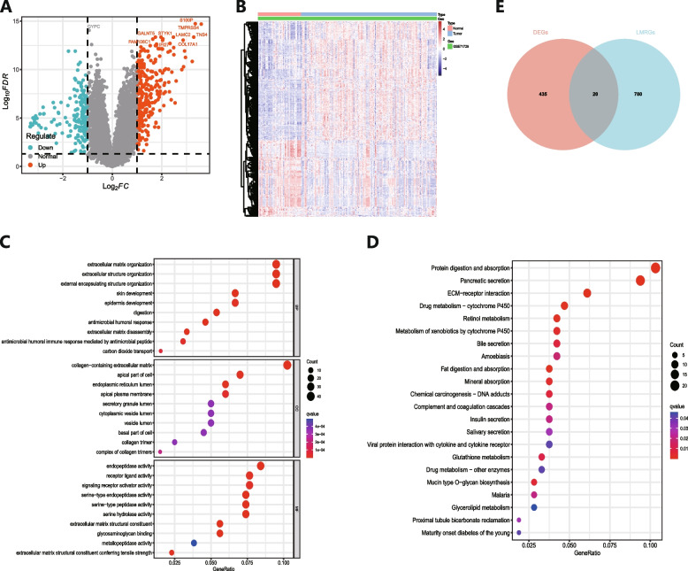
Background: Pancreatic adenocarcinoma (PAAD) is the most frequent kind of pancreatic cancer (PC). Recent studies suggest that lipid metabolism facilitates tumorigenesis, disease progression, and resistance to therapy by promoting lipid synthesis, accumulation, and breakdown. Thus, exploring the lipid metabolism network could unveil novel therapeutic avenues for early detection, precision medicine, and prognostication in PAAD. This project intends to develop new lipid metabolism-related biomarkers for PAAD diagnosis and investigate the link between important genes and immune cell infiltration (ICI).

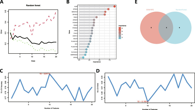
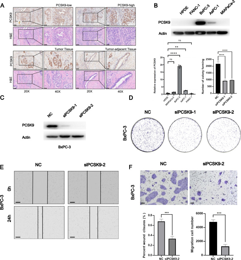
Methods: Tissue samples from 20 PAAD patients and 20 healthy controls were obtained. Analysis were focused on the datasets GSE71729 and GSE16515, which include samples of PAAD (n = 161) and those from healthy human tissue (n = 61), derived from the GEO database. Knockdown of PCSK9 on PC cells were conducted by si-RNA and sh-RNA. Migration and cell functional experiments were performed to assess the role of PCSK9 in cell multiplication. Furthermore, a xenograft mouse model was employed to confirm PCSK9's function in vivo.

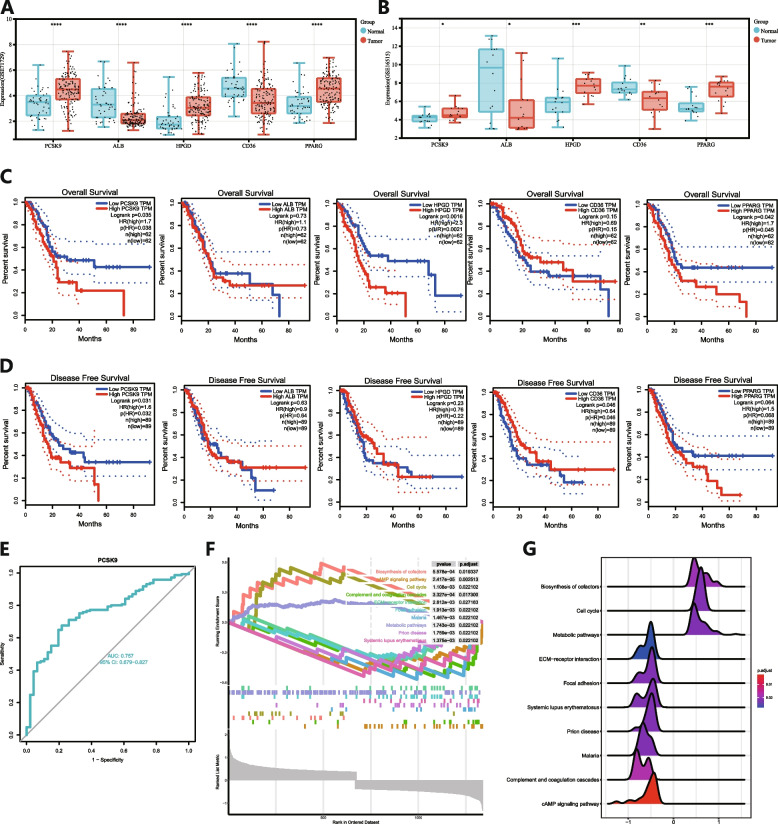
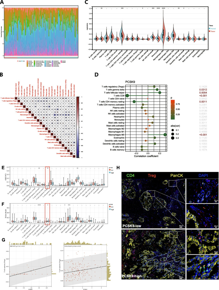
Results: The expression level of Proprotein convertase subtilisin/kexin type 9 (PCSK9) is significantly elevated in tissues affected by PAAD when compared to normal tissues. Survival analyses indicated that increased PCSK9 levels are inversely related to overall and disease-free survival (DFS). PCSK9's functional annotation associated it with the cell cycle and metabolism, especially energy metabolism. Examination of ICI data determined that PCSK9 expression demonstrated an unambiguous association with the M0 macrophages, T follicular helper cells (Tfh), gamma delta T cells and activated DC, and an inverse relationship with Monocytes, CD8+ T cells, memory B cells, resting CD4+ memory T cells, activated NK cells and resting DC abundance. PCSK9 expression knockdown has the ability to impede PC cells' migration and proliferation.

Conclusion: Our study identified PCSK9 as a critical gene in PAAD. Expression levels of PCSK9 varied between PAAD and normal samples. ROC analysis verified PCSK9's strong capacity to differentiate PC from normal samples. Importantly, PCSK9 expression was considerably elevated in PC cell lines and tissues. Furthermore, PCSK9 stimulates the migration and proliferation of tumor cells in vivo and vitro.

Keywords: Pancreatic cancer; Proprotein convertase subtilisin/kexin type 9; Tumor immunotherapy; Tumorigenesis and progression.


Conflict of interest statement

The authors declare no competing interests.

Figures

Fig. 1
Fig. 1
Discovery of 20 Potential Genes Associated with PC. A Volcano plot representing DEGs. B Heatmap depicting the differential expression patterns of DEGs between tumor and non-tumor tissues. C GO analysis outcomes for the DEGs. D KEGG pathway analysis results for the DEGs. E Overlap between the DEGs and LMRGs identified within the GEO cohort
Fig. 2
Fig. 2
Using machine learning to screen possible biomarkers for PC. A,B The random forest algorithm illustrates the inaccuracy in PC; the control group and genes are prioritized according to their importance score. C,D Plots of biological marker screening using SVM-RFE arithmetic. E The Venn diagram depicts the identification of five possible diagnostic genes using the two algorithms described above
Fig. 3
Fig. 3
Identification and validation of the major gene signature of PCSK9 in PAAD. A,B Expression differences in five genes between tumor and normal tissues in GEO datasets. ***P < 0.001, **P < 0.01, *P < 0.05. C,D OS and DFS analysis of five genes on PC using the GEPIA2 database. E The ROC curve for PCSK9. F,G Ridge plot and line graph from the GESA study of PCSK9
Fig. 4
Fig. 4
Comparison of ICI in PC and Normal Tissue. A A bar plot depicts the distribution of 22 types of immune cells across different samples. B A schematic illustrating the relationship between the 22 immune cell types. C A violin plot compares the proportions of 22 immune cell types in PC with normal tissue. D A graph depicts the relationship between PCSK9 expression and ICI in both PC and normal samples. E&F A violin plot compares the proportions of 22 immune cell types in PC with different expression of PCSK9 in GSE15471 and TCGA-PAAD databases. G Correlation between PCSK9 expression and Treg infiltration. H mIHC showed the Treg infiltration in different PCSK9 tissue samples
Fig. 5
Fig. 5
Elevated PCSK9 Expression in Human PAAD Cell Lines and Tissues, with Insights into PCSK9's Impact on PAAD Cell Growth and Movement. A IHC and H&E staining exhibits both weak and strong PCSK9 expression in normal and tumor tissues from three individuals. Scale bar: 20X-100 μm,40X-50 μm. B Comparative analysis of PCSK9 expression in HPDE normal pancreatic epithelial cells and various PC (PC) cell lines C Western blot analysis of PCSK9 expression in BxPC-3 cells. D A colony-formation assay demonstrates the effect of PCSK9 silencing (siPCSK9-1 and siPCSK9-2) on the proliferative capacity of BxPC-3 cells. E Wound-healing assays reveal the reduced migratory ability of BxPC-3 cells subjected to siPCSK9-2 treatment for 72 h. Scale bar: 200 μm. F Transwell assays quantify the decrease in migration and invasion capabilities of PC cells following 72-h siPCSK9 treatment. Scale bar: 100 μm
Fig. 6
Fig. 6
PCSK9's Role in Modulating PC Proliferation In Vivo. A Western blot analysis of PCSK9 expression in BxPC-3 cells. B Ten mice were divided into two groups, receiving treatments with shRNA-NC and shRNA-PCSK9, respectively. Post-euthanasia, the excised tumors were photographed. And a systematic record of each mouse's body weight was kept throughout the study period. D The tumor volumes extracted from each mouse were measured precisely. CE IHC staining showed the expression of PCSK9 and KI-67 in the tumor tissues from two groups. Scale bar: 250 μm

Similar articles

Cited by

References

    1. McGuigan A, Kelly P, Turkington RC, Jones C, Coleman HG, McCain RS. Pancreatic cancer: A review of clinical diagnosis, epidemiology, treatment and outcomes. World J Gastroenterol. 2018;24(43):4846–61. - PMC - PubMed
    1. Siegel RL, Miller KD, Wagle NS, Jemal A. Cancer statistics, 2023. CA Cancer J Clin. 2023;73(1):17–48. - PubMed
    1. Rahib L, Smith BD, Aizenberg R, Rosenzweig AB, Fleshman JM, Matrisian LM. Projecting cancer incidence and deaths to 2030: the unexpected burden of thyroid, liver, and pancreas cancers in the United States. Cancer Res. 2014;74(11):2913–21. - PubMed
    1. Yang J, Xu R, Wang C, Qiu J, Ren B, You L. Early screening and diagnosis strategies of pancreatic cancer: a comprehensive review. Cancer Commun (Lond). 2021;41(12):1257–74. - PMC - PubMed
    1. Vander Heiden MG, DeBerardinis RJ. Understanding the intersections between metabolism and cancer biology. Cell. 2017;168(4):657–69. - PMC - PubMed

MeSH terms