. 2024 Sep 28;57(1):70.
doi: 10.1186/s40659-024-00548-4.

Maternal stress during pregnancy alters circulating small extracellular vesicles and enhances their targeting to the placenta and fetus

Affiliations

Maternal stress during pregnancy alters circulating small extracellular vesicles and enhances their targeting to the placenta and fetus

Mario Sánchez-Rubio et al. Biol Res. .

Abstract

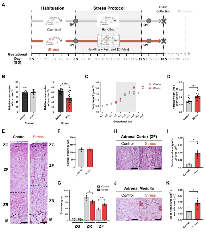
Background: Maternal psychological distress during pregnancy can negatively impact fetal development, resulting in long-lasting consequences for the offspring. These effects show a sex bias. The mechanisms whereby prenatal stress induces functional and/or structural changes in the placental-fetal unit remain poorly understood. Maternal circulating small extracellular vesicles (sEVs) are good candidates to act as "stress signals" in mother-to-fetus communication. Using a repetitive restraint-based rat model of prenatal stress, we examined circulating maternal sEVs under stress conditions and tested whether they could target placental-fetal tissues.

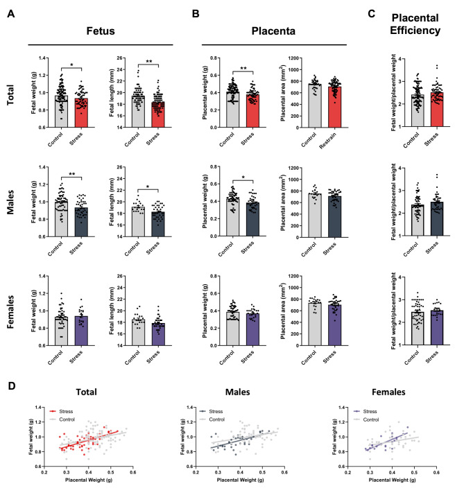
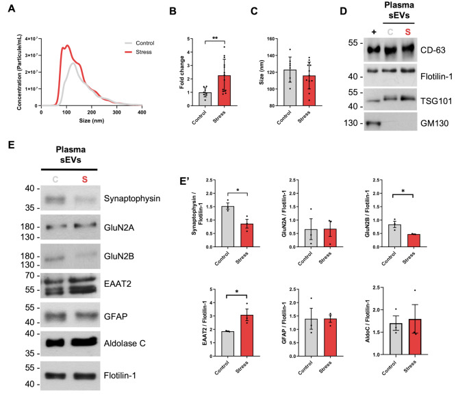
Results: Our mild chronic maternal stress during pregnancy paradigm induced anhedonic-like behavior in pregnant dams and led to intrauterine growth restriction (IUGR), particularly in male fetuses and placentas. The concentration and cargo of maternal circulating sEVs changed under stress conditions. Specifically, there was a significant reduction in neuron-enriched proteins and a significant increase in astrocyte-enriched proteins in blood-borne sEVs from stressed dams. To study the effect of repetitive restraint stress on the biodistribution of maternal circulating sEVs in the fetoplacental unit, sEVs from pregnant dams exposed to stress or control protocol were labeled with DiR fluorescent die and injected into pregnant females previously exposed to control or stress protocol. Remarkably, maternal circulating sEVs target placental/fetal tissues and, under stress conditions, fetal tissues are more receptive to sEVs.

Conclusion: Our results suggest that maternal circulating sEVs can act as novel mediators/modulators of mother-to-fetus stress communication. Further studies are needed to identify placental/fetal cellular targets of maternal sEVs and characterize their contribution to stress-induced sex-specific placental and fetal changes.

Keywords: Biodistribution; Exosomes; Fetus; Placenta; Prenatal stress; Restraint; Sex-bias.


Conflict of interest statement

The authors declare that they have no competing interests.

Figures

Fig. 1
Fig. 1
Repetitive restraint stress induces changes in maternal behavior and adrenal glands histology. (A) Schematic representation of the experimental restraint stress protocol. (B) Sucrose preference test (SPT) before (black bars) and after the restraint stress protocol (grey bar: control group; red bar: stress group). Relative sucrose versus water consumption is expressed as percentage (n = 12 control; n = 24 stress). (C) Maternal body weight gain expressed as percentage from gestational day (GD) 0.5. (D) Adrenal glands weight normalized by maternal body weight without uterus, placentas and fetuses/fetal membranes (n = 13 control, n = 9 stress). (E-K) Histological analysis of adrenal glands. (E) Hematoxylin-eosin staining of histological sections from adrenal glands. ZG: zona glomerulosa, ZF: zona fasciculata, ZR: zona reticularis, M: medulla. (F-G) Morphometric analysis of adrenal glands. Total cortical thickness (F) and thickness of different cortical layers (G) were measured in adrenal glands from control and stressed dams. (H-K) Representative images and quantification of the relative area of blood vessels in the adrenal cortex (zona fasciculata, ZF) (H-I) and in the adrenal medulla (J-K). Bars represent mean ± SEM (n = 4 control; n = 4 stress). * p < 0.05; ** p < 0.01; *** p < 0.001; **** p < 0.0001 (Student’s t-test). Scale bars: 100 μm
Fig. 2
Fig. 2
Repetitive restraint stress induces sex-biased effects in placental and fetal growth. (A) Fetal weight and length of total fetuses and separated by sex. (B) Placental weight and area of total placentas and separated by sex. (C) Placental efficiency. Bars represent mean ± SEM. In fetal/placental weight analyses: Control group: n = 114 fetuses/placentas (65 males and 49 females) from 8 different control dams; Stress group: n = 62 fetuses/placentas (42 males and 20 females) from 4 different stressed dams). In fetal length and placental area analyses: Control group: n = 38 fetuses/placentas (19 males and 19 females) from 4 different control dams; Stress group: 75 fetuses/placentas (38 males and 37 females) from 5 stressed dams). * p < 0.05; ** p < 0.01 (Statistical comparisons by mixed-effects modelling to control for litter effects; treatment (stress) was used as fixed effect and litter as random effect). (D) Correlation of fetal weight vs. placental weight in control (grey) and stress (red) groups. Total control group: y = 0.5783x + 0.73, r = 0.4045, r2 = 0.1636, p < 0.0001; Total Stress group: y = 0.8920x + 0.59; r = 0.6776, r2 = 0.4592, p < 0.0001; Males control group: y = 0.4874x + 0.7871, r = 0.3488, r2 = 0.1217, p = 0.0044; Male Stress group: y = 0.7761x + 0.6422, r = 0.6479, r2 = 0.4198, p = 0.0044; Female Control group: y = 0.4955x + 0.7388, r = 0.3430, r2 = 0.1177, p = 0.0158; Female Stress group: y = 1.258x + 0.4634, r = 0.7615, r2 = 0.58, p < 0.0001
Fig. 3
Fig. 3
Repetitive restraint stress induces changes in concentration and cargo of maternal circulating sEVs. (A) Size and concentration distribution profile of circulating blood plasma-borne sEVs from control and stressed pregnant dams. (B) Concentration (particles/ml) of blood plasma-borne sEVs in control and stressed pregnant dams. (C) Size (mode) of blood plasma-borne sEVs in control and stressed pregnant dams. Bars represent mean ± SEM (Control group: n = 9; Stress group: n = 13). (D) Representative images of Western blot analyses for characterization of blood plasma-borne sEVs from control (C) and stressed (S) pregnant dams. Positive (CD-63, flotillin-1, TSG-101) and negative (GM130) markers for sEVS were used for characterization. Rat brain protein homogenates were used as positive controls (+). (E-E’) Western blot analyses of maternal blood plasma-borne sEVs for brain neuronal-enriched (synaptophysin, GluN2A, Glun2B) and astrocyte-enriched (EAAT2, GFAP, Aldolase C) proteins. Representative images of Western blots (E) and densitometric quantification analyses (E’) are shown. Bars represent mean ± SEM (Control and Stress groups: n = 3 pools of plasma-borne sEVs; each pool is composed by plasma-derived sEVS from 4 different pregnant rats). * p < 0.05; ** p < 0.01 (Student’s t-test)
Fig. 4
Fig. 4
Repetitive restraint stress affect biodistribution of maternal circulating sEVS into placental and fetal tissues. (A) Schematic representation of the experimental design. Plasma-borne sEVs from 4 control rats or 4 stressed rats at GD17.5 were isolated, pooled, and stained with the lipophilic marker DiR. Labelled-sEVS from both stressed and control rats (Donors) were intravenously (tail vein) injected into pregnant stressed and pregnant control recipient rats at E17.5 and analyzed after 24 h (at 18.5). (B, D) Representative images of DiR fluorescent signal distribution in placentas (B) and fetuses (D) 24 h after labelled-sEVs injection. PBS-DiR was used as negative control. (C, E) Quantification of DiR fluorescent signals in placentas (C) and fetuses (E) from Group CC (control recipients that received control donor sEVs), Group SS (stressed recipients that received donor sEVs from stressed pregnant dams), Group CS (control recipients that received donor sEvs from stressed dams), and Group SC (stressed recipients that received control donor sEVs). Fluorescence intensity (arbitrary units, A.U.) was normalized by placental and fetal area, respectively. Data shown as scatter dot plots with mean ± SEM. n = 44 placentas/fetuses from 4 litters for Group CC; n = 36 placentas/fetuses from 3 litters for Group SS; n = 50 placentas/fetuses from 4 litters for Group CS; n = 62 placentas/fetuses from 4 litters for Group SC. * p < 0.05 (Statistical comparisons by mixed-effects modelling to control for litter effects; treatments were used as fixed effect and litter as random effect)

Similar articles

Cited by

References

    1. Barker DJ. The fetal and infant origins of adult disease. BMJ. 1990;301:1111. - PMC - PubMed
    1. Bock J, Rether K, Groger N, Xie L, Braun K. Perinatal programming of emotional brain circuits: an integrative view from systems to molecules. Front Neurosci. 2014;8:11. - PMC - PubMed
    1. Bock J, Wainstock T, Braun K, Segal M. Stress in Utero: prenatal programming of Brain plasticity and cognition. Biol Psychiatry. 2015;78:315–26. - PubMed
    1. Markham JA, Koenig JI. Prenatal stress: role in psychotic and depressive diseases. Psychopharmacology. 2011;214:89–106. - PMC - PubMed
    1. Weinstock M. The long-term behavioural consequences of prenatal stress. Neurosci Biobehav Rev. 2008;32:1073–86. - PubMed

LinkOut - more resources